A novel method for testing accuracy of bite registration using intraoral scanners
- Affiliations
-
- 1Department of Orthodontics, School of Dentistry, National and Kapodistrian University of Athens, Athens, Greece
- KMID: 2545041
- DOI: http://doi.org/10.4041/kjod22.199
Abstract
Objective
The evidence on the accuracy of bite registration using intraoral scanners is sparse. This study aimed to develop a new method for evaluating bite registration accuracy using intraoral scanners.
Methods
Two different types of models were used; 10 stone models and 10 with acrylic resin teeth. A triangular frame with cylindrical posts at each apex (one anterior and two posteriors) was digitally designed and manufactured using three-dimensional (3D) printing. Such a structure was fitted in the lingual space of each maxillary and mandibular model so that, in occlusion, the posts would contact their opposing counterparts, enforcing a small interocclusal gap between the two arches. This ensured no tooth interference and full contact between opposing posts. Bite registration accuracy was evaluated by measuring the distance between opposing posts, with small values indicating high-accuracy. Three intraoral scanners were used: Medit i500, Primescan, and Trios 4. Viewbox software was used to measure the distance between opposing posts and compute roll and pitch.
Results
The average maximum error in interocclusal registration exceeded 50 μm. Roll and pitch orientation errors ranged above 0.1 degrees, implying an additional interocclusal error of around 40 μm or more. The models with acrylic teeth exhibited higher errors.
Conclusions
A method that avoids the need for reference hardware and the imprecision of locating reference points on tooth surfaces, and offers simplicity in the assessment of bite registration with an intraoral scanner, was developed. These results suggest that intraoral scanners may exhibit clinically significant errors in reproducing the interocclusal relationships.
Keyword
Figure
-

Figure 1 Schematic and three-dimensional renderings of the triangular frames used for aligning and occluding the models; occlusal view and frames in ‘occlusion.’
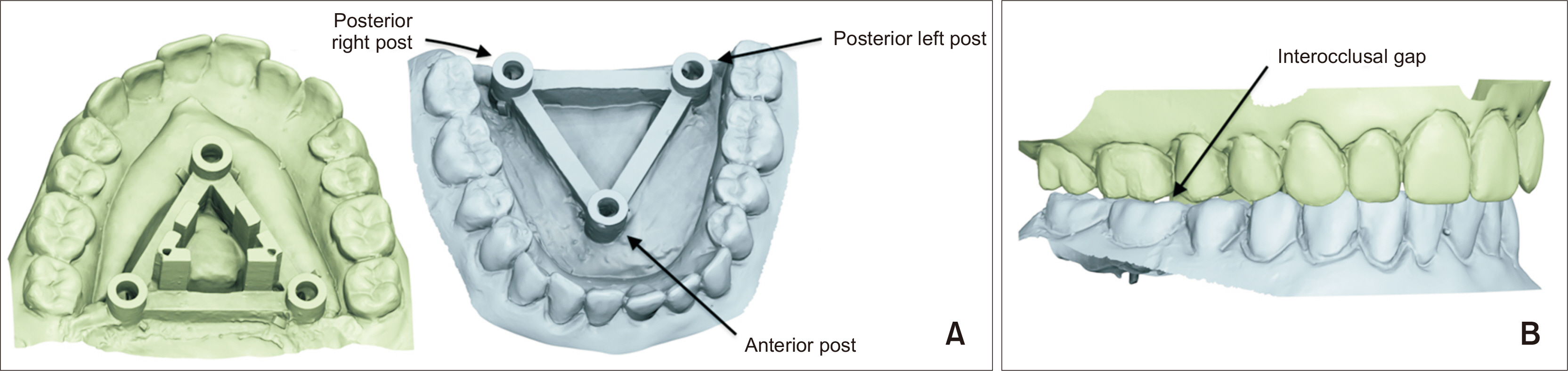
Figure 2 A, Digital models with frames in lingual and palatal space. B, Models in ‘occlusion’ showing interocclusal space.
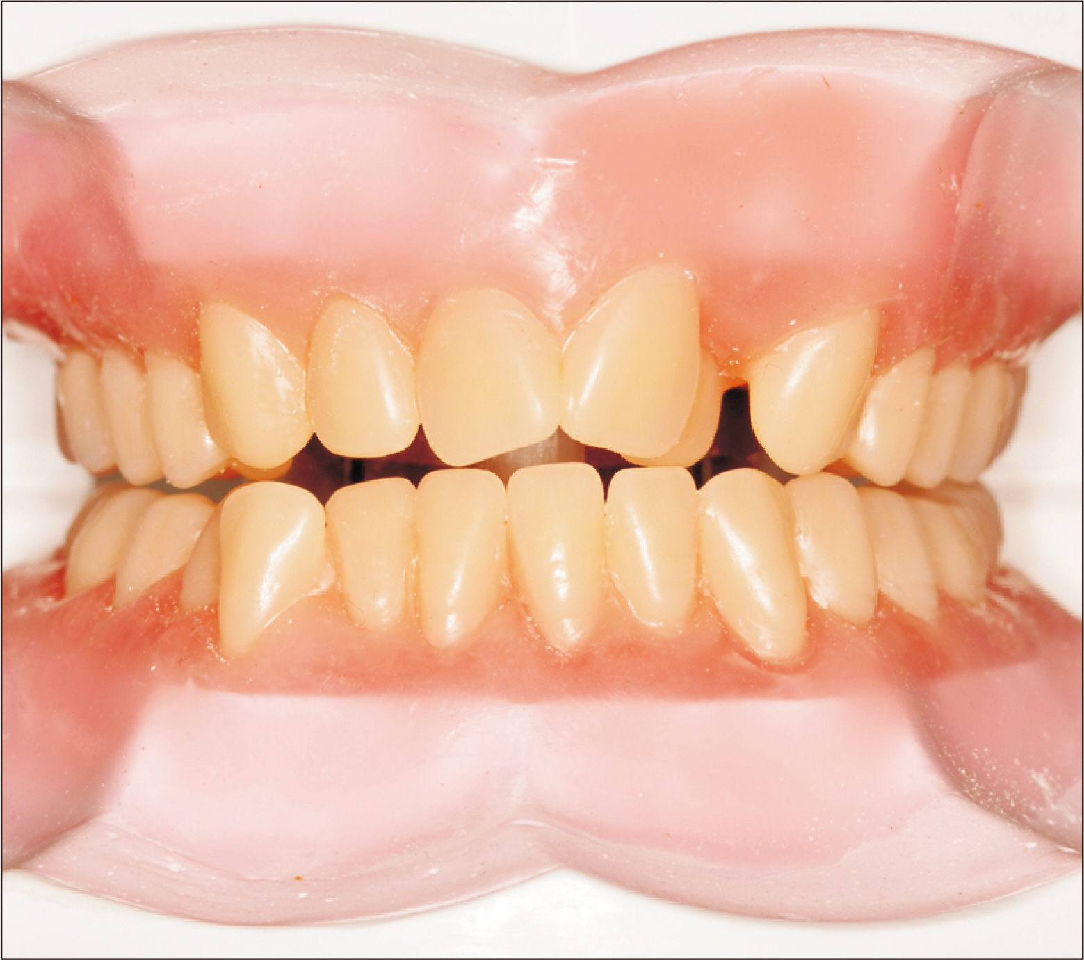
Figure 3 Acrylic teeth on pink wax dental bases.

Figure 4 Posterior view of digital models, as aligned by intraoral scanner software. Inset shows space between the upper and lower posts (here approximately 0.2 mm), even though the posts were in full contact during scanning.
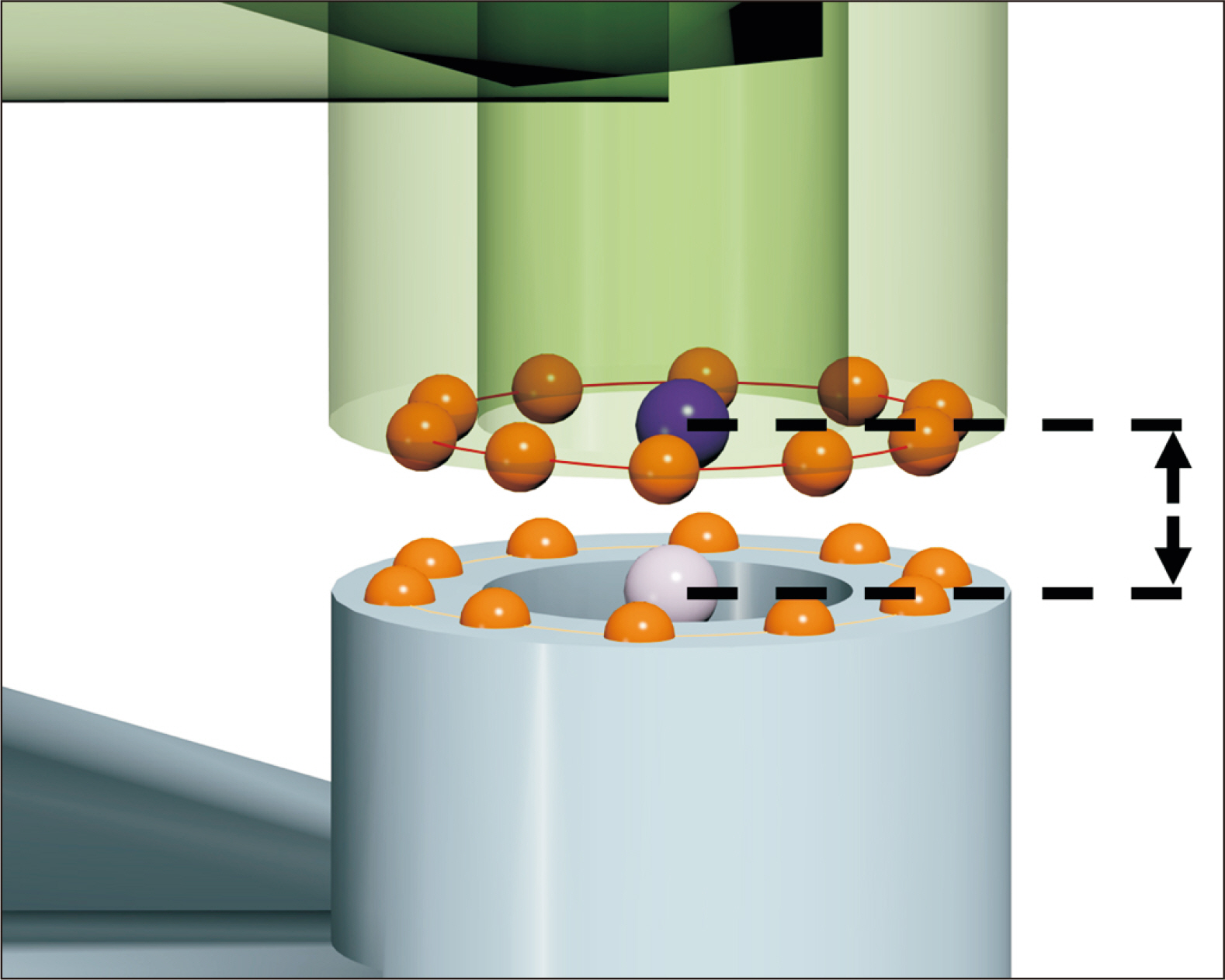
Figure 5 Measuring procedure. Ten points (orange) were digitized on the occluding surface of each post. The calculated average of these points (light and dark purple) was used to measure the inter-post distance, along a direction perpendicular to the plane of the 10 landmarks.
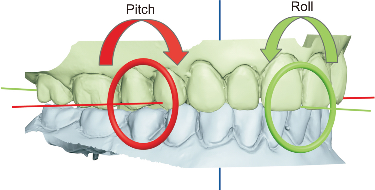
Figure 6 Roll and pitch. Arrows show positive direction.

Figure 7 Univariate plots of A, roll, B, pitch, C, AveErr, and D, MaxErr. Group A: stone models; Group B: acrylic teeth on wax bases. Scanning of the acrylic teeth on wax bases (Group B) was not possible with the Medit (MEDIT Co., Seoul, Korea) scanner. AveErr, the average of the three post measurements of each model; MaxErr, the maximum absolute value of the three measurements of each model.
Reference
-
1. Mangano F, Gandolfi A, Luongo G, Logozzo S. 2017; Intraoral scanners in dentistry: a review of the current literature. BMC Oral Health. 17:149. https://doi.org/10.1186/s12903-017-0442-x. DOI: 10.1186/s12903-017-0442-x. PMID: 29233132. PMCID: PMC5727697. PMID: 7b629d06d1744e679aa8123ce1b01b24.
Article2. Abduo J, Elseyoufi M. 2018; Accuracy of intraoral scanners: a systematic review of influencing factors. Eur J Prosthodont Restor Dent. 26:101–21. https://pubmed.ncbi.nlm.nih.gov/29989757/. DOI: 10.1922/EJPRD_01752Abduo21. PMID: 29989757.3. Kachhara S, Nallaswamy D, Ganapathy DM, Sivaswamy V, Rajaraman V. 2020; Assessment of intraoral scanning technology for multiple implant impressions- a systematic review and meta-analysis. J Indian Prosthodont Soc. 20:141–52. https://doi.org/10.4103/jips.jips_379_19. DOI: 10.4103/jips.jips_379_19. PMID: 32655218. PMCID: PMC7335030.
Article4. Goracci C, Franchi L, Vichi A, Ferrari M. 2016; Accuracy, reliability, and efficiency of intraoral scanners for full-arch impressions: a systematic review of the clinical evidence. Eur J Orthod. 38:422–8. https://doi.org/10.1093/ejo/cjv077. DOI: 10.1093/ejo/cjv077. PMID: 26487391.
Article5. Rossini G, Parrini S, Castroflorio T, Deregibus A, Debernardi CL. 2016; Diagnostic accuracy and measurement sensitivity of digital models for orthodontic purposes: a systematic review. Am J Orthod Dentofacial Orthop. 149:161–70. https://doi.org/10.1016/j.ajodo.2015.06.029. DOI: 10.1016/j.ajodo.2015.06.029. PMID: 26827972.
Article6. Ahlholm P, Sipilä K, Vallittu P, Jakonen M, Kotiranta U. 2018; Digital versus conventional impressions in fixed prosthodontics: a review. J Prosthodont. 27:35–41. https://doi.org/10.1111/jopr.12527. DOI: 10.1111/jopr.12527. PMID: 27483210.
Article7. International Organization for Standardization (ISO) 5725-1:1994. 1994. Accuracy (trueness and precision) of measurement methods and results - part 1: general principles and definitions [Internet]. ISO;Geneva: Available from: https://www.iso.org/standard/11833.html. cited 2022 Mar 20.8. Nedelcu R, Olsson P, Nyström I, Rydén J, Thor A. 2018; Accuracy and precision of 3 intraoral scanners and accuracy of conventional impressions: a novel in vivo analysis method. J Dent. 69:110–8. https://doi.org/10.1016/j.jdent.2017.12.006. DOI: 10.1016/j.jdent.2017.12.006. PMID: 29246490.9. Winkler J, Gkantidis N. 2020; Trueness and precision of intraoral scanners in the maxillary dental arch: an in vivo analysis. Sci Rep. 10:1172. https://doi.org/10.1038/s41598-020-58075-7. DOI: 10.1038/s41598-020-58075-7. PMID: 31980724. PMCID: PMC6981254.10. Ender A, Attin T, Mehl A. 2016; In vivo precision of conventional and digital methods of obtaining complete-arch dental impressions. J Prosthet Dent. 115:313–20. https://doi.org/10.1016/j.prosdent.2015.09.011. DOI: 10.1016/j.prosdent.2015.09.011. PMID: 26548890.11. Kuhr F, Schmidt A, Rehmann P, Wöstmann B. 2016; A new method for assessing the accuracy of full arch impressions in patients. J Dent. 55:68–74. https://doi.org/10.1016/j.jdent.2016.10.002. DOI: 10.1016/j.jdent.2016.10.002. PMID: 27717754.12. Edher F, Hannam AG, Tobias DL, Wyatt CCL. 2018; The accuracy of virtual interocclusal registration during intraoral scanning. J Prosthet Dent. 120:904–12. https://doi.org/10.1016/j.prosdent.2018.01.024. DOI: 10.1016/j.prosdent.2018.01.024. PMID: 29961618.13. Ayuso-Montero R, Mariano-Hernandez Y, Khoury-Ribas L, Rovira-Lastra B, Willaert E, Martinez-Gomis J. 2020; Reliability and validity of T-scan and 3D intraoral scanning for measuring the occlusal contact area. J Prosthodont. 29:19–25. https://doi.org/10.1111/jopr.13096. DOI: 10.1111/jopr.13096. PMID: 31270888.14. Gintaute A, Keeling AJ, Osnes CA, Zitzmann NU, Ferrari M, Joda T. 2020; Precision of maxillo-mandibular registration with intraoral scanners in vitro. J Prosthodont Res. 64:114–9. https://doi.org/10.1016/j.jpor.2019.05.006. DOI: 10.1016/j.jpor.2019.05.006. PMID: 31387847.15. Park JM, Jeon J, Heo SJ. 2018; Accuracy comparison of buccal bite scans by five intra-oral scanners. J Dent Rehabil Appl Sci. 34:17–31. https://doi.org/10.14368/jdras.2018.34.1.17. DOI: 10.14368/jdras.2018.34.1.17.16. Wong KY, Esguerra RJ, Chia VAP, Tan YH, Tan KBC. 2018; Three-dimensional accuracy of digital static interocclusal registration by three intraoral scanner systems. J Prosthodont. 27:120–8. https://doi.org/10.1111/jopr.12714. DOI: 10.1111/jopr.12714. PMID: 29160904.17. Botsford KP, Frazier MC, Ghoneima AAM, Utreja A, Bhamidipalli SS, Stewart KT. 2019; Precision of the virtual occlusal record. Angle Orthod. 89:751–7. https://doi.org/10.2319/092018-684.1. DOI: 10.2319/092018-684.1. PMID: 30920871. PMCID: PMC8111830.18. Abdulateef S, Edher F, Hannam AG, Tobias DL, Wyatt CCL. 2020; Clinical accuracy and reproducibility of virtual interocclusal records. J Prosthet Dent. 124:667–73. https://doi.org/10.1016/j.prosdent.2019.11.014. DOI: 10.1016/j.prosdent.2019.11.014. PMID: 32014284.19. Li H, Lyu P, Wang Y, Sun Y. 2017; Influence of object translucency on the scanning accuracy of a powder-free intraoral scanner: a laboratory study. J Prosthet Dent. 117:93–101. https://doi.org/10.1016/j.prosdent.2016.04.008. DOI: 10.1016/j.prosdent.2016.04.008. PMID: 27460324.20. Song J, Kim M. 2020; Accuracy on scanned images of full arch models with orthodontic brackets by various intraoral scanners in the presence of artificial saliva. Biomed Res Int. 2020:2920804. https://doi.org/10.1155/2020/2920804. DOI: 10.1155/2020/2920804. PMID: 32185200. PMCID: PMC7063212.21. Ryan EA, Tam LE, McComb D. 2010; Comparative translucency of esthetic composite resin restorative materials. J Can Dent Assoc. 76:a84. https://pubmed.ncbi.nlm.nih.gov/20719098/. PMID: 20719098.22. Nogueira AD, Della Bona A. 2013; The effect of a coupling medium on color and translucency of CAD-CAM ceramics. J Dent. 41 Suppl 3:e18–23. https://doi.org/10.1016/j.jdent.2013.02.005. DOI: 10.1016/j.jdent.2013.02.005. PMID: 23438417.23. Kurz M, Attin T, Mehl A. 2015; Influence of material surface on the scanning error of a powder-free 3D measuring system. Clin Oral Investig. 19:2035–43. https://doi.org/10.1007/s00784-015-1440-5. DOI: 10.1007/s00784-015-1440-5. PMID: 25743568.24. Im J, Cha JY, Lee KJ, Yu HS, Hwang CJ. 2014; Comparison of virtual and manual tooth setups with digital and plaster models in extraction cases. Am J Orthod Dentofacial Orthop. 145:434–42. https://doi.org/10.1016/j.ajodo.2013.12.014. DOI: 10.1016/j.ajodo.2013.12.014. PMID: 24703281.25. Hildebrand JC, Palomo JM, Palomo L, Sivik M, Hans M. 2008; Evaluation of a software program for applying the American Board of Orthodontics objective grading system to digital casts. Am J Orthod Dentofacial Orthop. 133:283–9. https://doi.org/10.1016/j.ajodo.2006.03.035. DOI: 10.1016/j.ajodo.2006.03.035. PMID: 18249296.26. Costalos PA, Sarraf K, Cangialosi TJ, Efstratiadis S. 2005; Evaluation of the accuracy of digital model analysis for the American Board of Orthodontics objective grading system for dental casts. Am J Orthod Dentofacial Orthop. 128:624–9. https://doi.org/10.1016/j.ajodo.2004.08.017. DOI: 10.1016/j.ajodo.2004.08.017. PMID: 16286210.27. Okunami TR, Kusnoto B, BeGole E, Evans CA, Sadowsky C, Fadavi S. 2007; Assessing the American Board of Orthodontics objective grading system: digital vs plaster dental casts. Am J Orthod Dentofacial Orthop. 131:51–6. https://doi.org/10.1016/j.ajodo.2005.04.042. DOI: 10.1016/j.ajodo.2005.04.042. PMID: 17208106.28. Besl PJ, McKay ND. 1992; A method for registration of 3-D shapes. IEEE Trans Pattern Anal Mach Intell. 14:239–56. https://doi.org/10.1109/34.121791. DOI: 10.1109/34.121791.29. Rajbhoj AA, Parchake P, Begnoni G, Willems G, Cadenas de Llano-Pérula M. 2021; Dental changes in humans with untreated normal occlusion throughout lifetime: a systematic scoping review. Am J Orthod Dentofacial Orthop. 160:340–62.e3. https://doi.org/10.1016/j.ajodo.2021.02.014. DOI: 10.1016/j.ajodo.2021.02.014. PMID: 34456004.30. Iwaki Y, Wakabayashi N, Igarashi Y. 2013; Dimensional accuracy of optical bite registration in single and multiple unit restorations. Oper Dent. 38:309–15. https://doi.org/10.2341/12-233-L. DOI: 10.2341/12-233-L. PMID: 23092147.31. Resnik RR, Misch CE. 2018. Misch's avoiding complications in oral implantology. Mosby;St. Louis: DOI: 10.1016/B978-0-323-37580-1.00017-2.32. Kim Y, Oh TJ, Misch CE, Wang HL. 2005; Occlusal considerations in implant therapy: clinical guidelines with biomechanical rationale. Clin Oral Implants Res. 16:26–35. https://doi.org/10.1111/j.1600-0501.2004.01067.x. DOI: 10.1111/j.1600-0501.2004.01067.x. PMID: 15642028.33. Owens S, Buschang PH, Throckmorton GS, Palmer L, English J. 2002; Masticatory performance and areas of occlusal contact and near contact in subjects with normal occlusion and malocclusion. Am J Orthod Dentofacial Orthop. 121:602–9. https://doi.org/10.1067/mod.2002.122829. DOI: 10.1067/mod.2002.122829. PMID: 12080313.34. Sakaguchi RL, Anderson GC, DeLong R. 1994; Digital imaging of occlusal contacts in the intercuspal position. J Prosthodont. 3:193–7. https://doi.org/10.1111/j.1532-849x.1994.tb00154.x. DOI: 10.1111/j.1532-849X.1994.tb00154.x. PMID: 7866500.
- Full Text Links
-
- Actions
-
Cited
- CITED
-
- Close
- Share
-
- Similar articles
-
- Accuracy comparison of buccal bite scans by five intra-oral scanners
- A comparison of the accuracy of intraoral scanners using an intraoral environment simulator
- Full-arch accuracy of five intraoral scanners:In vivo analysis of trueness and precision
- Effect of the volumetric dimensions of a complete arch on the accuracy of scanners
- Accuracy of 14 intraoral scanners for the All-on-4 treatment concept: a comparative in vitro study
