Korean J Radiol.
2019 Mar;20(3):513-521. 10.3348/kjr.2018.0409.
Solitary Nodular Invasive Mucinous Adenocarcinoma of the Lung: Imaging Diagnosis Using the Morphologic-Metabolic Dissociation Sign
- Affiliations
-
- 1Department of Radiology and Center for Imaging Science, Samsung Medical Center, Sungkyunkwan University School of Medicine, Seoul, Korea. kyungs.lee@samsung.com
- 2Department of Radiology, Chung-Ang University Hospital, Chung-Ang University College of Medicine, Seoul, Korea.
- 3Department of Nuclear Medicine, Samsung Medical Center, Sungkyunkwan University School of Medicine, Seoul, Korea.
- KMID: 2438282
- DOI: http://doi.org/10.3348/kjr.2018.0409
Abstract
OBJECTIVE
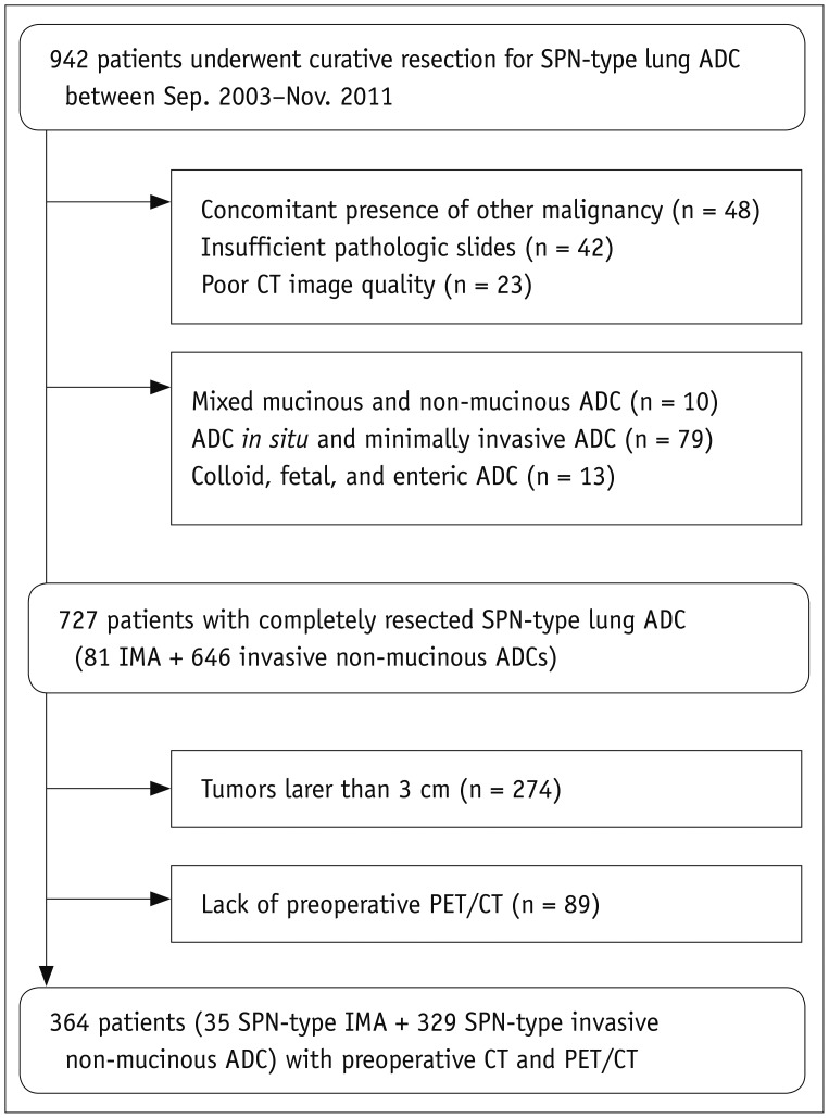
To evaluate the efficacy of the morphologic-metabolic (M-M) dissociation sign based on computed tomography (CT) and fluorine-18-fluorodeoxyglucose positron emission tomography (PET)/CT in discriminating invasive mucinous adenocarcinoma (IMA) from invasive non-mucinous adenocarcinomas (ADCs) of the lung.
MATERIALS AND METHODS
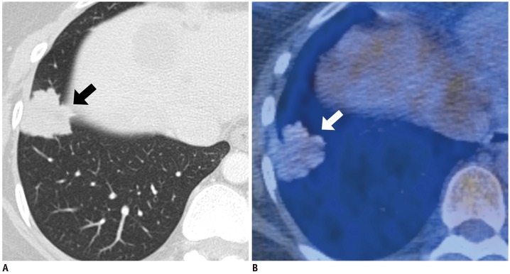
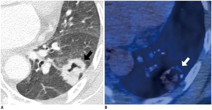
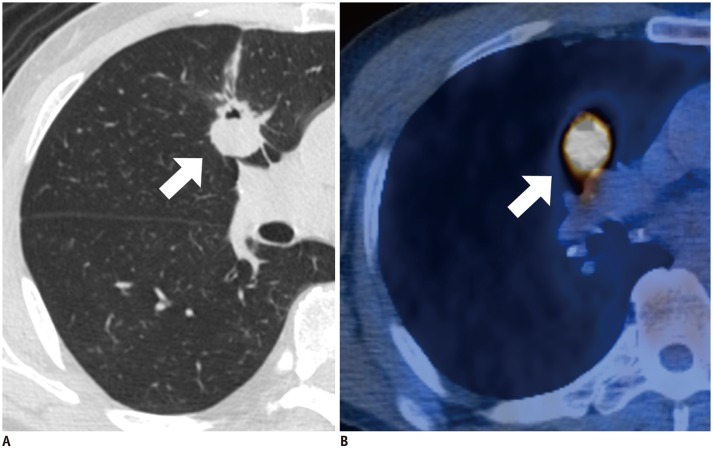
The Institutional Review Board approved this retrospective study. Among surgically resected solitary pulmonary nodule (SPN)-type ADCs (< 3 cm in diameter), 35 patients with IMAs and 329 with invasive non-mucinous ADCs were included. Morphologic malignancy was established if the tumor with lobulated or spiculated margin on CT presented a tumor shadow disappearance rate of < 0.5. The M-M dissociation sign was determined when a malignant-morphologic nodule on CT showed maximum standardized uptake value (SUVmax) < 3.5 on PET/CT.
RESULTS
Among 35 IMAs (size: 21 ± 7 mm, SUVmax: 1.8 ± 2.0) and 329 invasive non-mucinous ADCs (size: 21 ± 6 mm, SUVmax: 4.6 ± 4.2), the M-M dissociation sign was observed in 54% of IMAs (19/35) and 10% of invasive non-mucinous ADCs (34/329) (p < 0.001). The diagnostic performance of the sign in discriminating IMA from invasive non-mucinous ADCs showed a sensitivity of 54.3% (95% confidence interval [CI], 36.7-71.2), specificity 89.7% (95% CI, 85.9-92.7), positive predictive value 35.8% (95% CI, 26.5-46.5), and negative predictive value 94.9% (95% CI, 92.8-96.4). Multivariate analyses revealed metabolic benignity (odds ratio [OR] 2.99; 95% CI, 1.01-8.93; p = 0.047) and M-M dissociation sign (OR 6.35; 95% CI, 2.76-14.62; p < 0.001) to be significant predictors of SPN-type IMAs.
CONCLUSION
Identification of the absence of M-M dissociation sign is an accurate indicator for excluding IMA from SPN-type lung ADCs.
Keyword
MeSH Terms
-
Adenocarcinoma
Adenocarcinoma, Mucinous*
Diagnosis*
Ethics Committees, Research
Fluorodeoxyglucose F18
Humans
Lung*
Mucins*
Multivariate Analysis
Positron-Emission Tomography
Positron-Emission Tomography and Computed Tomography
Retrospective Studies
Sensitivity and Specificity
Solitary Pulmonary Nodule
Fluorodeoxyglucose F18
Mucins
Figure
Reference
-
1. Travis WD, Brambilla E, Noguchi M, Nicholson AG, Geisinger KR, Yatabe Y, et al. International Association for the Study of Lung Cancer/American Thoracic Society/European Respiratory Society international multidisciplinary classification of lung adenocarcinoma. J Thorac Oncol. 2011; 6:244–285. PMID: 21252716.2. Travis WD, Brambilla E, Burke AP, Marx A, Nicholson AG. Introduction to The 2015 World Health Organization Classification of tumors of the lung, pleura, thymus, and heart. J Thorac Oncol. 2015; 10:1240–1242. PMID: 26291007.
Article3. Travis WD, Brambilla E, Burke AP, Marx A, Nicholson AG. WHO classification of tumours of the lung, pleura, thymus and heart. 4th ed. Lyon: IARC Publications;2015.4. Shim HS, Kenudson M, Zheng Z, Liebers M, Cha YJ, Hoang Ho Q, et al. Unique genetic and survival characteristics of invasive mucinous adenocarcinoma of the lung. J Thorac Oncol. 2015; 10:1156–1162. PMID: 26200269.
Article5. Finberg KE, Sequist LV, Joshi VA, Muzikansky A, Miller JM, Han M, et al. Mucinous differentiation correlates with absence of EGFR mutation and presence of KRAS mutation in lung adenocarcinomas with bronchioloalveolar features. J Mol Diagn. 2007; 9:320–326. PMID: 17591931.
Article6. Kadota K, Yeh YC, D'Angelo SP, Moreira AL, Kuk D, Sima CS, et al. Associations between mutations and histologic patterns of mucin in lung adenocarcinoma: invasive mucinous pattern and extracellular mucin are associated with KRAS mutation. Am J Surg Pathol. 2014; 38:1118–1127. PMID: 25029118.7. Duruisseaux M, Antoine M, Rabbe N, Poulot V, Fleury-Feith J, Vieira T, et al. The impact of intracytoplasmic mucin in lung adenocarcinoma with pneumonic radiological presentation. Lung Cancer. 2014; 83:334–340. PMID: 24461303.
Article8. Lee HY, Cha MJ, Lee KS, Lee HY, Kwon OJ, Choi JY, et al. Prognosis in resected invasive mucinous adenocarcinomas of the lung: related factors and comparison with resected nonmucinous adenocarcinomas. J Thorac Oncol. 2016; 11:1064–1073. PMID: 27016260.
Article9. Yoshizawa A, Sumiyoshi S, Sonobe M, Kobayashi M, Fujimoto M, Kawakami F, et al. Validation of the IASLC/ATS/ERS lung adenocarcinoma classification for prognosis and association with EGFR and KRAS gene mutations: analysis of 440 Japanese patients. J Thorac Oncol. 2013; 8:52–61. PMID: 23242438.
Article10. Nakamura H, Saji H, Shinmyo T, Tagaya R, Kurimoto N, Koizumi H, et al. Close association of IASLC/ATS/ERS lung adenocarcinoma subtypes with glucose-uptake in positron emission tomography. Lung Cancer. 2015; 87:28–33. PMID: 25481488.
Article11. Casali C, Rossi G, Marchioni A, Sartori G, Maselli F, Longo L, et al. A single institution-based retrospective study of surgically treated bronchioloalveolar adenocarcinoma of the lung: clinicopathologic analysis, molecular features, and possible pitfalls in routine practice. J Thorac Oncol. 2010; 5:830–836. PMID: 20521350.
Article12. Watanabe H, Saito H, Yokose T, Sakuma Y, Murakami S, Kondo T, et al. Relation between thin-section computed tomography and clinical findings of mucinous adenocarcinoma. Ann Thorac Surg. 2015; 99:975–981. PMID: 25624054.
Article13. Lee HY, Lee KS, Han J, Kim BT, Cho YS, Shim YM, et al. Mucinous versus nonmucinous solitary pulmonary nodular bronchioloalveolar carcinoma: CT and FDG PET findings and pathologic comparisons. Lung Cancer. 2009; 65:170–175. PMID: 19111932.
Article14. Edge SB, Compton CC. The American Joint Committee on Cancer: the 7th edition of the AJCC cancer staging manual and the future of TNM. Ann Surg Oncol. 2010; 17:1471–1474. PMID: 20180029.
Article15. Erasmus JJ, Connolly JE, McAdams HP, Roggli VL. Solitary pulmonary nodules: part I. Morphologic evaluation for differentiation of benign and malignant lesions. Radiographics. 2000; 20:43–58. PMID: 10682770.
Article16. Lee HY, Han J, Lee KS, Koo JH, Jeong SY, Kim BT, et al. Lung adenocarcinoma as a solitary pulmonary nodule: prognostic determinants of CT, PET, and histopathologic findings. Lung Cancer. 2009; 66:379–385. PMID: 19299033.
Article17. Okada M, Tauchi S, Iwanaga K, Mimura T, Kitamura Y, Watanabe H, et al. Associations among bronchioloalveolar carcinoma components, positron emission tomographic and computed tomographic findings, and malignant behavior in small lung adenocarcinomas. J Thorac Cardiovasc Surg. 2007; 133:1448–1454. PMID: 17532938.
Article18. Aoki T, Tomoda Y, Watanabe H, Nakata H, Kasai T, Hashimoto H, et al. Peripheral lung adenocarcinoma: correlation of thin-section CT findings with histologic prognostic factors and survival. Radiology. 2001; 220:803–809. PMID: 11526285.
Article19. Kim EA, Johkoh T, Lee KS, Han J, Fujimoto K, Sadohara J, et al. Quantification of ground-glass opacity on high-resolution CT of small peripheral adenocarcinoma of the lung: pathologic and prognostic implications. AJR Am J Roentgenol. 2001; 177:1417–1422. PMID: 11717098.20. Shim SS, Lee KS, Kim BT, Chung MJ, Lee EJ, Han J, et al. Non-small cell lung cancer: prospective comparison of integrated FDG PET/CT and CT alone for preoperative staging. Radiology. 2005; 236:1011–1019. PMID: 16014441.
Article21. Hata A, Katakami N, Fujita S, Kaji R, Imai Y, Takahashi Y, et al. Frequency of EGFR and KRAS mutations in Japanese patients with lung adenocarcinoma with features of the mucinous subtype of bronchioloalveolar carcinoma. J Thorac Oncol. 2010; 5:1197–1200. PMID: 20661086.
Article22. O'Neill AC, Jagannathan JP, Ramaiya NH. Evolving cancer classification in the era of personalized medicine: a primer for radiologists. Korean J Radiol. 2017; 18:6–17. PMID: 28096714.23. Krajewski KM, Braschi-Amirfarzan M, DiPiro PJ, Jagannathan JP, Shinagare AB. Molecular targeted therapy in modern oncology: imaging assessment of treatment response and toxicities. Korean J Radiol. 2017; 18:28–41. PMID: 28096716.
Article24. Manning JT Jr, Spjut HJ, Tschen JA. Bronchioloalveolar carcinoma: the significance of two histopathologic types. Cancer. 1984; 54:525–534. PMID: 6329505.
Article25. Chang JM, Lee HJ, Goo JM, Lee HY, Lee JJ, Chung JK, et al. False positive and false negative FDG-PET scans in various thoracic diseases. Korean J Radiol. 2006; 7:57–69. PMID: 16549957.
Article26. Berger KL, Nicholson SA, Dehdashti F, Siegel BA. FDG PET evaluation of mucinous neoplasms: correlation of FDG uptake with histopathologic features. AJR Am J Roentgenol. 2000; 174:1005–1008. PMID: 10749239.27. Cha MJ, Lee KS, Kim HS, Lee SW, Jeong CJ, Kim EY, et al. Improvement in imaging diagnosis technique and modalities for solitary pulmonary nodules: from ground-glass opacity nodules to part-solid and solid nodules. Expert Rev Respir Med. 2016; 10:261–278. PMID: 26751340.
Article28. Jeong YJ, Lee KS, Jeong SY, Chung MJ, Shim SS, Kim H, et al. Solitary pulmonary nodule: characterization with combined wash-in and washout features at dynamic multi-detector row CT. Radiology. 2005; 237:675–683. PMID: 16244276.
Article29. Jeong SY, Lee KS, Shin KM, Bae YA, Kim BT, Choe BK, et al. Efficacy of PET/CT in the characterization of solid or partly solid solitary pulmonary nodules. Lung Cancer. 2008; 61:186–194. PMID: 18280613.
Article30. Lee KS, Yi CA, Jeong SY, Jeong YJ, Kim S, Chung MJ, et al. Solid or partly solid solitary pulmonary nodules: their characterization using contrast wash-in and morphologic features at helical CT. Chest. 2007; 131:1516–1525. PMID: 17494800.31. Ohde Y, Nagai K, Yoshida J, Nishimura M, Takahashi K, Suzuki K, et al. The proportion of consolidation to ground-glass opacity on high resolution CT is a good predictor for distinguishing the population of non-invasive peripheral adenocarcinoma. Lung Cancer. 2003; 42:303–310. PMID: 14644518.
Article32. Lee HY, Lee SW, Lee KS, Jeong JY, Choi JY, Kwon OJ, et al. Role of CT and PET imaging in predicting tumor recurrence and survival in patients with lung adenocarcinoma: a comparison with the International Association for the Study of Lung Cancer/American Thoracic Society/European Respiratory Society classification of lung adenocarcinoma. J Thorac Oncol. 2015; 10:1785–1794. PMID: 26473646.
- Full Text Links
- Actions
-
Cited
- CITED
-
- Close
- Share
- Similar articles
-
- Ovarian and Pituitary Metastasis from Adenocarcinoma of the Lung: A Case Report
- Pulmonary Invasive Mucinous Adenocarcinoma in an Asymptomatic 23-Year-Old Male: Presentation with a Large Cavitary Mass
- Metastatic adenocarcinoma of the pancreas presents as metastases to the axillary/supraclavicular region as the first sign of the disease
- A case of mucinous borderline tumour showing persistently elevated tumor markers progressed into invasive mucinous cystadenocarcinoma
- Utility of Magnetic Resonance Imaging in the Diagnosis of Lung Adenocarcinoma with Extensive Necrosis: a Case Report


