Yonsei Med J.
2019 Feb;60(2):158-162. 10.3349/ymj.2019.60.2.158.
Image Analysis of HER2 Immunohistochemical Staining of Surgical Breast Cancer Specimens
- Affiliations
-
- 1Department of Hospital Pathology, Uijeongbu St. Mary's Hospital, College of Medicine, The Catholic University of Korea, Uijeongbu, Korea.
- 2Department of Hospital Pathology, Seoul St. Mary's Hospital, College of Medicine, The Catholic University of Korea, Seoul, Korea. klee@catholic.ac.kr
- 3Cancer Research Institute, The Catholic University of Korea, Seoul, Korea.
- KMID: 2431632
- DOI: http://doi.org/10.3349/ymj.2019.60.2.158
Abstract
- PURPOSE
Trastuzumab is an effective treatment for human epidermal growth factor receptor 2 (HER2)-amplified breast cancers. We sought to develop a simple protocol for HER2 image analysis of breast cancer specimens.
MATERIALS AND METHODS
In a preliminary test, we found that at least 1000 tumor cells need to be examined in the most strongly stained areas. Next, we evaluated the clinical usefulness of this established protocol of image analysis in 555 breast cancer patients. Results of the HER2 immunohistochemical (IHC) staining were compared between manual scoring and image analysis.
RESULTS
The HER2 IHC results obtained by the image analysis method correlated well with those obtained by the manual scoring method (Cohen's kappa=0.830). Using the HER2 silver in situ hybridization (SISH) results as a gold standard, sensitivity values were 72.1% for manual scoring and 74.0% for image analysis; specificity values were 96.2% for manual scoring and 94.7% for image analysis; and accuracy values were 91.7% for manual scoring and 90.8% for image analysis. McNemar's test was applied to the results, and there were no statistically significant differences in sensitivity and specificity between the positive (p=0.688) and negative (p=0.118) SISH groups.
CONCLUSION
HER2 image analysis results were similar to those obtained via the manual scoring method, indicating that the use of image analysis can reduce assessment time and effort. We suggest that image analysis-based evaluation of 1000 tumor cells in the most strongly IHC-stained area, regardless of stroma content, is sufficient for determining HER2 expression levels in breast cancer specimens.
Keyword
MeSH Terms
Figure
-
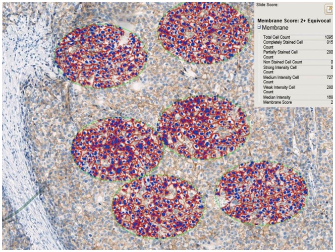
Fig. 1 Drawing of several ellipses, each with a constant area of 40457.64 µm2, until 500, 1000, or 2000 cells were included in the focal view, was used to determine the optimal interpretation numbers (×200).
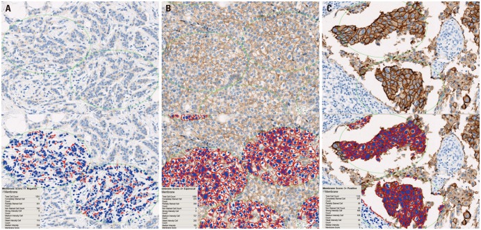
Fig. 2 Examples of image analysis scoring results. (A) HER2 IHC score 1, (B) HER2 IHC score 2, and (C) HER2 IHC score 3; ×200. HER2, human epidermal growth factor receptor 2; IHC, immunohistochemistry.
Reference
-
1. Wolff AC, Hammond ME, Hicks DG, Dowsett M, McShane LM, Allison KH, et al. Recommendations for human epidermal growth factor receptor 2 testing in breast cancer: American Society of Clinical Oncology/College of American Pathologists clinical practice guideline update. J Clin Oncol. 2013; 31:3997–4013. PMID: 24101045.
Article2. Kim S, Park HS, Kim JY, Ryu J, Park S, Kim SI. Comparisons of oncologic outcomes between triple-negative breast cancer (TNBC) and non-TNBC among patients treated with breast-conserving therapy. Yonsei Med J. 2016; 57:1192–1198. PMID: 27401651.
Article3. Goldenberg MM. Trastuzumab, a recombinant DNA-derived humanized monoclonal antibody, a novel agent for the treatment of metastatic breast cancer. Clin Ther. 1999; 21:309–318. PMID: 10211534.
Article4. Hudis CA. Trastuzumab--mechanism of action and use in clinical practice. N Engl J Med. 2007; 357:39–51. PMID: 17611206.5. Ross JS, Fletcher JA, Bloom KJ, Linette GP, Stec J, Symmans WF, et al. Targeted therapy in breast cancer: the HER-2/neu gene and protein. Mol Cell Proteomics. 2004; 3:379–398. PMID: 14762215.6. Dobson L, Conway C, Hanley A, Johnson A, Costello S, O'Grady A, et al. Image analysis as an adjunct to manual HER-2 immunohistochemical review: a diagnostic tool to standardize interpretation. Histopathology. 2010; 57:27–38. PMID: 20584089.
Article7. Helin HO, Tuominen VJ, Ylinen O, Helin HJ, Isola J. Free digital image analysis software helps to resolve equivocal scores in HER2 immunohistochemistry. Virchows Arch. 2016; 468:191–198. PMID: 26493985.
Article8. Minot DM, Voss J, Rademacher S, Lwin T, Orsulak J, Caron B, et al. Image analysis of HER2 immunohistochemical staining. Reproducibility and concordance with fluorescence in situ hybridization of a laboratory-validated scoring technique. Am J Clin Pathol. 2012; 137:270–276. PMID: 22261453.
- Full Text Links
-
- Actions
-
Cited
- CITED
-
- Close
- Share
-
- Similar articles
-
- Diagnosis and Treatment of HER2-Positive Breast Cancer
- Expression and Role of Epithelial Membrane Proteins in Tumorigenesis of Hormone Receptor-Positive Breast Cancer
- Differences in HER2-0 and HER2-low Breast Cancer: Androgen Receptor and Programmed Death Ligand 1 as Predictive Factors
- Analysis of the molecular subtypes of preoperative core needle biopsy and surgical specimens in invasive breast cancer
- Reliability of Core Needle Biopsy in Evaluating Estrogen Receptor, Progesterone Receptor, and Human Epidermal Growth Factor Receptor 2, and Ki-67 Status
