Small Red Blood Cell Fraction on the UF-1000i Urine Analyzer as a Screening Tool to Detect Dysmorphic Red Blood Cells for Diagnosing Glomerulonephritis
- Affiliations
-
- 1Department of Laboratory Medicine, College of Medicine, The Catholic University of Korea, Seoul, Korea. hkl@catholic.ac.kr
- 2Department of Internal Medicine, College of Medicine, The Catholic University of Korea, Seoul, Korea.
- 3Department of Pediatrics, College of Medicine, The Catholic University of Korea, Seoul, Korea.
- KMID: 2431604
- DOI: http://doi.org/10.3343/alm.2019.39.3.271
Abstract
- BACKGROUND
Dysmorphic red blood cells (dRBCs) are first-line biomarkers for detecting glomerulonephritis (GN) in patients with hematuria. The UF-1000i system (Sysmex, Kobe, Japan), based on flow cytometry, provides small red blood cell (RBC) values (UF-1000i [UF]-%sRBCs). We evaluated the clinical application of UF-%sRBCs for detecting %dRBCs and GN.
METHODS
Urine samples of 103 patients (47 with GN; 56 without GN [NGN]) were analyzed using UF-1000i urinalysis, phase-contrast microscopy (PCM), and urine chemistry. Serum creatinine (mg/dL), serum albumin (g/dL), serum protein (mg/dL), urine protein (mg/dL), and urea nitrogen (mg/dL) levels were measured using an automated chemical analyzer. To determine the cut-off level of predicting GN, ROC curve was analyzed.
RESULTS
UF-%sRBCs, %dRBCs, urine protein, serum creatinine, and estimated-glomerular filtration rate differed between the GN and NGN groups, with the greatest differences detected for UF-%sRBCs and %dRBCs (P < 0.0001). In ROC curve analysis, urine protein had the highest area under the curve (0.828), followed by %dRBCs (0.771) and UF-%sRBCs (0.745). To screen for GN, the best cut-off values of UF-%sRBCs and %dRBCs were >40.5% and >6.7%, respectively. %dRBCs (P=0.0001) and UF-%sRBCs (P=0.0006) differed between the GN and NGN groups in patients with isolated hematuria but without proteinuria.
CONCLUSIONS
UF-%sRBCs had similar diagnostic power to %dRBCs determined by PCM for identifying patients with GN. UF-%sRBCs may be more useful for diagnosing GN in patients with isolated hematuria. Predicting %dRBCs using UF-1000i will provide information on possible GN in patients presenting with asymptomatic hematuria.
Keyword
MeSH Terms
Figure
-
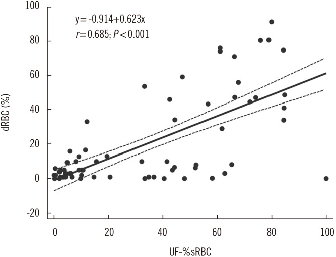
Fig. 1 Correlation between dysmorphic red blood cells (dRBCs) and UF-1000i small red blood cells (UF-%sRBCs) (r=0.685, P<0.0001). %dRBCs was counted by phase-contrast microscopy. %dRBCs <20% was used as the criterion to rule out non-glomerular nephritis.
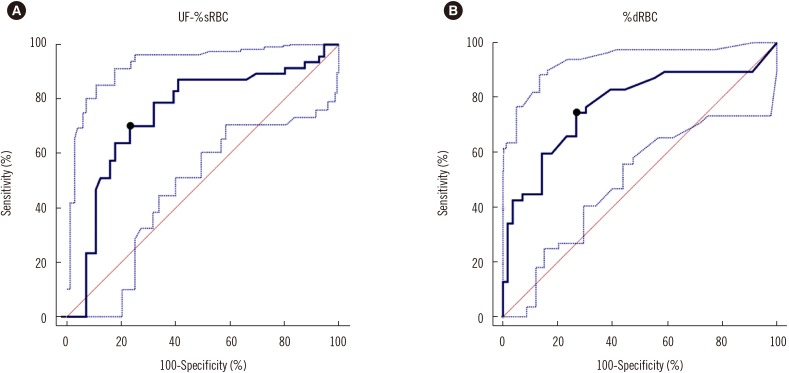
Fig. 2 ROC curves for UF-1000i small red blood cells (RBCs)% (UF-%sRBCs) and dysmorphic RBCs (%dRBCs) to differentiate glomerulonephritis from non-glomerulonephritis patients. The cut-off was determined to be (A) >40.5% with area under the curve (AUC)=0.745 for UF-%sRBCs (B) >6.7% with AUC=0.771 for %dRBCs. Dotted lines are 95% confidence intervals of ROC cuves.
Cited by 1 articles
-
Diagnostic Characteristics of Urinary Red Blood Cell Distribution Incorporated in UF-5000 for Differentiation of Glomerular and Non-Glomerular Hematuria
Hanwool Cho, Jaeeun Yoo, Hyunjung Kim, Hyunsik Jang, Yonggoo Kim, Hyojin Chae
Ann Lab Med. 2022;42(2):160-168. doi: 10.3343/alm.2022.42.2.160.
Reference
-
1. Chae DW. Current status of primary glomerulonephritis. Korean J Med. 2013; 84:1–5.2. Fairley KF, Birch DF. Hematuria: a simple method for identifying glomerular bleeding. Kidney Int. 1982; 21:105–108. PMID: 7077941.3. De Santo NG, Nuzzi F, Capodicasa G, Lama G, Caputo G, Rosati P, et al. Phase contrast microscopy of the urine sediment for the diagnosis of glomerular and nonglomerular bleeding-data in children and adults with normal creatinine clearance. Nephron. 1987; 45:35–39. PMID: 3543709.4. Shichiri M, Hosoda K, Nishio Y, Ogura M, Suenaga M, Saito H, et al. Red-cell-volume distribution curves in diagnosis of glomerular and non-glomerular haematuria. Lancet. 1988; 1:908–911. PMID: 2895832.5. Shichiri M, Oowada A, Nishio Y, Tomita K, Shiigai T. Use of autoanalyser to examine urinary-red-cell morphology in the diagnosis of glomerular haematuria. Lancet. 1986; 2:781–782. PMID: 2876237.6. Racki S, Grzetić M, Prodan-Merlak Z, Vuksanović-Mikulicić S, Sladoje-Martinović B, Zivcić-Cosić S. Clinical use of phase-contrast microscopy in the differential diagnosis of microhematuria. Acta Med Croatica. 2003; 57:11–16. PMID: 12876856.7. Ito CA, Pecoits-Filho R, Bail L, Wosiack MA, Afinovicz D, Hauser AB. Comparative analysis of two methodologies for the identification of urinary red blood cell casts. J Bras Nefrol. 2011; 33:402–407. PMID: 22189802.8. Chu-Su Y, Shukuya K, Yokoyama T, Lin WC, Chiang CK, Lin CW. Enhancing the detection of dysmorphic red blood cells and renal tubular epithelial cells with a modified urinalysis protocol. Sci Rep. 2017; 7:40521. PMID: 28074941.9. Fogazzi GB, Grignani S. Urine microscopic analysis–an art abandoned by nephrologists? Nephrol Dial Transplant. 1998; 13:2485–2487. PMID: 9794548.10. Davis R, Jones JS, Barocas DA, Castle EP, Lang EK, Leveillee RJ, et al. Diagnosis, evaluation and follow-up of asymptomatic microhematuria (AMH) in adults: AUA guideline. J Urol. 2012; 188:2473–2481. PMID: 23098784.11. Uno D, Kawakami S. UF-1000i/UF-500i clinical case study. 2nd ed. Kobe: Sysmex Corporation;2014. p. 1–35.12. Jiang T, Chen P, Ouyang J, Zhang S, Cai D. Urine particles analysis: performance evaluation of Sysmex UF-1000i and comparison among urine flow cytometer, dipstick, and visual microscopic examination. Scand J Clin Lab Invest. 2011; 71:30–37. PMID: 21091139.13. Hyodo T, Kumano K, Sakai T. Differential diagnosis between glomerular and nonglomerular hematuria by automated urinary flow cytometer. Kitasato University Kidney Center criteria. Kitasato University Kidney Center criteria. Nephron. 1999; 82:312–323. PMID: 10450033.14. Nguyen GK. Urine cytology in renal glomerular disease and value of G1 cell in the diagnosis of glomerular bleeding. Diagn Cytopathol. 2003; 29:67–73. PMID: 12889042.15. van der Zwet WC, Hessels J, Canbolat F, Deckers MM. Evaluation of the Sysmex UF-1000i® urine flow cytometer in the diagnostic work-up of suspected urinary tract infection in a Dutch general hospital. Clin Chem Lab Med. 2010; 48:1765–1771. PMID: 20726812.16. Oyaert M, Delanghe J. Progress in Automated Urinalysis. Ann Lab Med. 2019; 39:15–22. PMID: 30215225.17. CLSI. Urinalysis; approved guideline. CLSI GP16-A3. 3rd ed. Wayne, PA: Clinical and Laboratory Standards Institute;2009.18. Broeren MA, Bahçeci S, Vader HL, Arents NL. Screening for urinary tract infection with the Sysmex UF-1000i urine flow cytometer. J Clin Microbiol. 2011; 49:1025–1029. PMID: 21248088.19. Köhler H, Wandel E, Brunck B. Acanthocyturia–a characteristic marker for glomerular bleeding. Kidney Int. 1991; 40:115–120. PMID: 1921146.20. Levey AS, Stevens LA, Schmid CH, Zhang YL, Castro AF 3rd, Feldman HI, et al. A new equation to estimate glomerular filtration rate. Ann Intern Med. 2009; 150:604–612. PMID: 19414839.21. Pollock C, Liu PL, Györy AZ, Grigg R, Gallery ED, Caterson R, et al. Dysmorphism of urinary red blood cells–value in diagnosis. Kidney Int. 1989; 36:1045–1049. PMID: 2689749.22. Fuiano G, Mazza G, Comi N, Caglioti A, De Nicola L, Iodice C, et al. Current indications for renal biopsy: a questionnaire-based survey. Am J Kidney Dis. 2000; 35:448–457. PMID: 10692270.23. Sultana T, Sultana T, Rahman MQ, Ahmed ANN. Evaluation of haematuria and use of phase contrast microscope: a short review. J Dhaka Med Coll. 2011; 20:63–67.24. Santangelo L, Netti GS, Giordano P, Carbone V, Martino M, Torres DD, et al. Indications and results of renal biopsy in children: a 36-year experience. World J Pediatr. 2018; 14:127–133. PMID: 29569185.25. Jo YI. Diagnosis of primary glomerular diseases. Korean J Med. 2013; 84:6–12.26. Albani JM, Ciaschini MW, Streem SB, Herts BR, Angermeier KW. The role of computerized tomographic urography in the initial evaluation of hematuria. J Urol. 2007; 177:644–648. PMID: 17222650.27. Crop MJ, de Rijke YB, Verhagen PC, Cransberg K, Zietse R. Diagnostic value of urinary dysmorphic erythrocytes in clinical practice. Nephron Clin Pract. 2010; 115:c203–c212. PMID: 20413998.28. Stapleton FB. Morphology of urinary red blood cells: a simple guide in localizing the site of hematuria. Pediatr Clin North Am. 1987; 34:561–569. PMID: 3295717.
- Full Text Links
-
- Actions
-
Cited
- CITED
-
- Close
- Share
-
- Similar articles
-
- Comparison of YD URiSCAN PluScope Urine Microscopic Analyzer and Sysmex UF-1000i Flow Cytometry Systems
- Comparison of IRIS Iq200, UF-1000i, and Cobas u701 Module Automated Urine Sediment Analyzers
- Age-Specific Cutoffs of the Sysmex UF-1000i Automated Urine Analyzer for Rapid Screening of Urinary Tract Infections in Outpatients
- Evaluation of an Automated Urine Flow Cytometer for Screening of Bacterial Contamination in Platelet Concentrates
- Evaluation and Establishment of Reference Range of Automated Urine Cell Analyzer UF-100
