Ann Clin Microbiol.
2018 Sep;21(3):64-67. 10.5145/ACM.2018.21.3.64.
Nocardia abscessus Cutaneous Abscess: A Case Report and Review of the Literature
- Affiliations
-
- 1Department of Laboratory Medicine, Chungbuk National University Hospital, Cheongju, Korea.
- 2Department of Laboratory Medicine, Chungbuk National University College of Medicine, Cheongju, Korea. ksshin@chungbuk.ac.kr
- 3Department of Microbiology, Chungbuk National University College of Medicine, Cheongju, Korea.
- KMID: 2429657
- DOI: http://doi.org/10.5145/ACM.2018.21.3.64
Abstract
- We describe a cutaneous abscess caused by Nocardia abscessus in a previously healthy woman. A 74-year-old woman presented with recurrent bullae on her left forearm that developed 1 week prior and was initially suspected to be a cutaneous infection with Mycobacteria or Tinea corporis. Histopathologically, the skin lesion formed an abscess. A smear revealed a few branched Gram-positive filamentous microorganisms that formed a creamy white colony on a blood agar plate after incubation for 3 days. The colony tested negative on acid-fast bacilli (AFB) staining, but was positive on modified AFB staining. The isolate was confirmed to be N. abscessus by 16S rRNA sequencing analysis. The isolate was susceptible to trimethoprim-sulfamethoxazole, amikacin, cefotaxime and erythromycin but resistant to penicillin. The patient was treated with clarithromycin but subsequently lost to follow-up. To the best of our knowledge, this is the first report of a human cutaneous infection with N. abscessus in Korea.
MeSH Terms
Figure
-
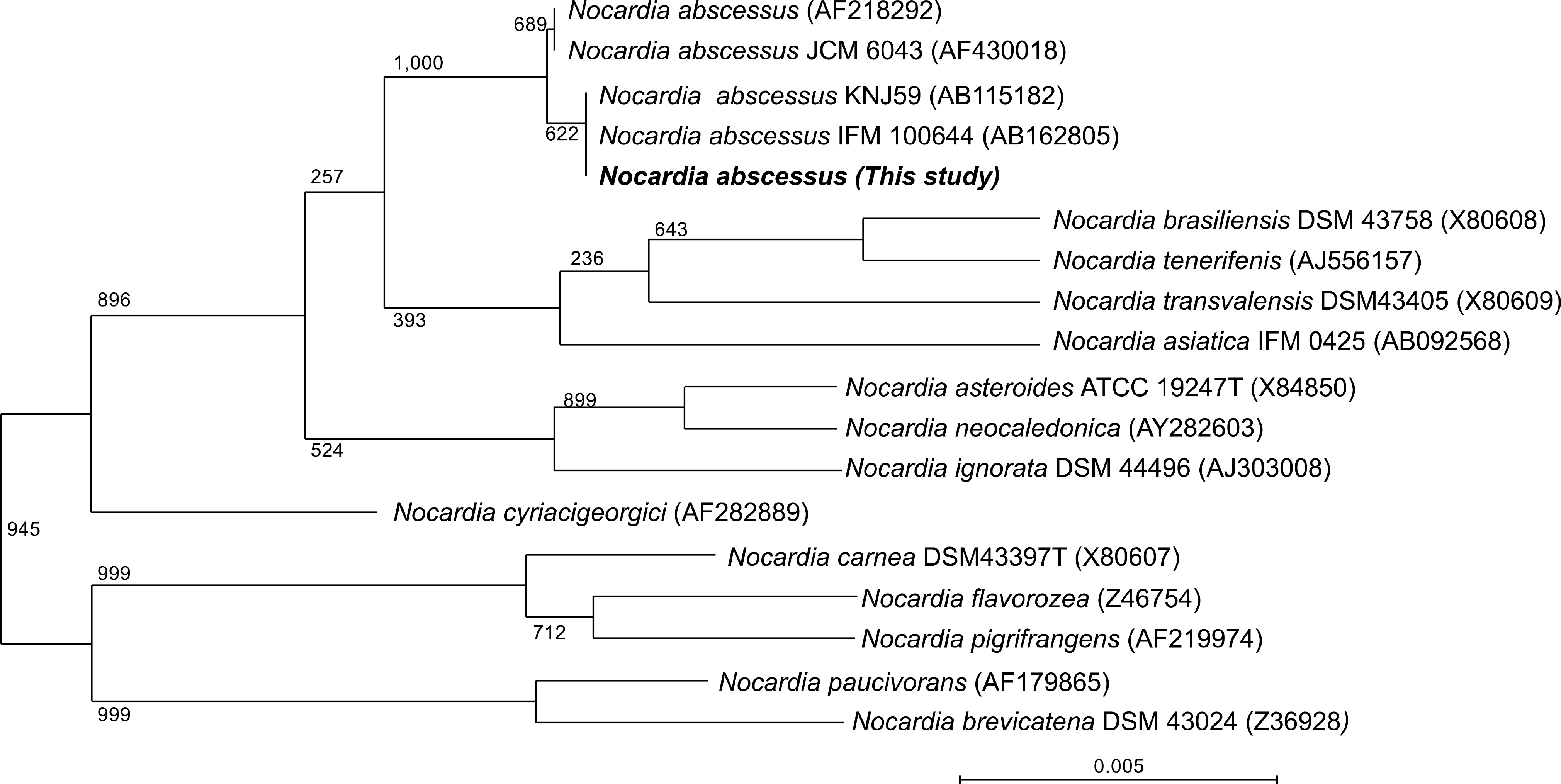
Fig. 1. Phylogenetic tree of the current isolate (CBU 05/1969: 1,362 bp) and Nocardia species. The 16S rRNA gene sequences of Nocardia species available in GenBank were aligned using CLUSTAL V and the phylogenetic tree was generated by the neighbor-joining method. Bootstrap values (%) are shown near their corresponding branches; ‘0.1’ indicates 0.1 nucleotide substitutions per site.
Reference
-
References
1. Beaman BL and Beaman L. Nocardia species: host-parasite relationships. Clin Microbiol Rev. 1994; 7:213–64.
Article2. Ambrosioni J, Lew D, Garbino J. Nocardiosis: updated clinical review and experience at a tertiary center. Infection. 2010; 38:89–97.
Article3. Al Tawfiq JA, Mayman T, Memish ZA. Nocardia abscessus brain abscess in an immunocompetent host. J Infect Public Health. 2013; 6:158–61.
Article4. Horre R, Schumacher G, Marklein G, Stratmann H, Wardelmann E, Gilges S, et al. Mycetoma due to Pseudallescheria boydii and co-isolation of Nocardia abscessus in a patient injured in road accident. Med Mycobiol. 2002; 40:525–7.5. Choe WH, Kang JO, Pai HJ, Choi TY. A case of Norcardia asteroids type I induced pneumonia. Ann Lab Med. 2005; 25:324–8.6. CLSI. Susceptibility testing of mycobacteria, nocardia, and other aerobic actinomycetes; approved standard. CLSI document M24-A. Wayne, PA: Clinical and Laboratory Standards Institute;2003.
- Full Text Links
-
- Actions
-
Cited
- CITED
-
- Close
- Share
-
- Similar articles
-
- Multiple Intramuscular Abscesses Caused by Nocardia abscessus in a Patient with Chronic Obstructive Lung Disease: Clinical Microbiology Considerations
- A Case of Disseminated Nocardiosis by Nocardia brasiliensis after Steroid Injection
- Primary Cutaneous Nocardiosis Caused by Nocardia niigatensis
- A Case of Nocardia farcinica Brain Abscess in a Focal Segmental Glomerulosclerosis Patient after Steroid Treatment
- A Case of Skin Infection Caused by Mycobacterium abscessus
