Korean J Orthod.
2018 Nov;48(6):349-356. 10.4041/kjod.2018.48.6.349.
Assessment of lower incisor alveolar bone width using cone-beam computed tomography images in skeletal Class III adults of different vertical patterns
- Affiliations
-
- 1Department of Orthodontics, Yonsei University College of Dentistry, Seoul, Korea.
- 2Department of Orthodontics, Gangnam Severance Dental Hospital, Yonsei University College of Dentistry, Seoul, Korea. khkim@yuhs.ac
- 3Institute of Craniofacial Deformity, Yonsei University College of Dentistry, Seoul, Korea.
- KMID: 2427772
- DOI: http://doi.org/10.4041/kjod.2018.48.6.349
Abstract
OBJECTIVE
This study was performed to investigate the alveolar bone of lower incisors in skeletal Class III adults of different vertical facial patterns and to compare it with that of Class I adults using cone-beam computed tomography (CBCT) images.
METHODS
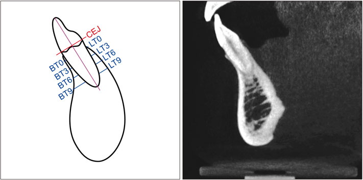
CBCT images of 90 skeletal Class III and 29 Class I patients were evaluated. Class III subjects were divided by mandibular plane angle: high (SN-MP > 38.0°), normal (30.0°< SN-MP < 37.0°), and low (SN-MP < 28.0°) groups. Buccolingual alveolar bone thickness was measured using CBCT images of mandibular incisors at alveolar crest and 3, 6, and 9 mm apical levels. Linear mixed model, Bonferroni post-hoc test, and Pearson correlation analysis were used for statistical significance.
RESULTS
Buccolingual alveolar bone in Class III high, normal and low angle subjects was not significantly different at alveolar crest and 3 mm apical level while lingual bone was thicker at 6 and 9 mm apical levels than on buccal side. Class III high angle group had thinner alveolar bone at all levels except at buccal alveolar crest and 9 mm apical level on lingual side compared to the Class I group. Class III high angle group showed thinner alveolar bone than the Class III normal or low angle groups in most regions. Mandibular plane angle showed negative correlations with mandibular anterior alveolar bone thickness.
CONCLUSIONS
Skeletal Class III subjects with high mandibular plane angles showed thinner mandibular alveolar bone in most areas compared to normal or low angle subjects. Mandibular plane angle was negatively correlated with buccolingual alveolar bone thickness.
Keyword
Figure
Reference
-
1. Jiang N, Guo W, Chen M, Zheng Y, Zhou J, Kim SG, et al. Periodontal ligament and alveolar bone in health and adaptation: tooth movement. Front Oral Biol. 2016; 18:1–8.
Article2. Wainwright WM. Faciolingual tooth movement: its influence on the root and cortical plate. Am J Orthod. 1973; 64:278–302.
Article3. Edwards JG. A study of the anterior portion of the palate as it relates to orthodontic therapy. Am J Orthod. 1976; 69:249–273.
Article4. Mulie RM, Hoeve AT. The limitations of tooth movement within the symphysis, studied with laminagraphy and standardized occlusal films. J Clin Orthod. 1976; 10:882–893. 886–889.5. Molina-Berlanga N, Llopis-Perez J, Flores-Mir C, Puigdollers A. Lower incisor dentoalveolar compensation and symphysis dimensions among Class I and III malocclusion patients with different facial vertical skeletal patterns. Angle Orthod. 2013; 83:948–955.
Article6. Handelman CS. The anterior alveolus: its importance in limiting orthodontic treatment and its influence on the occurrence of iatrogenic sequelae. Angle Orthod. 1996; 66:95–109. discussion 109-10.7. Kim YS, Cha JY, Yu HS, Hwang CJ. Comparison of mandibular anterior alveolar bone thickness in different facial skeletal types. Korean J Orthod. 2010; 40:314–324.
Article8. Chung CJ, Jung S, Baik HS. Morphological characteristics of the symphyseal region in adult skeletal Class III crossbite and openbite malocclusions. Angle Orthod. 2008; 78:38–43.
Article9. Choe HY, Park W, Jeon JK, Kim YH, Shon BW. Differences in mandibular anterior alveolar bone thickness according to age in a normal skeletal group. Korean J Orthod. 2007; 37:220–230.10. Janson GR, Metaxas A, Woodside DG. Variation in maxillary and mandibular molar and incisor vertical dimension in 12-year-old subjects with excess, normal, and short lower anterior face height. Am J Orthod Dentofacial Orthop. 1994; 106:409–418.
Article11. Lee KS, Chung KR. A cephalometric analysis of Korean adult normal occlusion. Korean J Orthod. 1987; 17:199–213.12. Kim JH, Gansukh O, Amarsaikhan B, Lee SJ, Kim TW. Comparison of cephalometric norms between Mongolian and Korean adults with normal occlusions and well-balanced profiles. Korean J Orthod. 2011; 41:42–50.
Article13. Gargiulo AW, Wentz FM, Orban B. Dimensions and relations of the dentogingival junction in humans. J Periodontol. 1961; 32:261–267.
Article14. Morad G, Behnia H, Motamedian SR, Shahab S, Gholamin P, Khosraviani K, et al. Thickness of labial alveolar bone overlying healthy maxillary and mandibular anterior teeth. J Craniofac Surg. 2014; 25:1985–1991.
Article15. Deguchi T, Nasu M, Murakami K, Yabuuchi T, Kamioka H, Takano-Yamamoto T. Quantitative evaluation of cortical bone thickness with computed tomographic scanning for orthodontic implants. Am J Orthod Dentofacial Orthop. 2006; 129:721.e7–721.e12.
Article16. Gracco A, Luca L, Bongiorno MC, Siciliani G. Computed tomography evaluation of mandibular incisor bony support in untreated patients. Am J Orthod Dentofacial Orthop. 2010; 138:179–187.
Article17. Esenlik E, Sabuncuoglu FA. Alveolar and symphysis regions of patients with skeletal class II division 1 anomalies with different vertical growth patterns. Eur J Dent. 2012; 6:123–132.
Article18. Wehrbein H, Bauer W, Diedrich P. Mandibular incisors, alveolar bone, and symphysis after orthodontic treatment. A retrospective study. Am J Orthod Dentofacial Orthop. 1996; 110:239–246.
Article19. Han JY, Jung GU. Labial and lingual/palatal bone thickness of maxillary and mandibular anteriors in human cadavers in Koreans. J Periodontal Implant Sci. 2011; 41:60–66.
Article20. Wingard CE, Bowers GM. The effects of facial bone from facial tipping of incisors in monkeys. J Periodontol. 1976; 47:450–454.21. Steiner GG, Pearson JK, Ainamo J. Changes of the marginal periodontium as a result of labial tooth movement in monkeys. J Periodontol. 1981; 52:314–320.
Article22. Artun J, Krogstad O. Periodontal status of mandibular incisors following excessive proclination. A study in adults with surgically treated mandibular prognathism. Am J Orthod Dentofacial Orthop. 1987; 91:225–232.
Article23. Allais D, Melsen B. Does labial movement of lower incisors influence the level of the gingival margin? A case-control study of adult orthodontic patients. Eur J Orthod. 2003; 25:343–352.
Article24. Choi YJ, Chung CJ, Kim KH. Periodontal consequences of mandibular incisor proclination during presurgical orthodontic treatment in Class III malocclusion patients. Angle Orthod. 2015; 85:427–433.
Article25. Graber L, Vanarsdall R, Vig K. Orthodontics: current principles and techniques. 5th ed. Philadelphia: Mosby, Inc.;2012.26. Yamada C, Kitai N, Kakimoto N, Murakami S, Furukawa S, Takada K. Spatial relationships between the mandibular central incisor and associated alveolar bone in adults with mandibular prognathism. Angle Orthod. 2007; 77:766–772.
Article27. Kuitert R, Beckmann S, van Loenen M, Tuinzing B, Zentner A. Dentoalveolar compensation in subjects with vertical skeletal dysplasia. Am J Orthod Dentofacial Orthop. 2006; 129:649–657.
Article28. Ingervall B, Janson T. The value of clinical lip strength measurements. Am J Orthod. 1981; 80:496–507.
Article29. Latif A. An electromyographic study of the temporalis muscle in normal persons during selected positions and movements of the mandible. Am J Orthod Dentofacial Orthop. 1957; 43:577–591.
Article30. Björk A. Prediction of mandibular growth rotation. Am J Orthod. 1969; 55:585–599.
Article31. Kook YA, Kim G, Kim Y. Comparison of alveolar bone loss around incisors in normal occlusion samples and surgical skeletal class III patients. Angle Orthod. 2012; 82:645–652.
Article32. Silva MJ, Wang C, Keaveny TM, Hayes WC. Direct and computed tomography thickness measurements of the human, lumbar vertebral shell and endplate. Bone. 1994; 15:409–414.
Article33. Spoor CF, Zonneveld FW, Macho GA. Linear measurements of cortical bone and dental enamel by computed tomography: applications and problems. Am J Phys Anthropol. 1993; 91:469–484.
Article34. Panzarella FK, Junqueira JL, Oliveira LB, de Araújo NS, Costa C. Accuracy assessment of the axial images obtained from cone beam computed tomography. Dentomaxillofac Radiol. 2011; 40:369–378.
Article35. Patcas R, Müller L, Ullrich O, Peltomäki T. Accuracy of cone-beam computed tomography at different resolutions assessed on the bony covering of the mandibular anterior teeth. Am J Orthod Dentofacial Orthop. 2012; 141:41–50.
Article36. Durbar US. Racial variations in different skulls. J Pharm Sci Res. 2014; 6:370–372.
- Full Text Links
- Actions
-
Cited
- CITED
-
- Close
- Share
- Similar articles
-
- Comparison of interradicular distances and cortical bone thickness in Thai patients with Class I and Class II skeletal patterns using cone-beam computed tomography
- Crown-root angulations of the maxillary anterior teeth according to malocclusions: A cone-beam computed tomography study in Korean population
- Reproducibility of cone-beam computed tomographic measurements of bone plates and the interdental septum in the anterior mandible
- A study on the morphological changes of lower incisor and symphysis during surgical-orthodontic treatment in skeletal class III malocclusion
- Location and shape of the mandibular lingula: Comparison of skeletal class I and class III patients using panoramic radiography and cone-beam computed tomography


