Korean J Orthod.
2022 Nov;52(6):432-438. 10.4041/kjod22.126.
Crown-root angulations of the maxillary anterior teeth according to malocclusions: A cone-beam computed tomography study in Korean population
- Affiliations
-
- 1Department of Orthodontics, College of Dentistry, Gangneung-Wonju National University, Gangneung, Korea
- KMID: 2536336
- DOI: http://doi.org/10.4041/kjod22.126
Abstract
Objective
To compare crown-root angulations of the permanent maxillary anterior teeth in skeletal Class I, Class II, and Class III Korean malocclusion patients using cone-bean computed tomography (CBCT) images.
Methods
Sixty CBCT images were collected from orthodontic patients archive based on skeletal Class I (0˚< A point-nasion-B point angle [ANB] < 4˚), Class II (ANB ≥ 4˚), and Class III (ANB ≤ 0˚) to have 20 samples in each group. Mesiodistal crown-root angulation (MDCRA) and labiolingual crown-root angulation (LLCRA) were evaluated after orientation of images. Crown-root angulations were compared among Class I, Class II, and Class III groups and among the maxillary anterior teeth in each group.
Results
LLCRAs of the maxillary central incisor and the lateral incisor were significantly lower in Class III group than those in Class I group. However, those of the canine showed no significant differences among groups. MDCRAs of the maxillary anterior teeth did not significantly differ among groups either.
Conclusions
Our results suggest that skeletal Class III malocclusion might affect LLCRA of the maxillary incisors, especially the central incisor.
Keyword
Figure
-
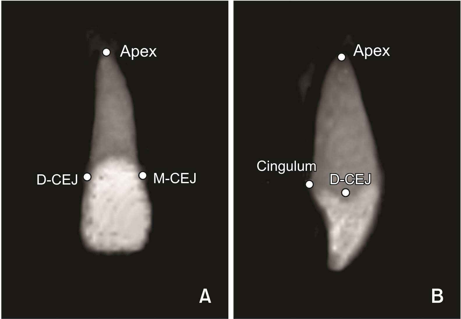
Figure 1 Four reference points: apex, mesial cementoenamel junction (M-CEJ), distal cementoenamle junction (D-CEJ), and cingulum. A, Coronal view. B, Proximal view.

Figure 2 Three-dimensional orientation of tooth. A, X-axis is the line passing through mesial cementoenamel junction (M-CEJ) and distal cementoenamle junction (D-CEJ). Y-axis is the line passing through the projected distal CEJ (D-CEJ’) and cingulum point (Cingulum’) on the sagittal reference plane. B, Z-axis is the cross product of the X-axis and Y-axis.
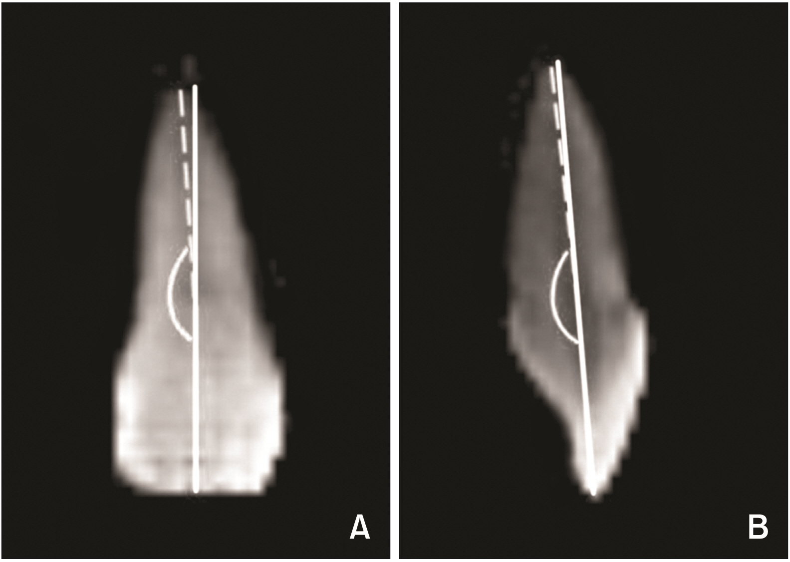
Figure 3 Measurement of the crown-root angulation (CRA). A, Mesiodistal CRA (MDCRA) in the coronal plane. B, Labiolingual CRA (LLCRA) in the sagittal plane.
Reference
-
1. Bryant RM, Sadowsky PL, Hazelrig JB. 1984; Variability in three morphologic features of the permanent maxillary central incisor. Am J Orthod. 86:25–32. DOI: 10.1016/0002-9416(84)90273-2. PMID: 6588757. PMID: https://www.scopus.com/inward/record.uri?partnerID=HzOxMe3b&scp=0021464910&origin=inward.2. Andrews LF. 1976; The straight-wire appliance, origin, controversy, commentary. J Clin Orthod. 10:99–114. PMID: 1074876. PMID: https://www.scopus.com/inward/record.uri?partnerID=HzOxMe3b&scp=0016922634&origin=inward.3. Carlson SK, Johnson E. 2001; Bracket positioning and resets: five steps to align crowns and roots consistently. Am J Orthod Dentofacial Orthop. 119:76–80. DOI: 10.1067/mod.2001.111220. PMID: 11174544. PMID: https://www.scopus.com/inward/record.uri?partnerID=HzOxMe3b&scp=0035220077&origin=inward.4. Taylor NG, Cook PA. 1992; The reliability of positioning pre-adjusted brackets: an in vitro study. Br J Orthod. 19:25–34. DOI: 10.1179/bjo.19.1.25. PMID: 1562576. PMID: https://www.scopus.com/inward/record.uri?partnerID=HzOxMe3b&scp=0026811899&origin=inward.5. Meyer M, Nelson G. 1978; Preadjusted edgewise appliances: theory and practice. Am J Orthod. 73:485–98. DOI: 10.1016/0002-9416(78)90239-7. PMID: 354404. PMID: https://www.scopus.com/inward/record.uri?partnerID=HzOxMe3b&scp=0017884304&origin=inward.6. Germane N, Bentley B, Isaacson RJ, Revere JH Jr. 1990; The morphology of canines in relation to preadjusted appliances. Angle Orthod. 60:49–54. DOI: 10.1043/0003-3219(1990)060<0049:TMOCIR>2.0.CO;2. PMID: 2316904.7. Balut N, Klapper L, Sandrik J, Bowman D. 1992; Variations in bracket placement in the preadjusted orthodontic appliance. Am J Orthod Dentofacial Orthop. 102:62–7. DOI: 10.1016/0889-5406(92)70015-3. PMID: 1626532. PMID: https://www.scopus.com/inward/record.uri?partnerID=HzOxMe3b&scp=0026889604&origin=inward.8. Creekmore TD, Kunik RL. 1993; Straight wire: the next generation. Am J Orthod Dentofacial Orthop. 104:8–20. Erratum in: Am J Orthod Dentofacial Orthop 1993;104:20. DOI: 10.1016/0889-5406(93)70023-H. PMID: 8257493. PMID: https://www.scopus.com/inward/record.uri?partnerID=HzOxMe3b&scp=0027632979&origin=inward.9. Shroff B. 2001; Root correction during orthodontic therapy. Semin Orthod. 7:50–8. DOI: 10.1053/sodo.2001.21074. PMID: https://www.scopus.com/inward/record.uri?partnerID=HzOxMe3b&scp=0006632714&origin=inward.10. Mavroskoufis F, Ritchie GM. 1980; Variation in size and form between left and right maxillary central incisor teeth. J Prosthet Dent. 43:254–7. DOI: 10.1016/0022-3913(80)90398-4. PMID: 6928193. PMID: https://www.scopus.com/inward/record.uri?partnerID=HzOxMe3b&scp=0018990259&origin=inward.11. van Loenen M, Degrieck J, De Pauw G, Dermaut L. 2005; Anterior tooth morphology and its effect on torque. Eur J Orthod. 27:258–62. DOI: 10.1093/ejo/cji007. PMID: 15947225. PMID: https://www.scopus.com/inward/record.uri?partnerID=HzOxMe3b&scp=22144475486&origin=inward.12. Harris EF, Hassankiadeh S, Harris JT. 1993; Maxillary incisor crown-root relationships in different angle malocclusions. Am J Orthod Dentofacial Orthop. 103:48–53. DOI: 10.1016/0889-5406(93)70104-V. PMID: 8422031. PMID: https://www.scopus.com/inward/record.uri?partnerID=HzOxMe3b&scp=0027344545&origin=inward.13. McIntyre GT, Millett DT. 2003; Crown-root shape of the permanent maxillary central incisor. Angle Orthod. 73:710–5. DOI: 10.1043/0003-3219(2003)073<0710:CSOTPM>2.0.CO;2. PMID: 14719737.14. Delivanis HP, Kuftinec MM. 1980; Variation in morphology of the maxillary central incisors found in class II, division 2 malocclusions. Am J Orthod. 78:438–43. DOI: 10.1016/0002-9416(80)90024-X. PMID: 6933852. PMID: https://www.scopus.com/inward/record.uri?partnerID=HzOxMe3b&scp=0019074747&origin=inward.15. Carlsson R, Rönnerman A. 1973; Crown-root angles of upper central incisors. Am J Orthod. 64:147–54. DOI: 10.1016/S0002-9416(73)90306-0. PMID: 4515887. PMID: https://www.scopus.com/inward/record.uri?partnerID=HzOxMe3b&scp=0015653238&origin=inward.16. Sicher H, DuBrul EL. 1970. Oral anatomy. 5th ed. Mosby;St. Louis: DOI: 10.1016/s0002-9416(73)90306-0. PMID: https://www.scopus.com/inward/record.uri?partnerID=HzOxMe3b&scp=0015653238&origin=inward.17. Taylor RM. 1969; Variation in form of human teeth: I. An anthropologic and forensic study of maxillary incisors. J Dent Res. 48:5–16. DOI: 10.1177/00220345690480012501. PMID: 5252101. PMID: https://www.scopus.com/inward/record.uri?partnerID=HzOxMe3b&scp=0014440950&origin=inward.18. Hong HS, Baik HS. 1995; Maxillary incisor crown-root angle (collum angle) in different malocclusions. Korean J Orthod. 25:453–63. PMID: https://www.scopus.com/inward/record.uri?partnerID=HzOxMe3b&scp=1442356460&origin=inward.19. Tong H, Enciso R, Van Elslande D, Major PW, Sameshima GT. 2012; A new method to measure mesiodistal angulation and faciolingual inclination of each whole tooth with volumetric cone-beam computed tomography images. Am J Orthod Dentofacial Orthop. 142:133–43. DOI: 10.1016/j.ajodo.2011.12.027. PMID: 22748999. PMID: https://www.scopus.com/inward/record.uri?partnerID=HzOxMe3b&scp=84860457170&origin=inward.20. Chohayeb AA. 1983; Dilaceration of permanent upper lateral incisors: frequency, direction, and endodontic treatment implications. Oral Surg Oral Med Oral Pathol. 55:519–20. DOI: 10.1016/0030-4220(83)90239-6. PMID: 6575342. PMID: https://www.scopus.com/inward/record.uri?partnerID=HzOxMe3b&scp=0020758490&origin=inward.21. Smith BG, Knight JK. 1984; An index for measuring the wear of teeth. Br Dent J. 156:435–8. DOI: 10.1038/sj.bdj.4805394. PMID: 6590081. PMID: https://www.scopus.com/inward/record.uri?partnerID=HzOxMe3b&scp=0021769350&origin=inward.22. Little RM. 1975; The irregularity index: a quantitative score of mandibular anterior alignment. Am J Orthod. 68:554–63. DOI: 10.1016/0002-9416(75)90086-X. PMID: 1059332. PMID: https://www.scopus.com/inward/record.uri?partnerID=HzOxMe3b&scp=0016571462&origin=inward.23. Backlund E. 1960; Tooth form and overbite. Trans Eur Orthod Soc. 36:97–104. PMID: https://www.scopus.com/inward/record.uri?partnerID=HzOxMe3b&scp=1442356460&origin=inward.24. Heravi F, Salari S, Tanbakuchi B, Loh S, Amiri M. 2013; Effects of crown-root angle on stress distribution in the maxillary central incisors' PDL during application of intrusive and retraction forces: a three-dimensional finite element analysis. Prog Orthod. 14:26. DOI: 10.1186/2196-1042-14-26. PMID: 24326061. PMCID: PMC4384911. PMID: https://www.scopus.com/inward/record.uri?partnerID=HzOxMe3b&scp=84884563347&origin=inward.25. Logan W. 1959; Deckbiss-a clinical evaluation. Trans Eur Orthod Soc. 35:313–7. PMID: https://www.scopus.com/inward/record.uri?partnerID=HzOxMe3b&scp=1442356460&origin=inward.26. Macri V, Athanasiou AE. Athanasiou AE, editor. 1997. Sources of error in lateral cephalometry. Orthodontic cephalometry. Mosby-Wolfe;London: p. 125–4. PMID: https://www.scopus.com/inward/record.uri?partnerID=HzOxMe3b&scp=1442356460&origin=inward.27. Houston WJ, Maher RE, McElroy D, Sherriff M. 1986; Sources of error in measurements from cephalometric radiographs. Eur J Orthod. 8:149–51. DOI: 10.1093/ejo/8.3.149. PMID: 3464438. PMID: https://www.scopus.com/inward/record.uri?partnerID=HzOxMe3b&scp=0022764043&origin=inward.
- Full Text Links
-
- Actions
-
Cited
- CITED
-
- Close
- Share
-
- Similar articles
-
- Cone-beam computed tomography for the assessment of root–crown ratios of the maxillary and mandibular incisors in a Korean population
- Crown and root lengths of incisors, canines, and premolars measured by cone-beam computed tomography in patients with malocclusions
- Detection of maxillary second molar with two palatal roots using cone beam computed tomography: a case report
- Root surface areas of maxillary permanent teeth in anterior normal overbite and anterior open bite assessed using cone-beam computed tomography
- Fused roots of maxillary molars: characterization and prevalence in a Latin American sub-population: a cone beam computed tomography study
