Imaging Sci Dent.
2017 Dec;47(4):241-246. 10.5624/isd.2017.47.4.241.
Root surface areas of maxillary permanent teeth in anterior normal overbite and anterior open bite assessed using cone-beam computed tomography
- Affiliations
-
- 1Department of Orthodontics, Faculty of Dentistry, Chiang Mai University, Chiang Mai, Thailand.
- 2Department of Orthodontics and Pediatric Dentistry, Faculty of Dentistry, Chiang Mai University, Chiang Mai, Thailand. dhirawat.j@gmail.com
- 3Department of Radiology, Faculty of Medicine Siriraj Hospital, Mahidol University, Bangkok, Thailand.
- KMID: 2397838
- DOI: http://doi.org/10.5624/isd.2017.47.4.241
Abstract
- PURPOSE
The aim of this study was to compare the root surface areas of the maxillary permanent teeth in Thai patients exhibiting anterior normal overbite and in those exhibiting anterior open bite, using cone-beam computed tomography (CBCT).
MATERIALS AND METHODS
CBCT images of maxillary permanent teeth from 15 patients with anterior normal overbite and 18 patients with anterior open bite were selected. Three-dimensional tooth models were constructed using Mimics Research version 17.0. The cementoenamel junction was marked manually. The root surface area was calculated automatically by 3-Matic Research version 9.0. The root surface areas of each tooth type from both types of bite were compared using the independent t-test (P < .05). The intraclass correlation coefficient was used to assess intraobserver reliability.
RESULTS
The mean root surface areas of the maxillary central and lateral incisors in individuals with anterior open bite were significantly less than those in those with normal bite. The mean root surface area of the maxillary second premolar in individuals with anterior open bite was significantly greater than in those with normal bite.
CONCLUSION
Anterior open-bite malocclusion might affect the root surface area, so orthodontic force magnitudes should be carefully determined.
MeSH Terms
Figure
-
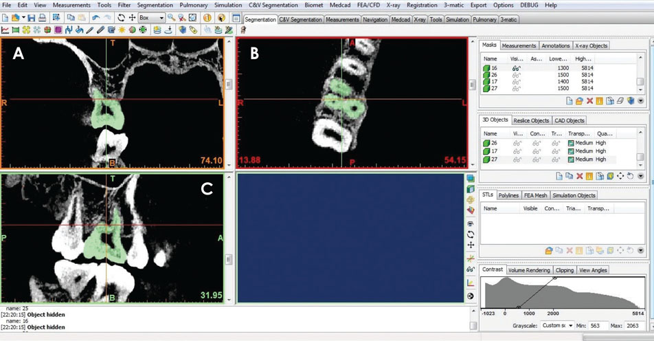
Fig. 1 Identification of tooth morphology on 2-dimensional images of each slice orientation. A. Coronal view. B. Axial view. C. Sagittal view.
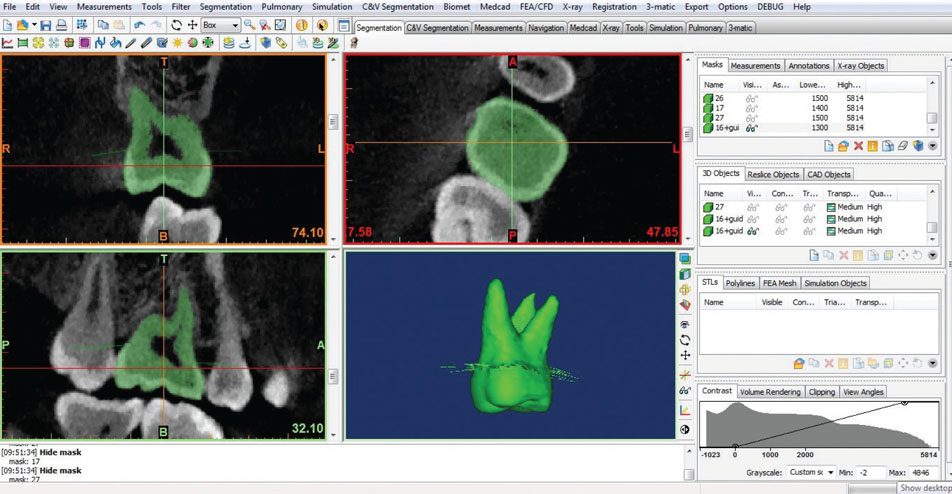
Fig. 2 Construction of intentional extension spine markings by marking the cementoenamel junction on each section.
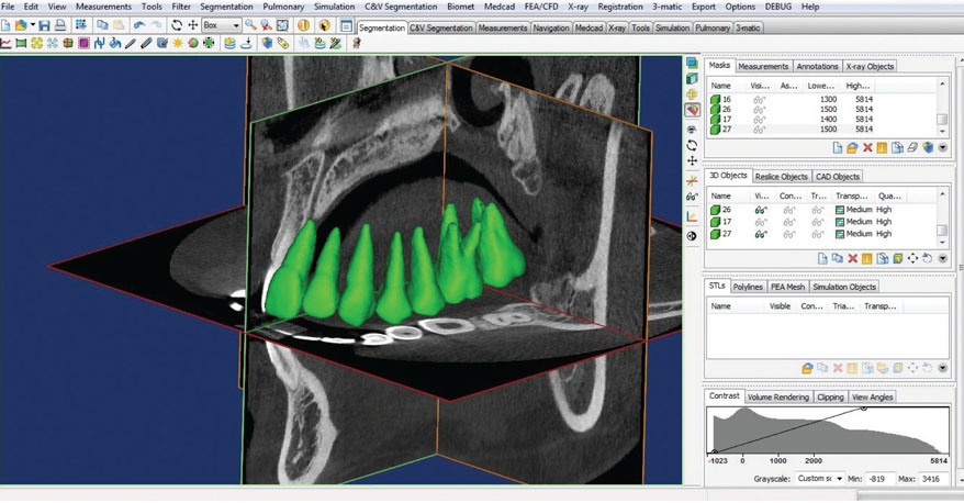
Fig. 3 Construction of the 3-dimensional tooth models.
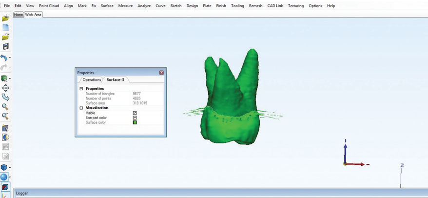
Fig. 4 Identification of the cementoenamel junction and automatic calculation of the root surface area using 3-Matic Research version 9.0.
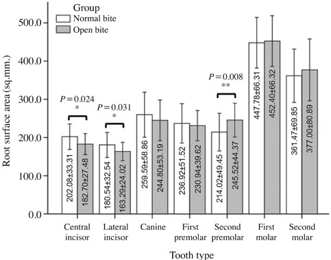
Fig. 5 Bar graph demonstrates a comparison of the root surface areas of the maxillary permanent teeth.
Reference
-
1. Hujoel P. A meta-analysis of normal ranges for root surface areas of the permanent dentition. J Clin Periodontol. 1994; 21:225–229.
Article2. Gu Y, Tang Y, Zhu Q, Feng X. Measurement of root surface area of permanent teeth with root variations in a Chinese population-A micro-CT analysis. Arch Oral Biol. 2016; 63:75–81.
Article3. Tasanapanont J, Apisariyakul J, Wattanachai T, Sriwilas P, Midtbø M, Jotikasthira D. Comparison of 2 root surface area measurement methods: 3-dimensional laser scanning and cone-beam computed tomography. Imaging Sci Dent. 2017; 47:117–122.
Article4. Enokida M, Kaneko S, Yanagishita M, Soma K. Influence of occlusal stimuli on the remodelling of alveolar bone in a rat hypofunction-recovery model. J Oral Biosci. 2005; 47:321–334.
Article5. Hayashi Y, Iida J, Warita H, Soma K. Effects of occlusal hypofunction on the microvasculature and endothelin expression in the periodontal ligaments of rat molars. Orthod Waves. 2001; 60:373–380.6. Tanaka A, Iida J, Soma K. Effect of hypofunction on the microvasculature in the periodontal ligament of the rat molar. Orthod Waves. 1998; 57:180–188.7. Karavade R, Kalia A, Nene S, Khandekar S, Patil V. Comparison of root-crown lengths and occlusal contacts in patients with class-III skeletal relationship, anterior open-bite and high mandibular plane angle. Int J Dent Med Spec. 2015; 2:7–13.8. Uehara S, Maeda A, Tomonari H, Miyawaki S. Relationships between the root-crown ratio and the loss of occlusal contact and high mandibular plane angle in patients with open bite. Angle Orthod. 2012; 83:36–42.
Article9. de Freitas MR, Beltrão RT, Janson G, Henriques JF, Cançado RH. Long-term stability of anterior open bite extraction treatment in the permanent dentition. Am J Orthod Dentofacial Orthop. 2004; 125:78–87.
Article10. Harris EF, Butler ML. Patterns of incisor root resorption before and after orthodontic correction in cases with anterior open bites. Am J Orthod Dentofacial Orthop. 1992; 101:112–119.
Article11. Cobourne MT, DiBiase AT. Handbook of orthodontics. 2nd ed. New York: Elsevier;2015.12. Shimomoto Y, Chung C, Iwasaki-Hayashi Y, Muramoto T, Soma K. Effects of occlusal stimuli on alveolar/jaw bone formation. J Dent Res. 2007; 86:47–51.
Article13. Linge BO, Linge L. Apical root resorption in upper anterior teeth. Eur J Orthod. 1983; 5:173–183.
Article14. Linge L, Linge BO. Patient characteristics and treatment variables associated with apical root resorption during orthodontic treatment. Am J Orthod Dentofacial Orthop. 1991; 99:35–43.
Article
- Full Text Links
-
- Actions
-
Cited
- CITED
-
- Close
- Share
-
- Similar articles
-
- Distances from the root apices of posterior teeth to the maxillary sinus and mandibular canal in patients with skeletal open bite: A cone-beam computed tomography study
- Cone-beam computed tomography for the assessment of root–crown ratios of the maxillary and mandibular incisors in a Korean population
- The effect of mesiodistal crown widths of anterior teeth on incisor relationship
- Crown-root angulations of the maxillary anterior teeth according to malocclusions: A cone-beam computed tomography study in Korean population
- A case report of orthodontic treatment of anterior open bite
