Prospective Validation of Intra- and Interobserver Reproducibility of a New Point Shear Wave Elastographic Technique for Assessing Liver Stiffness in Patients with Chronic Liver Disease
- Affiliations
-
- 1Department of Radiology, Seoul National University Hospital, Seoul 03080, Korea. jmsh@snu.ac.kr
- 2Institute of Radiation Medicine, Seoul National University College of Medicine, Seoul 03080, Korea.
- KMID: 2427202
- DOI: http://doi.org/10.3348/kjr.2017.18.6.926
Abstract
OBJECTIVE
To assess intra- and inter-observer reproducibility of a new point shear wave elastography technique (pSWE, S-Shearwave, Samsung Medison) and compare its accuracy in assessing liver stiffness (LS) with an established pSWE technique (Virtual Touch Quantification, VTQ).
MATERIALS AND METHODS
Thirty-three patients were enrolled in this Institutional Review Board-approved prospective study. LS values were measured by VTQ on an Acuson S2000 system (Siemens Healthineer) and S-Shearwave on an RS-80A (Samsung Medison) in the same session, followed by two further S-Shearwave sessions for inter- and intra-observer variation at 8-hour intervals. The technical success rate (SR) and reliability of the measurements of both pSWE techniques were compared. The intra- and inter-observer reproducibility of S-Shearwave was determined by intraclass correlation coefficients (ICCs). LS values were measured by both methods of pSWE. The diagnostic performance in severe fibrosis (F ≥ 3) and cirrhosis (F = 4) was evaluated using the receiver operating characteristics curve analysis and the Obuchowski measure with the LS values of transient elastography as the referenced standard.
RESULTS
The VTQ (100%, 33/33) and S-Shearwave (96.9%, 32/33) techniques did not display a significant difference in technical SR (p = 0.63) or reliability of LS measurements (96.9%, 32/33; 93.9%, 30/32, respectively, p = 0.61). The inter- and intra-observer agreement for LS measurements using the S-Shearwave technique was excellent (ICC = 0.98 and 0.99, respectively). The mean LS values of both pSWE techniques were not significantly different and exhibited a good correlation (r = 0.78). To detect F ≥ 3 and F = 4, VTQ and S-Shearwave showed comparable diagnostic accuracy as indicated by the following outcomes: areas under receiver operating characteristics curve (AUROC) = 0.87 (95% confidence intervals [CI] 0.70-0.96), 0.89 for VTQ (95% CI 0.74-0.97), respectively; and AUROC = 0.84 (95% CI 0.67-0.94), 0.94 (95% CI 0.80-0.99) for S-Shearwave (p > 0.48), respectively. The Obuchowski measures were similarly high for S-Shearwave and VTQ (0.94 vs. 0.95).
CONCLUSION
S-Shearwave shows excellent inter- and intra-observer agreement and diagnostic effectiveness comparable to VTQ in detecting LS.
MeSH Terms
-
Adult
Aged
Aged, 80 and over
Area Under Curve
Chronic Disease
*Elasticity Imaging Techniques
Female
Humans
Liver/diagnostic imaging/*physiopathology
Liver Cirrhosis/diagnosis/pathology
Liver Diseases/*diagnosis/diagnostic imaging
Male
Middle Aged
Prospective Studies
ROC Curve
Reproducibility of Results
Severity of Illness Index
Figure
-
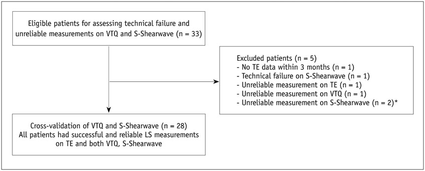
Fig. 1 Flow diagram of study population. *One population with unreliable measurements for S-Shearwave also had unreliable measurements for VTQ. LS = liver stiffness, S-Shearwave = S-Shearwave elastography, TE = transient elastography, VTQ = virtual touch quantification
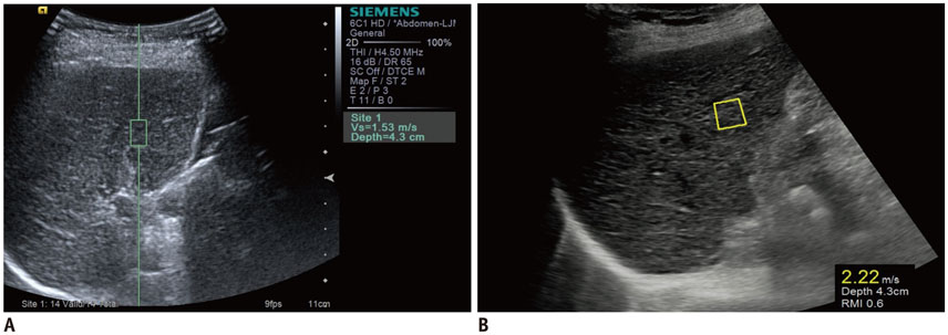
Fig. 2 LS measurement by two pSWE techniques, VTQ (A) and S-Shearwave (B). For both measurements, operator sited predefined measurement box in liver. pSWE = point shear wave elastography
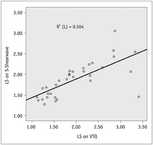
Fig. 3 Correlation between VTQ and S-Shearwave in same individuals. Linear correlation analysis of LS values seen with VTQ and S-Shearwave in same individuals indicates significant correlation between two techniques.

Fig. 4 Bland–Altman plot showing inter- (A) and intraobserver agreement (B) in S-Shearwave measurement. X-axis shows means of repeated LS measurements, and y-axis displays difference between repeat S-Shearwave LS measurements. Red line = mean difference, green line = 95% limit of agreement
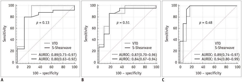
Fig. 5 Comparison of ROC curves for VTQ and S-Shearwave for different fibrosis thresholds. (A) F0–1 vs. F2–4, (B) F0–F2 vs. F3–F4, (C) F0–F3 vs. F4. In parentheses, 95% CI are shown. p values of differences between AUROCs are given. AUROC = areas under receiver operating characteristics curve, CI = confidence intervals, ROC = receiver operating characteristics
Cited by 3 articles
-
Age of Data in Contemporary Research Articles Published in Representative General Radiology Journals
Ji Hun Kang, Dong Hwan Kim, Seong Ho Park, Jung Hwan Baek
Korean J Radiol. 2018;19(6):1172-1178. doi: 10.3348/kjr.2018.19.6.1172.Histogram Analysis of Diffusion Kurtosis Magnetic Resonance Imaging for Diagnosis of Hepatic Fibrosis
Ruo-Fan Sheng, Kai-Pu Jin, Li Yang, He-Qing Wang, Hao Liu, Yuan Ji, Cai-Xia Fu, Meng-Su Zeng
Korean J Radiol. 2018;19(5):916-922. doi: 10.3348/kjr.2018.19.5.916.Validation of a New Point Shear-Wave Elastography Method for Noninvasive Assessment of Liver Fibrosis: A Prospective Multicenter Study
Ijin Joo, So Yeon Kim, Hee Sun Park, Eun Sun Lee, Hyo Jeong Kang, Jeong Min Lee
Korean J Radiol. 2019;20(11):1527-1535. doi: 10.3348/kjr.2019.0109.
Reference
-
1. Barrera F, George J. The role of diet and nutritional intervention for the management of patients with NAFLD. Clin Liver Dis. 2014; 18:91–112.2. Ferraioli G, Parekh P, Levitov AB, Filice C. Shear wave elastography for evaluation of liver fibrosis. J Ultrasound Med. 2014; 33:197–203.3. Patel K, Bedossa P, Castera L. Diagnosis of liver fibrosis: present and future. Semin Liver Dis. 2015; 35:166–183.4. Grant A, Neuberger J. Guidelines on the use of liver biopsy in clinical practice. British Society of Gastroenterology. Gut. 1999; 45:Suppl 4. IV1–IV11.5. Rockey DC, Caldwell SH, Goodman ZD, Nelson RC, Smith AD. American Association for the Study of Liver Diseases. Liver biopsy. Hepatology. 2009; 49:1017–1044.6. Piccinino F, Sagnelli E, Pasquale G, Giusti G. Complications following percutaneous liver biopsy. A multicentre retrospective study on 68,276 biopsies. J Hepatol. 1986; 2:165–1736.7. The French METAVIR Cooperative Study Group. Intraobserver and interobserver variations in liver biopsy interpretation in patients with chronic hepatitis C. The French METAVIR Cooperative Study Group. Hepatology. 1994; 20(1 Pt 1):15–20.8. Guha IN, Parkes J, Roderick P, Chattopadhyay D, Cross R, Harris S, et al. Noninvasive markers of fibrosis in nonalcoholic fatty liver disease: Validating the European Liver Fibrosis Panel and exploring simple markers. Hepatology. 2008; 47:455–460.9. Srinivasa Babu A, Wells ML, Teytelboym OM, Mackey JE, Miller FH, Yeh BM, et al. Elastography in chronic liver disease: modalities, techniques, limitations, and future directions. Radiographics. 2016; 36:1987–2006.10. Jeong WK, Lim HK, Lee HK, Jo JM, Kim Y. Principles and clinical application of ultrasound elastography for diffuse liver disease. Ultrasonography. 2014; 33:149–160.11. Yu JH, Lee JI. Current role of transient elastography in the management of chronic hepatitis B patients. Ultrasonography. 2017; 36:86–94.12. Chon YE, Choi EH, Song KJ, Park JY, Kim DY, Han KH, et al. Performance of transient elastography for the staging of liver fibrosis in patients with chronic hepatitis B: a meta-analysis. PLoS One. 2012; 7:e44930.13. Castéra L, Foucher J, Bernard PH, Carvalho F, Allaix D, Merrouche W, et al. Pitfalls of liver stiffness measurement: a 5-year prospective study of 13,369 examinations. Hepatology. 2010; 51:828–835.14. Friedrich-Rust M, Nierhoff J, Lupsor M, Sporea I, Fierbinteanu-Braticevici C, Strobel D, et al. Performance of Acoustic Radiation Force Impulse imaging for the staging of liver fibrosis: a pooled meta-analysis. J Viral Hepat. 2012; 19:e212–e219.15. Ferraioli G, Tinelli C, Dal Bello B, Zicchetti M, Filice G, Filice C. Liver Fibrosis Study Group. Accuracy of real-time shear wave elastography for assessing liver fibrosis in chronic hepatitis C: a pilot study. Hepatology. 2012; 56:2125–2133.16. Ferraioli G, Filice C, Castera L, Choi BI, Sporea I, Wilson SR, et al. WFUMB guidelines and recommendations for clinical use of ultrasound elastography: Part 3: liver. Ultrasound Med Biol. 2015; 41:1161–1179.17. Bavu E, Gennisson JL, Couade M, Bercoff J, Mallet V, Fink M, et al. Noninvasive in vivo liver fibrosis evaluation using supersonic shear imaging: a clinical study on 113 hepatitis C virus patients. Ultrasound Med Biol. 2011; 37:1361–1373.18. Ling W, Lu Q, Quan J, Ma L, Luo Y. Assessment of impact factors on shear wave based liver stiffness measurement. Eur J Radiol. 2013; 82:335–341.19. Ma JJ, Ding H, Mao F, Sun HC, Xu C, Wang WP. Assessment of liver fibrosis with elastography point quantification technique in chronic hepatitis B virus patients: a comparison with liver pathological results. J Gastroenterol Hepatol. 2014; 29:814–819.20. Choi KW, Kong DG, Hah ZG, Lee HK. A reliability index of shear wave speed measurement for shear wave elastography. Taipei. IEEE;2015. p. 1–4.21. Bamber J, Cosgrove D, Dietrich CF, Fromageau J, Bojunga J, Calliada F, et al. EFSUMB guidelines and recommendations on the clinical use of ultrasound elastography. Part 1: basic principles and technology. Ultraschall Med. 2013; 34:169–184.22. Musana KA, Yale SH, Abdulkarim AS. Tests of liver injury. Clin Med Res. 2004; 2:129–131.23. Tsochatzis EA, Gurusamy KS, Ntaoula S, Cholongitas E, Davidson BR, Burroughs AK. Elastography for the diagnosis of severity of fibrosis in chronic liver disease: a meta-analysis of diagnostic accuracy. J Hepatol. 2011; 54:650–659.24. Shiina T, Nightingale KR, Palmeri ML, Hall TJ, Bamber JC, Barr RG, et al. WFUMB guidelines and recommendations for clinical use of ultrasound elastography: Part 1: basic principles and terminology. Ultrasound Med Biol. 2015; 41:1126–1147.25. Sporea I, Raţiu I, Bota S, Sirli R, Jurchiş A. Are different cut-off values of liver stiffness assessed by transient elastography according to the etiology of liver cirrhosis for predicting significant esophageal varices? Med Ultrason. 2013; 15:111–115.26. Adebajo CO, Talwalkar JA, Poterucha JJ, Kim WR, Charlton MR. Ultrasound-based transient elastography for the detection of hepatic fibrosis in patients with recurrent hepatitis C virus after liver transplantation: a systematic review and meta-analysis. Liver Transpl. 2012; 18:323–331.27. Bota S, Sporea I, Sirli R, Popescu A, Gradinaru-Tascau O. How useful are ARFI elastography cut-off values proposed by meta-analysis for predicting the significant fibrosis and compensated liver cirrhosis? Med Ultrason. 2015; 17:200–205.28. Zou KH, Tuncali K, Silverman SG. Correlation and simple linear regression. Radiology. 2003; 227:617–622.29. Bland JM, Altman DG. Statistical methods for assessing agreement between two methods of clinical measurement. Lancet. 1986; 1:307–310.30. Kim SY, Lee SS, Byun JH, Park SH, Kim JK, Park B, et al. Malignant hepatic tumors: short-term reproducibility of apparent diffusion coefficients with breath-hold and respiratory-triggered diffusion-weighted MR imaging. Radiology. 2010; 255:815–823.31. Hanley JA, McNeil BJ. A method of comparing the areas under receiver operating characteristic curves derived from the same cases. Radiology. 1983; 148:839–843.32. Obuchowski NA. Estimating and comparing diagnostic tests’ accuracy when the gold standard is not binary. Acad Radiol. 2005; 12:1198–1204.33. Hollerieth K, Gaßmann B, Wagenpfeil S, Moog P, Vo-Cong MT, Heemann U, et al. Preclinical evaluation of acoustic radiation force impulse measurements in regions of heterogeneous elasticity. Ultrasonography. 2016; 35:345–352.34. Youk JH, Gweon HM, Son EJ. Shear-wave elastography in breast ultrasonography: the state of the art. Ultrasonography. 2017; [Epub]. DOI: 10.14366/usg.17024.35. Yoo H, Lee JM, Yoon JH, Lee DH, Chang W, Han JK. Prospective comparison of liver stiffness measurements between two point shear wave elastography methods: virtual touch quantification and elastography point quantification. Korean J Radiol. 2016; 17:750–757.36. Bota S, Sporea I, Sirli R, Popescu A, Danila M, Costachescu D. Intra- and interoperator reproducibility of acoustic radiation force impulse (ARFI) elastography-preliminary results. Ultrasound Med Bio. 2012; 38:1103–1108.37. Ferraioli G, Tinelli C, Zicchetti M, Above E, Poma G, Di Gregorio M, et al. Reproducibility of real-time shear wave elastography in the evaluation of liver elasticity. Eur J Radiol. 2012; 81:3102–3106.38. Ferraioli G, Tinelli C, Lissandrin R, Zicchetti M, Dal Bello B, Filice G, et al. Point shear wave elastography method for assessing liver stiffness. World J Gastroenterol. 2014; 20:4787–4796.39. Kircheis G, Sagir A, Vogt C, Vom Dahl S, Kubitz R, Häussinger D. Evaluation of acoustic radiation force impulse imaging for determination of liver stiffness using transient elastography as a reference. World J Gastroenterol. 2012; 18:1077–1084.40. Mun HS, Choi SH, Kook SH, Choi Y, Jeong WK, Kim Y. Validation of intra- and interobserver reproducibility of shearwave elastography: Phantom study. Ultrasonics. 2013; 53:1039–1043.41. Friedrich-Rust M, Poynard T, Castera L. Critical comparison of elastography methods to assess chronic liver disease. Nat Rev Gastroenterol Hepatol. 2016; 13:402–411.42. Sporea I, Bota S, Grădinaru-Taşcău O, Sirli R, Popescu A. Comparative study between two point shear wave elastographic techniques: acoustic radiation force impulse (ARFI) elastography and ElastPQ. Med Ultrason. 2014; 16:309–314.43. European Association for Study of Liver. Asociacion Latinoamericana para el Estudio del Higado. EASL-ALEH Clinical Practice Guidelines: non-invasive tests for evaluation of liver disease severity and prognosis. J Hepatol. 2015; 63:237–264.44. AASLD/IDSA HCV Guidance Panel. Hepatitis C guidance: AASLD-IDSA recommendations for testing, managing, and treating adults infected with hepatitis C virus. Hepatology. 2015; 62:932–954.45. WHO Guidelines Approved by the Guidelines Review Committee. Guidelines for the prevention, care and treatment of persons with chronic hepatitis B infection. Geneva: World Health Organization;2015.46. Cosgrove D, Piscaglia F, Bamber J, Bojunga J, Correas JM, Gilja OH, et al. EFSUMB guidelines and recommendations on the clinical use of ultrasound elastography. Part 2: clinical applications. Ultraschall Med. 2013; 34:238–253.47. Kim G, Kim MY, Baik SK. Transient elastography versus hepatic venous pressure gradient for diagnosing portal hypertension: a systematic review and meta-analysis. Clin Mol Hepatol. 2017; 23:34–41.48. Park SH, Kim SY, Suh CH, Lee SS, Kim KW, Lee SJ, et al. What we need to know when performing and interpreting US elastography. Clin Mol Hepatol. 2016; 22:406–414.49. Yoon JH, Lee JM, Joo I, Lee ES, Sohn JY, Jang SK, et al. Hepatic fibrosis: prospective comparison of MR elastography and US shear-wave elastography for evaluation. Radiology. 2014; 273:772–782.
- Full Text Links
-
- Actions
-
Cited
- CITED
-
- Close
- Share
-
- Similar articles
-
- Assessment of liver fibrosis using 2-dimensional shear wave elastography: a prospective study of intra- and inter-observer repeatability and comparison with point shear wave elastography
- Ultrasound Elastography for Liver Disease with Focus on Hepatic Fibrosis
- Inter-platform reproducibility of liver stiffness measured with two different point shear wave elastography techniques and 2-dimensional shear wave elastography using the comb-push technique
- Ultrasound-based Liver Elastography: Recent Advances
- Intra-individual comparison of two-dimensional shear wave elastography techniques using plane wave imaging and the multi-beam technique: are they interchangeable in measuring liver fibrosis?
