Ductal Breast Carcinoma Metastatic to the Stomach Resembling Primary Linitis Plastica in a Male Patient
- Affiliations
-
- 1Department of Medical Oncology, Santa Maria della Misericordia Hospital, Perugia, Italy. biagio.ricciuti@gmail.com
- 2Department of Experimental Medicine and Biochemical Sciences, Section of Anatomic Pathology and Histology, University of Perugia, Perugia, Italy.
- 3Department of Diagnostic Cytology and Histology Unit, Santa Maria della Misericordia Hospital, University of Perugia, Perugia, Italy.
- 4Department of Diagnostic Imaging, Santa Maria della Misericordia Hospital, University of Perugia, Perugia, Italy.
- KMID: 2413958
- DOI: http://doi.org/10.4048/jbc.2016.19.3.324
Abstract
- Breast cancer metastases to the gastrointestinal tract are very rare occurrences. Among the histological subtypes of breast cancer, invasive lobular carcinomas have a high capacity of metastasis to uncommon sites including the stomach. Conversely, there has not been sufficient evidence supporting the gastric metastasis of invasive ductal carcinoma. Herein, we report a unique case of metastatic ductal breast carcinoma mimicking primary linitis plastica in a male patient, particularly focusing on the clinical and pathological features of presentation. Moreover, we propose a immunohistochemical panel of selected antibodies including those for cytokeratin 20, cytokeratin 7, estrogen receptor, progesterone receptor, E-cadherin, gross cystic disease fluid protein 15, and GATA binding protein 3 for an accurate differential diagnosis.
Keyword
MeSH Terms
-
Antibodies
Biomarkers
Breast Neoplasms*
Breast*
Cadherins
Carcinoma, Ductal
Carcinoma, Lobular
Carrier Proteins
Diagnosis, Differential
Estrogens
Gastrointestinal Tract
Humans
Keratin-20
Keratin-7
Linitis Plastica*
Male*
Neoplasm Metastasis
Receptors, Progesterone
Stomach*
Antibodies
Biomarkers
Cadherins
Carrier Proteins
Estrogens
Keratin-20
Keratin-7
Receptors, Progesterone
Figure
-
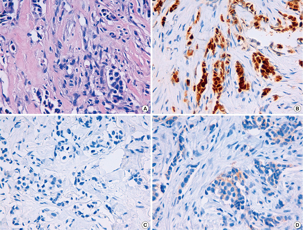
Figure 1 Pathologic findings of primary breast carcinoma. (A) Microphotograph showing invasive ductal carcinoma of the breast growing within a desmoplastic stroma (H&E stain, ×400). (B) Immunohistochemical stain showing positivity for estrogen receptor (90%) (×400). (C) Immunohistochemical stain showing absence of expression of progesterone receptor (×400). (D) Immunohistochemical stain showing negativity for human epidermal growth factor receptor 2 expression (1+) (×400). The aforementioned histologic features and the immunohistochemical expression pattern of the tumor were consistent with a diagnosis of invasive breast ductal carcinoma.
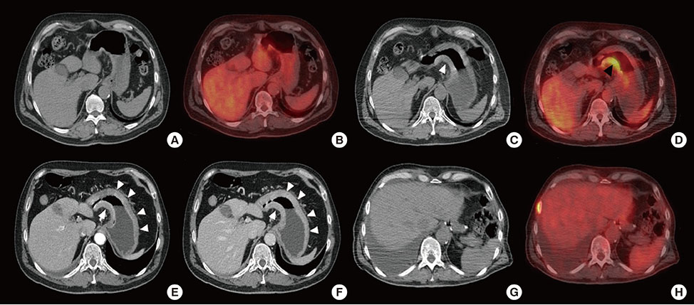
Figure 2 Pre- and postoperative positron emission tomography/computed tomography (PET/CT) finding. (A) Unenhanced CT and (B) Fusion PET/CT axial images showing no gastric fluorodeoxyglucose (FDG) uptake neither gastric wall thickening. (C, D) PET/CT showing the wall thickening of the gastric antrum (white arrow head) with a significant FDG uptake (black arrow head). (E) CT axial arterial and (F) venous phase, respectively at 35 and 80 seconds after intravenous iodinate contrast medium injection, showing the progression of the wall thickening, now involving the antrum and the body of the stomach, figuring out a linitis plastic (white arrow heads). (G, H) PET/CT axial images performed after the gastric resection showing no FDG uptake at the level of Roux-en-Y esophagojejunal anastomosis.
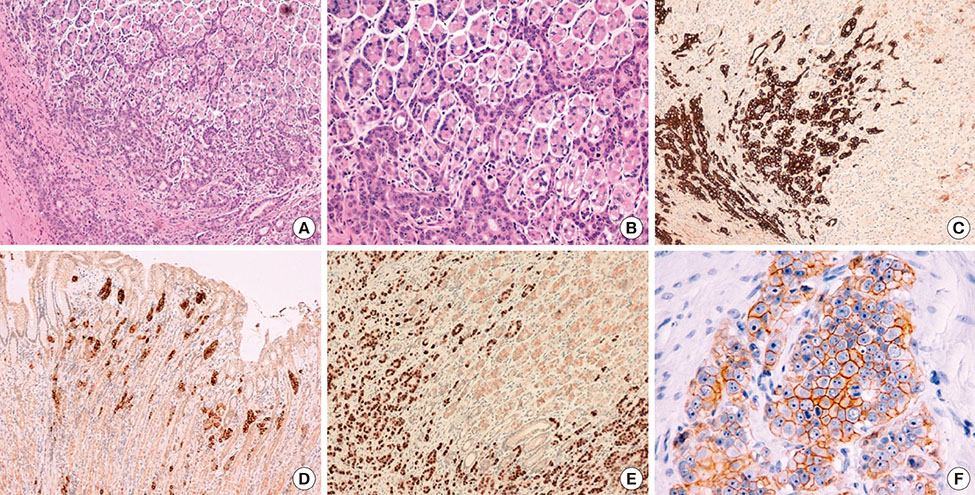
Figure 3 Pathologic findings of gastric recurrence. (A) microphotograph showing the stomach wall infiltrated by the neoplasm, arranged in tubular and solid pattern of growth (H&E stain, ×100). (B) Note the infiltration of tumor cells in the normal gastric mucosa, which have large vesicular nuclei with prominent nucleoli (H&E stain, ×200). (C) Immunohistochemical stain showing immunoreactivity for cytokeratin 7 (×100). (D) Immunohistochemical stain showing immunoreactivity for gross cystic disease fluid protein 15 (×100). (E) Immunohistochemical stain showing immunoreactivity for GATA binding protein 3 (GATA3) (×100). (F) Immunohistochemical stain showing positivity for human epidermal growth factor receptor 2 expression (×400). In the lacking of immunohistochemistry positivity to hormone receptors, the combined use of gross cystic disease fluid protein 15, GATA3 and cytokeratin 7 allowed for correct diagnosis of metastatic ductal breast cancer to the stomach.
Reference
-
1. Siegel R, Ma J, Zou Z, Jemal A. Cancer statistics, 2014. CA Cancer J Clin. 2014; 64:9–29.
Article2. Deb S, Lakhani SR, Ottini L, Fox SB. The cancer genetics and pathology of male breast cancer. Histopathology. 2016; 68:110–118.
Article3. Cummings MC, Simpson PT, Reid LE, Jayanthan J, Skerman J, Song S, et al. Metastatic progression of breast cancer: insights from 50 years of autopsies. J Pathol. 2014; 232:23–31.
Article4. Taal BG, den Hartog Jager FC, Steinmetz R, Peterse H. The spectrum of gastrointestinal metastases of breast carcinoma: I. stomach. Gastrointest Endosc. 1992; 38:130–135.
Article5. Ferri LE, Onerheim R, Emond C. Linitis plastica as the first indication of metastatic lobular carcinoma of the breast: case report and literature review. Can J Surg. 1999; 42:466–469.6. Ciulla A, Castronovo G, Tomasello G, Maiorana AM, Russo L, Daniele E, et al. Gastric metastases originating from occult breast lobular carcinoma: diagnostic and therapeutic problems. World J Surg Oncol. 2008; 6:78.
Article7. Madeya S, Börsch G. Gastrointestinal metastases of breast carcinoma. Gastrointest Endosc. 1993; 39:103–104.
Article8. Almubarak MM, Laé M, Cacheux W, de Cremoux P, Pierga JY, Reyal F, et al. Gastric metastasis of breast cancer: a single centre retrospective study. Dig Liver Dis. 2011; 43:823–827.
Article9. Nazareno J, Taves D, Preiksaitis HG. Metastatic breast cancer to the gastrointestinal tract: a case series and review of the literature. World J Gastroenterol. 2006; 12:6219–6224.
Article10. Yagi Y, Sasaki S, Yoshikawa A, Tsukioka Y, Fukushima W, Fujimura T, et al. Metastatic gastric carcinoma from breast cancer mimicking primary linitis plastica: a case report. Oncol Lett. 2015; 10:3483–3487.
Article11. Matsui M, Kojima O, Kawakami S, Uehara Y, Takahashi T. The prognosis of patients with gastric cancer possessing sex hormone receptors. Surg Today. 1992; 22:421–425.
Article12. van Velthuysen ML, Taal BG, van der Hoeven JJ, Peterse JL. Expression of oestrogen receptor and loss of E-cadherin are diagnostic for gastric metastasis of breast carcinoma. Histopathology. 2005; 46:153–157.
Article13. O'Connell FP, Wang HH, Odze RD. Utility of immunohistochemistry in distinguishing primary adenocarcinomas from metastatic breast carcinomas in the gastrointestinal tract. Arch Pathol Lab Med. 2005; 129:338–347.14. Davis DG, Siddiqui MT, Oprea-Ilies G, Stevens K, Osunkoya AO, Cohen C, et al. GATA-3 and FOXA1 expression is useful to differentiate breast carcinoma from other carcinomas. Hum Pathol. 2016; 47:26–31.
Article
- Full Text Links
-
- Actions
-
Cited
- CITED
-
- Close
- Share
-
- Similar articles
-
- Metastatic Gastric Linitis Plastica from Bladder Cancer Mimicking a Primary Gastric Carcinoma: a Case Report
- Primary Linitis Plastica of the Rectum: A Clinico-Pathologic Analysis of Five Cases with Special Reference to Comparison with Gastric Form
- Primary Linitis Plastica of the Colon with Mucinous Adenocarcinoma in Young Woman
- Breast Cancer Metastasis to the Stomach Resembling Early Gastric Cancer
- Synchronously Diagnosed Gastric Metastasis from Invasive Lobular Breast Carcinoma, Mimicking Primary Gastric Carcinoma
