Early Evaluation of Inflammatory Focus and Treatment Response Using ¹â¸F-Sodium Fluoride Bone Positron Emission Tomography/Computed Tomography in Patients with Metallic Implants: A Case Report
- Affiliations
-
- 1Department of Radiology, Soonchunhyang University Cheonan Hospital, Cheonan, Korea.
- 2Department of Nuclear Medicine, Soonchunhyang University Cheonan Hospital, Cheonan, Korea. gareen@naver.com
- 3Department of Orthopedic Surgery, Soonchunhyang University Cheonan Hospital, Cheonan, Korea.
- 4Department of Nuclear Medicine, Catholic Kwandong University College of Medicine, International St. Mary's Hospital, Incheon, Korea.
- KMID: 2412359
- DOI: http://doi.org/10.3348/jksr.2018.78.6.408
Abstract
- We report a case of a 76-year-old woman with elevated serum C-reactive protein levels for two weeks after spinal surgery. â¹â¹mTc-labeled white blood cell scan and dual ¹â¸F-sodium-fluoride (¹â¸F-NaF) bone positron emission tomography/computed tomography (PET/CT) were used to detect the site of inflammation. Using non-attenuation corrected images, soft tissue inflammation was detected without metal artifact. Antibiotic treatment attenuated inflammation as seen in an early-phase scan using follow-up ¹â¸F-NaF bone PET/CT. This case demonstrates the role of ¹â¸F-NaF bone PET/CT in the detection of inflammation sites, and can be used to evaluate treatment response in patients with metallic implants.
MeSH Terms
Figure
-
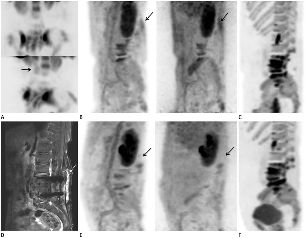
Fig. 1 A 76-year-old woman with perioperative inflammation detected on 18F-NaF bone PET/CT. A. MIP images of WBC SPECT. Upper row shows anterior view and lower row displays oblique view. Uptake of the soft tissue area in the early phase of 18F-NaF bone PET/CT is also observed in the WBC scan (arrow). B. MIP of early-phase 18F-NaF bone PET/CT scan shows increased radiotracer uptake around the hematoma removal site (arrows) and the spinal operation site, both on attenuation corrected (left) and non-attenuation corrected (right) images. C. MIP of standard 18F-NaF bone PET/CT scan shows an increase in radiotracer uptake at the operation site of spine. D. T2-weighted sagittal image of L-spine magnetic resonance imaging, using the Dixon technique. Image shows post-operative hematoma (arrow) and low signal intensity around the operation site due to metal artifact. E. The follow-up images of MIP of early-phase 18F-NaF bone PET/CT scan show decreased radiotracer uptake around the hematoma removal site (arrows), both on attenuation-corrected (left) and non-attenuation corrected (right) images. F. The follow-up image of MIP in standard 18F-NaF bone PET/CT scan shows diffuse radiotracer uptake due to post-operative changes without any significant difference compared with previous images. 18F-NaF = 18F-sodium-fluoride, MIP = maximum intensity projection, PET/CT = positron emission tomography/computed tomography, SPECT = single-photon emission computed tomography, WBC = white blood cell
Reference
-
1. Basu S, Zhuang H, Torigian DA, Rosenbaum J, Chen W, Alavi A. Functional imaging of inflammatory diseases using nuclear medicine techniques. Semin Nucl Med. 2009; 39:124–145.
Article2. El-Maghraby TA, Moustafa HM, Pauwels EK. Nuclear medicine methods for evaluation of skeletal infection among other diagnostic modalities. Q J Nucl Med Mol Imaging. 2006; 50:167–192.3. Bastawrous S, Bhargava P, Behnia F, Djang DS, Haseley DR. Newer PET application with an old tracer: role of 18F-NaF skeletal PET/CT in oncologic practice. Radiographics. 2014; 34:1295–1316.4. Sureshbabu W, Mawlawi O. PET/CT imaging artifacts. J Nucl Med Technol. 2005; 33:156–161.5. Shim JJ, Lee JW, Jeon MH, Lee SM. Recurrent surgical site infection of the spine diagnosed by dual 18F-NaF-bone PET/CT with early-phase scan. Skeletal Radiol. 2016; 45:1313–1316.6. Wong KK, Piert M. Dynamic bone imaging with 99mTc-labeled diphosphonates and 18F-NaF: mechanisms and applications. J Nucl Med. 2013; 54:590–599.7. Freesmeyer M, Stecker FF, Schierz JH, Hofmann GO, Winkens T. First experience with early dynamic 18F-NaF-PET/CT in patients with chronic osteomyelitis. Ann Nucl Med. 2014; 28:314–321.8. Palestro CJ, Love C, Tronco GG, Tomas MB, Rini JN. Combined labeled leukocyte and technetium 99m sulfur colloid bone marrow imaging for diagnosing musculoskeletal infection. Radiographics. 2006; 26:859–870.
Article9. Kransdorf MJ, Murphey MD. Radiologic evaluation of soft-tissue masses: a current perspective. AJR Am J Roentgenol. 2000; 175:575–587.10. Hargreaves BA, Worters PW, Pauly KB, Pauly JM, Koch KM, Gold GE. Metal-induced artifacts in MRI. AJR Am J Roentgenol. 2011; 197:547–555.
Article
- Full Text Links
-
- Actions
-
Cited
- CITED
-
- Close
- Share
-
- Similar articles
-
- Fluorine-18 Fluorodeoxyglucose Positron Emission Tomography/Computed Tomography Findings of Post Traumatic Lymphangioma in a Young Adult Male
- â¶â¸Gallium-Arginine-Glycine-Aspartic Acid and ¹â¸F-Fluorodeoxyglucose Positron Emission Tomography/Computed Tomography in Chondroblastic Osteosarcoma of the Skull
- Bone and Calcified Soft Tissue Metastases of Medullary Thyroid Carcinoma Better Characterized on ¹â¸F-Fluoride PET/CT than on â¶â¸Ga-Dotatate PET/CT
- F-18 Sodium Fluoride Positron Emission Tomography/Computed Tomography for Detection of Thyroid Cancer Bone Metastasis Compared with Bone Scintigraphy
- Oral cancer diagnosed using PET/CT: A case report
