Restor Dent Endod.
2018 Feb;43(1):e11. 10.5395/rde.2018.43.e11.
Mineral content analysis of root canal dentin using laser-induced breakdown spectroscopy
- Affiliations
-
- 1Department of Endodontics, Faculty of Dentistry, Hacettepe University, Ankara, Turkey. selenkkkaya@yahoo.com
- 2Department of Food Engineering, Faculty of Engineering, Hacettepe University, Ankara, Turkey.
- KMID: 2406096
- DOI: http://doi.org/10.5395/rde.2018.43.e11
Abstract
OBJECTIVES
This study aimed to introduce the use of laser-induced breakdown spectroscopy (LIBS) for evaluation of the mineral content of root canal dentin, and to assess whether a correlation exists between LIBS and scanning electron microscopy/energy dispersive spectroscopy (SEM/EDS) methods by comparing the effects of irrigation solutions on the mineral content change of root canal dentin.
MATERIALS AND METHODS
Forty teeth with a single root canal were decoronated and longitudinally sectioned to expose the canals. The root halves were divided into 4 groups (n = 10) according to the solution applied: group NaOCl, 5.25% sodium hypochlorite (NaOCl) for 1 hour; group EDTA, 17% ethylenediaminetetraacetic acid (EDTA) for 2 minutes; group NaOCl+EDTA, 5.25% NaOCl for 1 hour and 17% EDTA for 2 minutes; a control group. Each root half belonging to the same root was evaluated for mineral content with either LIBS or SEM/EDS methods. The data were analyzed statistically.
RESULTS
In groups NaOCl and NaOCl+EDTA, the calcium (Ca)/phosphorus (P) ratio decreased while the sodium (Na) level increased compared with the other groups (p < 0.05). The magnesium (Mg) level changes were not significant among the groups. A significant positive correlation was found between the results of LIBS and SEM/EDS analyses (r = 0.84, p < 0.001).
CONCLUSIONS
Treatment with NaOCl for 1 hour altered the mineral content of dentin, while EDTA application for 2 minutes had no effect on the elemental composition. The LIBS method proved to be reliable while providing data for the elemental composition of root canal dentin.
Keyword
MeSH Terms
Figure
-
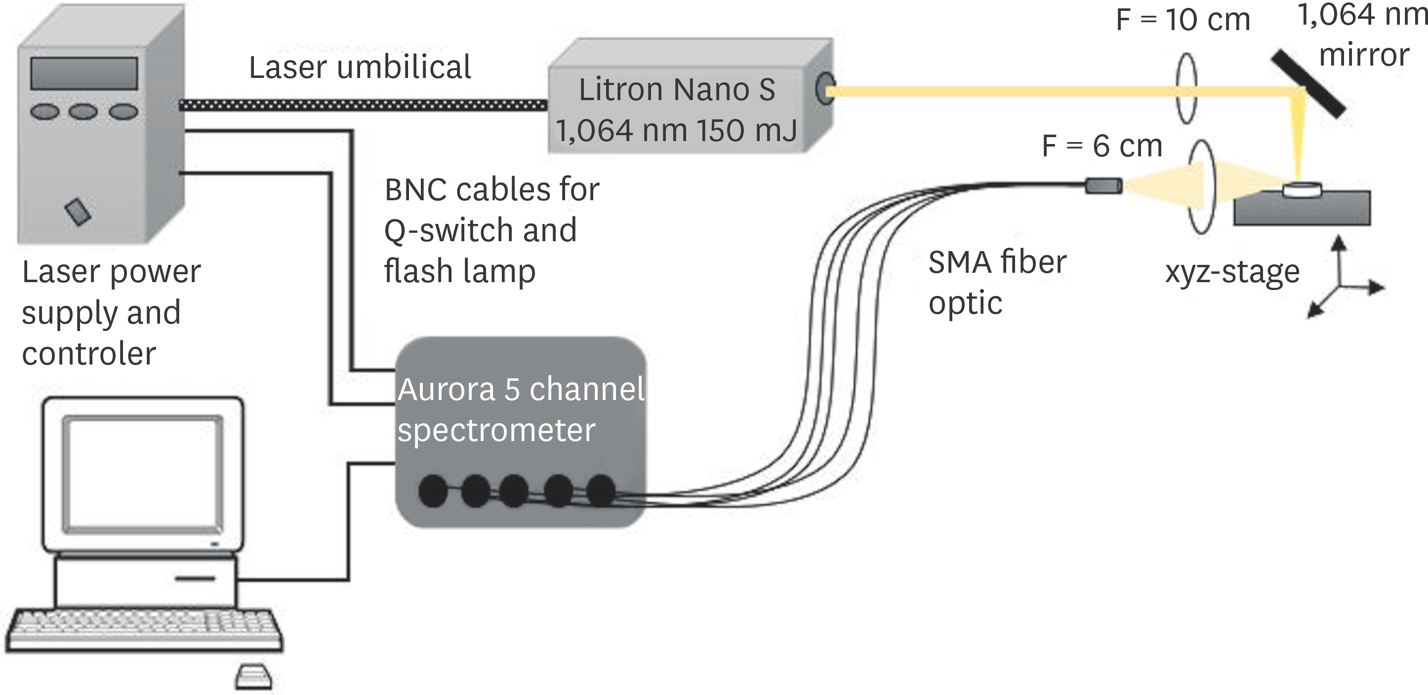
Figure 1. Schematic diagram of experimental setup for the laser-induced breakdown spectroscopy (LIBS) system. BNC, Bayonet Neill–Concelman; SMA, shape memory alloy.
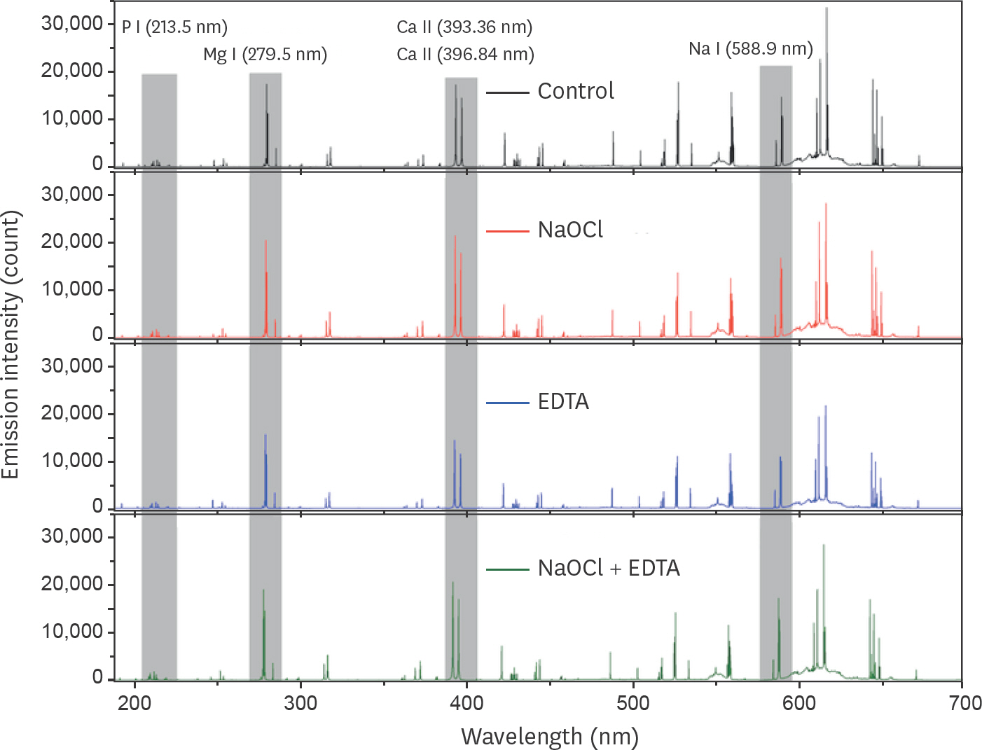
Figure 2. Laser-induced breakdown spectroscopy (LIBS) spectra for the groups. Ca, calcium; P, phosphorus; Mg, magnesium; Na, sodium; NaOCl, sodium hypochlorite; EDTA, ethylenediaminetetraacetic acid.
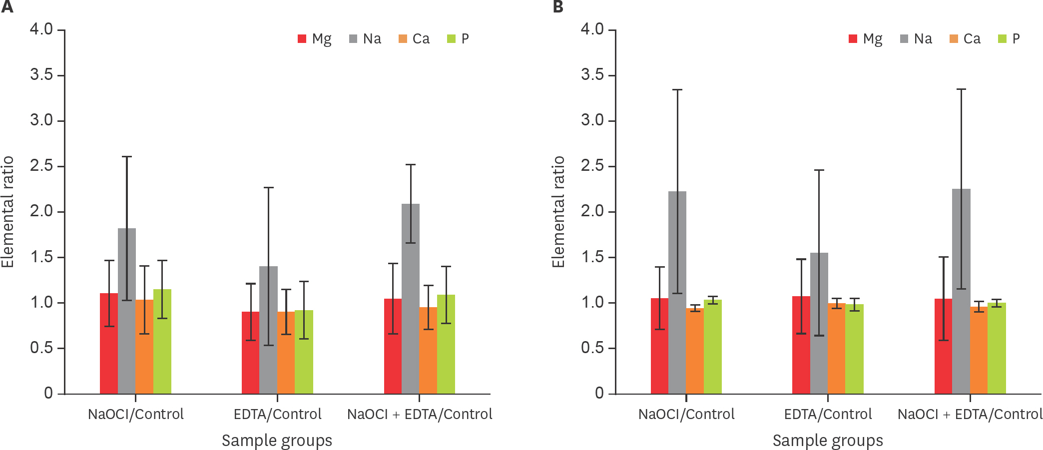
Figure 3. The change in the elemental distribution of the experimental groups compared with the control group. (A) Laser-induced breakdown spectroscopy (LIBS) analysis, (B) scanning electron microscopy/energy dispersive spectroscopy (SEM/EDS) analysis. Ca, calcium; P, phosphorus; Mg, magnesium; Na, sodium; NaOCl, sodium hypochlorite; EDTA, ethylenediaminetetraacetic acid.
Reference
-
References
1. Ørstavik D, Haapasalo M. Disinfection by endodontic irrigants and dressings of experimentally infected dentinal tubules. Endod Dent Traumatol. 1990; 6:142–149.2. Sen BH, Wesselink PR, Türkün M. The smear layer: a phenomenon in root canal therapy. Int Endod J. 1995; 28:141–148.
Article3. Doğ an H, Qalt S. Effects of chelating agents and sodium hypochlorite on mineral content of root dentin. J Endod. 2001; 27:578–580.4. Ari H, Erdemir A. Effects of endodontic irrigation solutions on mineral content of root canal dentin using ICP-AES technique. J Endod. 2005; 31:187–189.
Article5. Cohen M, Garnick JJ, Ringle RD, Hanes PJ, Thompson WO. Calcium and phosphorus content of roots exposed to the oral environment. J Clin Periodontol. 1992; 19:268–273.
Article6. Marshall GW Jr. Dentin: microstructure and characterization. Quintessence Int. 1993; 24:606–617.7. Rotstein I, Dankner E, Goldman A, Heling I, Stabholz A, Zalkind M. Histochemical analysis of dental hard tissues following bleaching. J Endod. 1996; 22:23–25.
Article8. Sayin TC, Serper A, Cehreli ZC, Otlu HG. The effect of EDTA, EGTA, EDTAC, and tetracycline-HCl with and without subsequent NaOCl treatment on the microhardness of root canal dentin. Oral Surg Oral Med Oral Pathol Oral Radiol Endod. 2007; 104:418–424.
Article9. Perdigão J, Eiriksson S, Rosa BT, Lopes M, Gomes G. Effect of calcium removal on dentin bond strengths. Quintessence Int. 2001; 32:142–146.10. Zhang K, Kim YK, Cadenaro M, Bryan TE, Sidow SJ, Loushine RJ, Ling JQ, Pashley DH, Tay FR. Effects of different exposure times and concentrations of sodium hypochlorite/ethylenediaminetetraacetic acid on the structural integrity of mineralized dentin. J Endod. 2010; 36:105–109.
Article11. Ozdemir HO, Buzoglu HD, Calt S, Cehreli ZC, Varol E, Temel A. Chemical and ultramorphologic effects of ethylenediaminetetraacetic acid and sodium hypochlorite in young and old root canal dentin. J Endod. 2012; 38:204–208.
Article12. Gurbuz T, Ozdemir Y, Kara N, Zehir C, Kurudirek M. Evaluation of root canal dentin after Nd: YAG laser irradiation and treatment with five different irrigation solutions: a preliminary study. J Endod. 2008; 34:318–321.
Article13. Ballal NV, Mala K, Bhat KS. Evaluation of decalcifying effect of maleic acid and EDTA on root canal dentin using energy dispersive spectrometer. Oral Surg Oral Med Oral Pathol Oral Radiol Endod. 2011; 112:e78–e84.
Article14. Arends J, ten Bosch JJ. Demineralization and remineralization evaluation techniques. J Dent Res. 1992; 71:924–928.
Article15. Alvira FC, Ramirez Rozzi F, Bilmes GM. Laser-induced breakdown spectroscopy microanalysis of trace elements in Homo sapiens teeth. Appl Spectrosc. 2010; 64:313–319.
Article16. Singh VK, Rai AK. Prospects for laser-induced breakdown spectroscopy for biomedical applications: a review. Lasers Med Sci. 2011; 26:673–687.
Article17. Khalid A, Bashir S, Akram M, Hayat A. Laser-induced breakdown spectroscopy analysis of human deciduous teeth samples. Lasers Med Sci. 2015; 30:2233–2238.
Article18. Anabitarte F, Cobo A, Lopez-Higuera JM. Laser-induced breakdown spectroscopy: fundamentals, applications, and challenges. ISRN Spectrosc. 2012; 2012:285240.
Article19. Alvira F, Ramirez Rozzi F, Torchia G, Roso L, Bilmes G. A new method for relative Sr determination in human teeth enamel. J Anthropol Sci. 2011; 89:153–160.20. National Institute of Standards and Technology: Atomic Spectra Database. Available from:. http://www.nist.gov/pml/atomic-spectra-database. (updated 2017 Nov 3).21. Samek LM, Liska M, Kaiser J, Beddows DC, Telle HH, Kukhlevsky SV. Clinical application of laser-induced breakdown spectroscopy to the analysis of teeth and dental materials. J Clin Laser Med Surg. 2000; 18:281–289.
Article22. Andrade DF, Pereira-Filho ER, Konieczynski P. Comparison of ICP OES and LIBS analysis of medicinal herbs rich in flavonoids from Eastern Europe. J Braz Chem Soc. 2017; 28:838–847.
Article23. El-Deftar MM, Robertson J, Foster S, Lennard C. Evaluation of elemental profiling methods, including laser-induced breakdown spectroscopy (LIBS), for the differentiation of Cannabis plant material grown in different nutrient solutions. Forensic Sci Int. 2015; 251:95–106.
Article24. Mehder AO, Habibullah YB, Gondal MA, Baig U. Qualitative and quantitative spectro-chemical analysis of dates using UV-pulsed laser induced breakdown spectroscopy and inductively coupled plasma mass spectrometry. Talanta. 2016; 155:124–132.
Article25. Hennequin M, Pajot J, Avignant D. Effects of different pH values of citric acid solutions on the calcium and phosphorus contents of human root dentin. J Endod. 1994; 20:551–554.
Article26. Bilge G, Sezer B, Eseller KE, Berberoglu H, Topcu A, Boyaci IH. Determination of whey adulteration in milk powder by using laser induced breakdown spectroscopy. Food Chem. 2016; 212:183–188.
Article27. Marending M, Paqué F, Fischer J, Zehnder M. Impact of irrigant sequence on mechanical properties of human root dentin. J Endod. 2007; 33:1325–1328.
Article28. Sim TP, Knowles JC, Ng YL, Shelton J, Gulabivala K. Effect of sodium hypochlorite on mechanical properties of dentine and tooth surface strain. Int Endod J. 2001; 34:120–132.
Article29. Marending M, Luder HU, Brunner TJ, Knecht S, Stark WJ, Zehnder M. Effect of sodium hypochlorite on human root dentine–mechanical, chemical and structural evaluation. Int Endod J. 2007; 40:786–793.30. Inaba D, Ruben J, Takagi O, Arends J. Effect of sodium hypochlorite treatment on remineralization of human root dentine in vitro. Caries Res. 1996; 30:218–224.31. Sayin TC, Serper A, Cehreli ZC, Kalayci S. Calcium loss from root canal dentin following EDTA, EGTA, EDTAC, and tetracycline-HCl treatment with or without subsequent NaOCl irrigation. J Endod. 2007; 33:581–584.
Article32. Cury JA, Bragotto C, Valdrighi L. The demineralizing efficiency of EDTA solutions on dentin. I. Influence of pH. Oral Surg Oral Med Oral Pathol. 1981; 52:446–448.33. Cobankara FK, Erdogan H, Hamurcu M. Effects of chelating agents on the mineral content of root canal dentin. Oral Surg Oral Med Oral Pathol Oral Radiol Endod. 2011; 112:e149–e154.
Article34. Verdelis K, Eliades G, Oviir T, Margelos J. Effect of chelating agents on the molecular composition and extent of decalcification at cervical, middle and apical root dentin locations. Endod Dent Traumatol. 1999; 15:164–170.
Article35. Apostolopoulos AX, Buonocore MG. Comparative dissolution rates of enamel, dentin, and bone. I. Effect of the organic matter. J Dent Res. 1966; 45:1093–1100.
Article
- Full Text Links
-
- Actions
-
Cited
- CITED
-
- Close
- Share
-
- Similar articles
-
- Optimizing experimental conditions of femtosecond laser-induced breakdown spectroscopy for differentiating melanoma from the normal dermis in Republic of Korea: an experimental study
- Age-dependent root canal instrumentation techniques: a comprehensive narrative review
- A comparative evaluation of CO2 and erbium-doped yttrium aluminium garnet laser therapy in the management of dentin hypersensitivity and assessment of mineral content
- Morphological characteristics of the mesiobuccal root in the presence of a second mesiobuccal canal: a micro-CT study
- The efficacy of ultrasonic irrigation technique on debris removal during root canal treatment
