A comparative evaluation of CO2 and erbium-doped yttrium aluminium garnet laser therapy in the management of dentin hypersensitivity and assessment of mineral content
- Affiliations
-
- 1Department of Oral Medicine, Periodontology & Oral Diagnosis, Faculty of Oral & Dental Medicine, Fayoum University, Al-Fayoum, Egypt. mahmoud_672000@yahoo.com
- 2Department of Public Health & Community Medicine, Faculty of Medicine, Tanta University, Tanta, Egypt.
- KMID: 2027820
- DOI: http://doi.org/10.5051/jpis.2014.44.5.227
Abstract
- PURPOSE
Dentin hypersensitivity is a potential threat to oral health. Laser irradiation may provide reliable and reproducible treatment but remains controversial. The present study aimed to evaluate the effects of CO2 or erbium-doped yttrium aluminium garnet (Er:YAG) laser therapy, and to assess mineral content.
METHODS
Eighteen human single-rooted teeth affected with advanced periodontitis were obtained. Buccal and lingual surfaces were planed to form 36 specimens. Ethylenediaminetetraacetic acid gel (24%) was applied to remove the smear layer and simulate hypersensitive teeth. The experimental groups were: group 1, control (no irradiation); group 2, CO2 laser (repetitive pulsed mode, 2 W, 2.7 J/cm2); and group 3, Er:YAG laser (slight contact mode, 40 mJ/pulse and 10 Hz). To evaluate dentinal tubule occlusion, six specimens per group (2-mm thickness) were prepared and observed using scanning electron microscopy (SEM) for calculation of the occlusion percentage. To evaluate the mineral content, six specimens per group (0.6-mm thickness) were used, and then the levels of Ca, K, Mg, Na, and P were measured by inductively coupled plasma-atomic emission spectrometry. In addition, the surface temperature of the specimens during laser irradiation was analyzed by a thermograph.
RESULTS
The SEM photomicrographs indicated melted areas around exposed dentinal tubules and a significantly greater percentage of tubular occlusion in the CO2 and Er:YAG laser groups than the control, and in the Er:YAG group than the CO2 laser group. In addition, no significant differences were noted among the experimental groups for the mineral elements analyzed. The CO2 laser group showed an evident thermal effect compared to the Er:YAG group.
CONCLUSIONS
CO2 and Er:YAG laser are effective in treating dentin hypersensitivity and reducing its symptoms. However, the Er:YAG laser has a more significant effect; thus, it may constitute a useful conditioning item. Furthermore, neither CO2 nor Er:YAG lasers affected the compositional structure of the mineral content.
Keyword
MeSH Terms
Figure
-
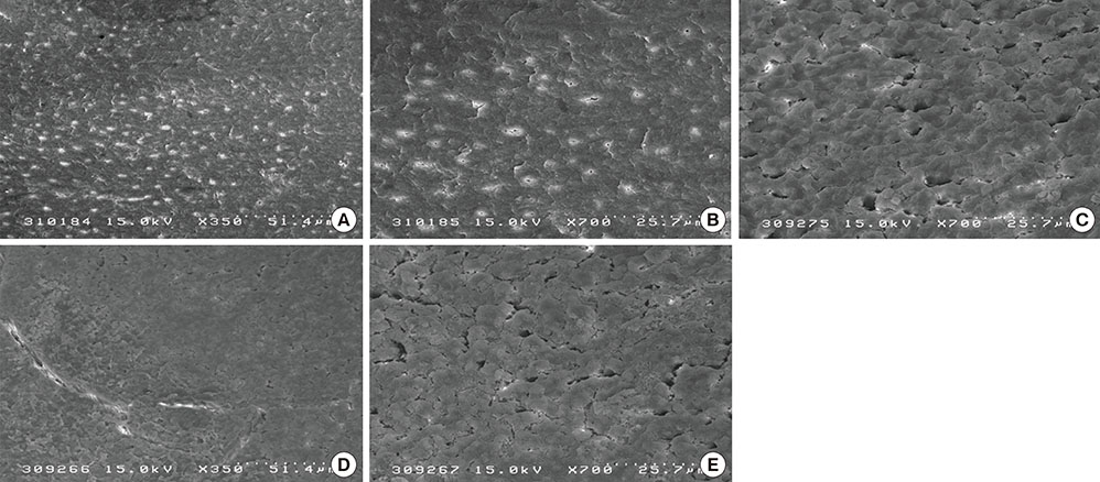
Figure 1 Scanning electron microscopy image of the root surfaces for the different experimental groups: (A-C) Group 1: control, clearly showed openings of the dentinal tubules with wide and funnel shaped orifices; (D and E) Group 2: the CO2 laser group, which generally showed a reduction in open dentinal tubules and reduced diameters when compared to the controls (×350 and ×700).
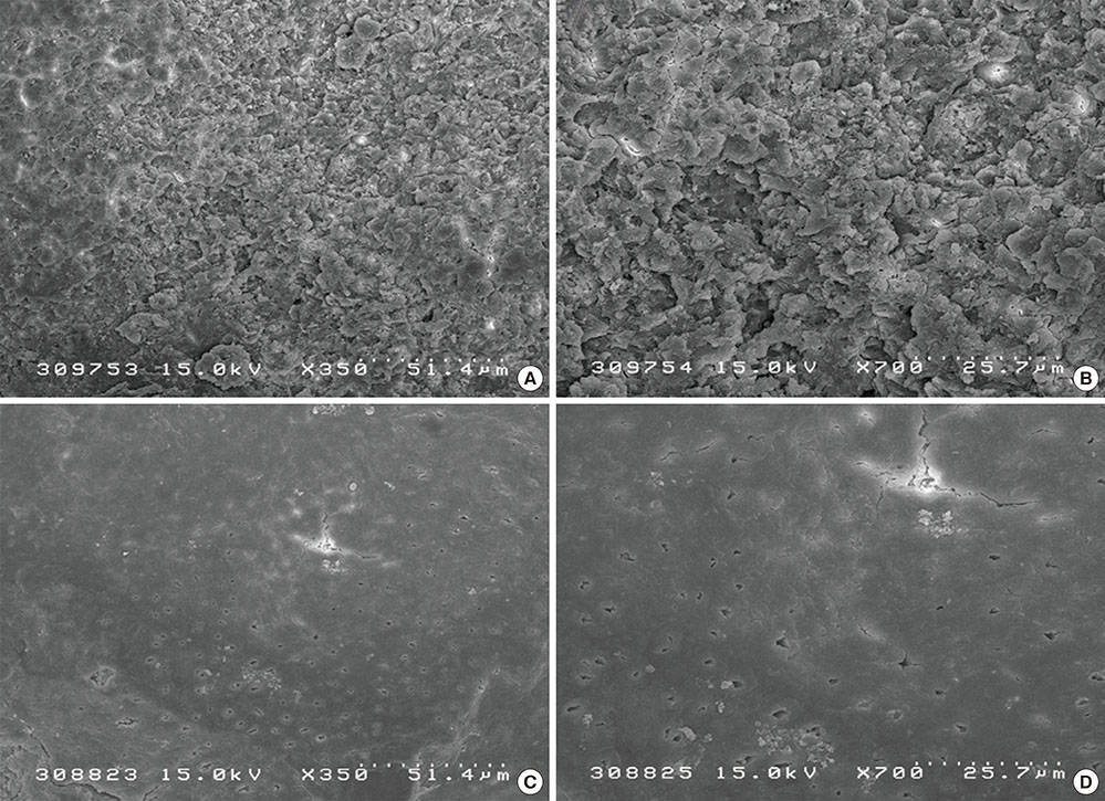
Figure 2 Scanning electron microscopy image of the root surfaces of group 3, which was treated by erbium-doped yttrium aluminium garnet laser. Narrowing of the orifices and a reduction in open dentinal tubules (A and B) as well as melting of the tubule orifices (C and D) can be noted (×350 and ×700).

Figure 3 Pie chart of the mean percentage of occluded tubules among the experimental groups. Er:YAG, erbium-doped yttrium aluminium garnet laser.
Reference
-
1. Petersson LG. The role of fluoride in the preventive management of dentin hypersensitivity and root caries. Clin Oral Investig. 2013; 17:Suppl 1. S63–S71.
Article2. Tonguc MO, Ozat Y, Sert T, Sonmez Y, Kirzioglu FY. Tooth sensitivity in fluorotic teeth. Eur J Dent. 2011; 5:273–280.
Article3. Barbour ME, Rees GD. The role of erosion, abrasion and attrition in tooth wear. J Clin Dent. 2006; 17:88–93.4. Lee BS, Chang CW, Chen WP, Lan WH, Lin CP. In vitro study of dentin hypersensitivity treated by Nd:YAP laser and bioglass. Dent Mater. 2005; 21:511–519.
Article5. Markowitz K, Pashley DH. Discovering new treatments for sensitive teeth: the long path from biology to therapy. J Oral Rehabil. 2008; 35:300–315.
Article6. Brannstrom M, Astrom A. The hydrodynamics of the dentine; its possible relationship to dentinal pain. Int Dent J. 1972; 22:219–227.7. Arrais CA, Micheloni CD, Giannini M, Chan DC. Occluding effect of dentifrices on dentinal tubules. J Dent. 2003; 31:577–584.
Article8. Pashley DH, Galloway SE. The effects of oxalate treatment on the smear layer of ground surfaces of human dentine. Arch Oral Biol. 1985; 30:731–737.
Article9. Kerns DG, Scheidt MJ, Pashley DH, Horner JA, Strong SL, Van Dyke TE. Dentinal tubule occlusion and root hypersensitivity. J Periodontol. 1991; 62:421–428.
Article10. Jacobsen PL, Bruce G. Clinical dentin hypersensitivity: understanding the causes and prescribing a treatment. J Contemp Dent Pract. 2001; 2:1–12.
Article11. Matsumoto K, Funai H, Shirasuka T, Wakabayashi H. Effects of Nd: YAG- laser in treatment of cervical hypersensitive dentine. Jpn J Conserv Dent. 1985; 28:760–765.12. Rohanizadeh R, LeGeros RZ, Fan D, Jean A, Daculsi G. Ultrastructural properties of laser-irradiated and heat-treated dentin. J Dent Res. 1999; 78:1829–1835.
Article13. Zhang C, Matsumoto K, Kimura Y, Harashima T, Takeda FH, Zhou H. Effects of CO2 laser in treatment of cervical dentinal hypersensitivity. J Endod. 1998; 24:595–597.
Article14. Aranha AC, Domingues FB, Franco VO, Gutknecht N, Eduardo Cde P. Effects of Er:YAG and Nd:YAG lasers on dentin permeability in root surfaces: a preliminary in vitro study. Photomed Laser Surg. 2005; 23:504–508.15. Kimura Y, Wilder-Smith P, Yonaga K, Matsumoto K. Treatment of dentine hypersensitivity by lasers: a review. J Clin Periodontol. 2000; 27:715–721.
Article16. Kantorowitz Z, Featherstone JD, Fried D. Caries prevention by CO2 laser treatment: dependency on the number of pulses used. J Am Dent Assoc. 1998; 129:585–591.
Article17. Ten Cate AR. Composition formation and structure of dentin. In : Nanci A, Ten Cate AR, editors. Ten Cate's oral histology: development, structure, and function. 7th ed. St. Louis: Mosby Elsevier;2008. p. 166–168.18. Ari H, Erdemir A. Effects of endodontic irrigation solutions on mineral content of root canal dentin using ICP-AES technique. J Endod. 2005; 31:187–189.
Article19. Pulido MT, Wefel JS, Hernandez MM, Denehy GE, Guzman-Armstrong S, Chalmers JM, et al. The inhibitory effect of MI paste, fluoride and a combination of both on the progression of artificial caries-like lesions in enamel. Oper Dent. 2008; 33:550–555.
Article20. Wiesmann HP, Tkotz T, Joos U, Zierold K, Stratmann U, Szuwart T, et al. Magnesium in newly formed dentin mineral of rat incisor. J Bone Miner Res. 1997; 12:380–383.
Article21. Deutsch AS, Cohen BI, Musikant BL. Inductively coupled plasma-emission spectroscopy and atomic absorption for the use of elemental analysis of a root canal after lasing with a holmium:YAG laser. J Endod. 2003; 29:404–406.
Article22. Secilmis A, Altintas S, Usumez A, Berk G. Evaluation of mineral content of dentin prepared by erbium, chromium:yttrium scandium gallium garnet laser. Lasers Med Sci. 2008; 23:421–425.
Article23. Khabbaz MG, Makropoulou MI, Serafetinides AA, Papadopoulos D, Papagiakoumou E. Q-switched versus free-running Er:YAG laser efficacy on the root canal walls of human teeth: a SEM study. J Endod. 2004; 30:585–588.
Article24. Hamachi T, Iwamoto Y, Hirofuji T, Kabashima H, Maeda K. Clinical evaluation of GaAlAs-semicondoctor laser in the treatment of cervical hypersensitivity dentin. Jpn J Conserv Dent. 1992; 35:12–17.25. Absi EG, Addy M, Adams D. Dentine hypersensitivity. A study of the patency of dentinal tubules in sensitive and non-sensitive cervical dentine. J Clin Periodontol. 1987; 14:280–284.
Article26. Schwarz F, Arweiler N, Georg T, Reich E. Desensitizing effects of an Er:YAG laser on hypersensitive dentine. J Clin Periodontol. 2002; 29:211–215.
Article27. Lan WH, Lee BS, Liu HC, Lin CP. Morphologic study of Nd:YAG laser usage in treatment of dentinal hypersensitivity. J Endod. 2004; 30:131–134.
Article28. Bonin P, Boivin R, Poulard J. Dentinal permeability of the dog canine after exposure of a cervical cavity to the beam of a CO2 laser. J Endod. 1991; 17:116–118.
Article29. Moritz A, Gutknecht N, Schoop U, Wernisch J, Lampert F, Sperr W. Effects of CO2 laser irradiation on treatment of hypersensitive dental necks: results of an in vitro study. J Clin Laser Med Surg. 1995; 13:397–400.
Article30. Moritz A, Gutknecht N, Schoop U, Goharkhay K, Ebrahim D, Wernisch J, et al. The advantage of CO2-treated dental necks, in comparison with a standard method: results of an in vivo study. J Clin Laser Med Surg. 1996; 14:27–32.
Article31. Aoki A, Ando Y, Watanabe H, Ishikawa I. In vitro studies on laser scaling of subgingival calculus with an erbium:YAG laser. J Periodontol. 1994; 65:1097–1106.
Article32. Walsh JT Jr, Flotte TJ, Deutsch TF. Er:YAG laser ablation of tissue: effect of pulse duration and tissue type on thermal damage. Lasers Surg Med. 1989; 9:314–326.
Article33. Matsui S, Kozuka M, Takayama J, Ueda K, Nakamura H, Ito K, et al. Stimulatory Effects of CO(2) Laser, Er:YAG Laser and Ga-Al-As Laser on Exposed Dentinal Tubule Orifices. J Clin Biochem Nutr. 2008; 42:138–143.
Article34. Takeda FH, Harashima T, Kimura Y, Matsumoto K. A comparative study of the removal of smear layer by three endodontic irrigants and two types of laser. Int Endod J. 1999; 32:32–39.
Article35. Rösing CK, Fiorini T, Liberman DN, Cavagni J. Dentine hypersensitivity: analysis of self-care products. Braz Oral Res. 2009; 23:Suppl 1. 56–63.
Article36. Dilber E, Malkoc MA, Ozturk AN, Ozturk F. Effect of various laser irradiations on the mineral content of dentin. Eur J Dent. 2013; 7:74–80.37. Kashima-Tanaka M, Tsujimoto Y, Kawamoto K, Senda N, Ito K, Yamazaki M. Generation of free radicals and/or active oxygen by light or laser irradiation of hydrogen peroxide or sodium hypochlorite. J Endod. 2003; 29:141–143.
Article38. Lee BS, Lin CP, Hung YL, Lan WH. Structural changes of Er:YAG laser-irradiated human dentin. Photomed Laser Surg. 2004; 22:330–334.
Article
- Full Text Links
-
- Actions
-
Cited
- CITED
-
- Close
- Share
-
- Similar articles
-
- Microshear bond strength according to dentin cleansing methods before recementation
- Erbium-doped Yttrium Aluminum Garnet Laser to Treat Familial Acanthosis Nigricans
- Treatment with the Pinhole Technique Using Erbium-Doped Yttrium Aluminium Garnet Laser for a Cafe au Lait Macule and Carbon Dioxide Laser for Facial Telangiectasia
- Effect of erbium-doped: yttrium, aluminium and garnet laser irradiation on the surface microstructure and roughness of sand-blasted, large grit, acid-etched implants
- Acquired Dermal Melanocytosis of the Nose Successfully Treated with Neodymium-doped Yttrium Aluminum Garnet 1064 nm Laser
