Microshear bond strength according to dentin cleansing methods before recementation
- Affiliations
-
- 1Department of Prosthodontics, Faculty of Dentistry, Near East University, Nicosia, Mersin, Turkey. gokcemeric@yahoo.com
- KMID: 2233334
- DOI: http://doi.org/10.4047/jap.2014.6.2.79
Abstract
- PURPOSE
The aim of this study was to determine the efficiency of Erbium, Chromium: Yttrium-Scandium-Gallium-Garnet laser in different output powers for removing permanent resin cement residues and therefore its influence on microshear bond strength compared to other cleaning methods.
MATERIALS AND METHODS
90 extracted human molars were sectioned in 1 mm thickness. Resin cement was applied to surface of sliced teeth. After the removal of initial cement, 6 test groups were prepared by various dentin surface treatment methods as follows: no treatment (Group 1), ethylene diamine tetra acetic acid application (Group 2), Endosolv R application (Group 3), 1.25 W Erbium, Chromium:Yttrium-Scandium-Gallium-Garnet laser irradiation (Group 4), 2 W Erbium, Chromium:Yttrium-Scandium-Gallium-Garnet laser irradiation (Group 5) and 3.5 W Erbium, Chromium:Yttrium-Scandium-Gallium-Garnet laser irradiation (Group 6). The topography and morphology of the treated dentin surfaces were investigated by scanning electron microscopy (n=2 for each group). Following the repetitive cementation, microshear bond strength between dentin and cement (n=26 in per group) were measured with universal testing machine and the data were analyzed by Kruskal Wallis H Test with Bonferroni correction (P<.05). Fracture patterns were investigated by light microscope.
RESULTS
Mean microshear bond strength +/- SD (MPa) for each group was 34.9 +/- 17.7, 32.1 +/- 15.8, 37.8 +/- 19.3, 31.3 +/- 12.7, 44.4 +/- 13.6, 40.2 +/- 13.2 respectively. Group 5 showed significantly difference from Group 1, Group 2 and Group 4. Also, Group 6 was found statistically different from Group 4.
CONCLUSION
2 W and 3.5 W Erbium, Chromium: Yttrium-Scandium-Gallium-Garnet laser application were found efficient in removing resin residues.
MeSH Terms
Figure
-
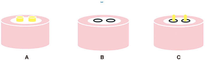
Fig. 1 Stages of sample preparation for initial and repetitive cement application. (A) Two parallel resin cylinders located on teeth (which was fixed on acrylic resin blocks) for initial cementation, (B) Debonded cement areas on dentin surface which were defined with permanent marker, (C) Two parallel resin cylinders located on teeth (which was fixed on acrylic resin blocks) for repetitive cementation.
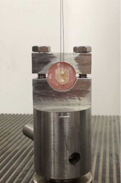
Fig. 2 Wire loop loading of samples during microshear bond test.
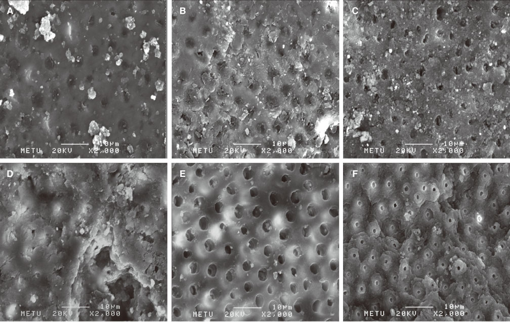
Fig. 3 (A) SEM view of untreated dentin surface with smear layer and plugs (Control group), (B) SEM view of EDTA applied dentin surface with some smear (Group 2), (C) SEM view of Endosolv R treated dentin with sporadically closed dentin tubules (Group 3), (D) SEM view of dentin irradiated with 1.25 W Er, Cr:YSGG laser which contains diffuse cement layer with locally perforated cement areas (Group 4), (E) SEM view of dentin irradiated with 2 W Er, Cr:YSGG laser with even dentin surface and open dentin tubules (Group 5), (F) SEM view of dentin irradiated with 3.5 W Er, Cr:YSGG laser with few dentin tubules (Group 6).

Fig. 4 Fracture modes of specimens after µSBS test.
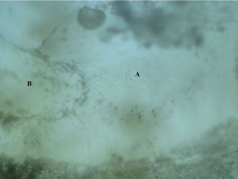
Fig. 5 Mixed fracture pattern of the dentin resin interface which occurred after microshear bond strength test. (A) Adhesive fracture pattern between tooth and resin, (B) Cohesive fracture pattern within tooth.
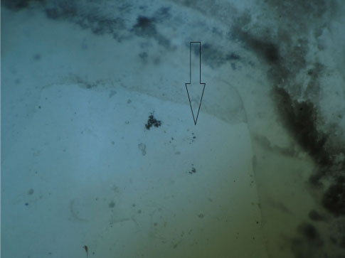
Fig. 6 Cohesive fracture within tooth which formed after microshear bond strength test. The blank arrow indicates cohesive fracture within tooth which formed after microshear bond strength test.
Cited by 1 articles
-
The effect of resin cement type and cleaning method on the shear bond strength of resin cements for recementing restorations
Roodabeh Koodaryan, Ali Hafezeqoran, Amin Khakpour Maleki
J Adv Prosthodont. 2017;9(2):110-117. doi: 10.4047/jap.2017.9.2.110.
Reference
-
1. Prata RA, de Oliveira VP, de Menezes FC, Borges GA, de Andrade OS, Gonçalves LS. Effect of 'Try-in' paste removal method on bond strength to lithium disilicate ceramic. J Dent. 2011; 39:863–870.2. Tanumiharja M, Burrow MF, Tyas MJ. Microtensile bond strengths of seven dentin adhesive systems. Dent Mater. 2000; 16:180–187.3. Sümer E, Değer Y. Contemporary permanent luting agents used in dentistry: A literature review. Int Dent Res. 2011; 1:26–31.4. Langer A, Ilie N. Dentin infiltration ability of different classes of adhesive systems. Clin Oral Investig. 2013; 17:205–216.5. Lohbauer U, Nikolaenko SA, Petschelt A, Frankenberger R. Resin tags do not contribute to dentin adhesion in self-etching adhesives. J Adhes Dent. 2008; 10:97–103.6. Giachetti L, Bertini F, Scaminaci Russo D. Investigation into the nature of dentin resin tags: a scanning electron microscopic morphological analysis of demineralized bonded dentin. J Prosthet Dent. 2004; 92:233–238.7. Tao L, Pashley DH. Shear bond strengths to dentin: effects of surface treatments, depth and position. Dent Mater. 1988; 4:371–378.8. Van Meerbeek B, Inokoshi S, Braem M, Lambrechts P, Vanherle G. Morphological aspects of the resin-dentin interdiffusion zone with different dentin adhesive systems. J Dent Res. 1992; 71:1530–1540.9. Gwinnett AJ. Quantitative contribution of resin infiltration/hybridization to dentin bonding. Am J Dent. 1993; 6:7–9.10. Nakabayashi N. Bonding of restorative materials to dentine: the present status in Japan. Int Dent J. 1985; 35:145–154.11. Obeidi A, Liu PR, Ramp LC, Beck P, Gutknecht N. Acid-etch interval and shear bond strength of Er,Cr:YSGG laser-prepared enamel and dentin. Lasers Med Sci. 2010; 25:363–369.12. Altintas SH, Tak O, Secilmis A, Usumez A. Effect of provisional cements on shear bond strength of porcelain laminate veneers. Eur J Dent. 2011; 5:373–379.13. Santos MJ, Bapoo H, Rizkalla AS, Santos GC. Effect of dentin-cleaning techniques on the shear bond strength of self-adhesive resin luting cement to dentin. Oper Dent. 2011; 36:512–520.14. Adebayo OA, Burrow MF, Tyas MJ. Dentine bonding after CPP-ACP paste treatment with and without conditioning. J Dent. 2008; 36:1013–1024.15. Munirathinam D, Mohanaj D, Beganam M. Efficacy of various cleansing techniques on dentin wettability and its influence on shear bond strength of a resin luting agent. J Adv Prosthodont. 2012; 4:139–145.16. Grasso CA, Caluori DM, Goldstein GR, Hittelman E. In vivo evaluation of three cleansing techniques for prepared abutment teeth. J Prosthet Dent. 2002; 88:437–441.17. Roberts S, Kim JR, Gu LS, Kim YK, Mitchell QM, Pashley DH, Tay FR. The efficacy of different sealer removal protocols on bonding of self-etching adhesives to AH plus-contaminated dentin. J Endod. 2009; 35:563–567.18. Usumez A, Aykent F. Bond strengths of porcelain laminate veneers to tooth surfaces prepared with acid and Er,Cr: YSGG laser etching. J Prosthet Dent. 2003; 90:24–30.19. Chou JC, Chen CC, Ding SJ. Effect of Er,Cr:YSGG laser parameters on shear bond strength and microstructure of dentine. Photomed Laser Surg. 2009; 27:481–486.20. Jaberi Ansari Z, Fekrazad R, Feizi S, Younessian F, Kalhori KA, Gutknecht N. The effect of an Er,Cr:YSGG laser on the micro-shear bond strength of composite to the enamel and dentin of human permanent teeth. Lasers Med Sci. 2012; 27:761–765.21. Gurgan S, Kiremitci A, Cakir FY, Gorucu J, Alpaslan T, Yazici E, Gutknecht N. Shear bond strength of composite bonded to Er,Cr:YSGG laser-prepared dentin. Photomed Laser Surg. 2008; 26:495–500.22. Cvikl B, Moser G, Wernisch J, Raabe M, Gruber R, Moritz A. The impact of Er,Cr:YSGG laser on the shear strength of the bond between dentin and ceramic is dependent on the adhesive material. Lasers Med Sci. 2012; 27:717–722.23. Lin S, Caputo AA, Eversole LR, Rizoiu I. Topographical characteristics and shear bond strength of tooth surfaces cut with a laser-powered hydrokinetic system. J Prosthet Dent. 1999; 82:451–455.24. Ishida K, Endo T, Shinkai K, Katoh Y. Shear bond strength of rebonded brackets after removal of adhesives with Er,Cr:YSGG laser. Odontology. 2011; 99:129–134.25. Ahrari F, Basafa M, Fekrazad R, Mokarram M, Akbari M. The efficacy of Er,Cr:YSGG laser in reconditioning of metallic orthodontic brackets. Photomed Laser Surg. 2012; 30:41–46.26. Ahrari F, Fekrazad R, Kalhori KA, Ramtin M. Reconditioning of ceramic orthodontic brackets with an Er,Cr:YSGG laser. Lasers Med Sci. 2013; 28:223–228.27. Bishara SE, VonWald L, Laffoon JF, Warren JJ. The effect of repeated bonding on the shear bond strength of a composite resin orthodontic adhesive. Angle Orthod. 2000; 70:435–441.28. Andrade AM, Garcia E, Moura SK, Reis A, Loguercio A, Silva LM, Pimentel GH, Grande RH. Do the microshear test variables affect the bond strength values? Int J Dent. 2012; 2012:618960.29. Armstrong S, Geraldeli S, Maia R, Raposo LH, Soares CJ, Yamagawa J. Adhesion to tooth structure: a critical review of "micro" bond strength test methods. Dent Mater. 2010; 26:e50–e62.30. Toba S, Veerapravati W, Shimada Y, Nikaido T, Tagami J. Micro-shear bond strengths of adhesive resins to coronal dentin versus the floor of the pulp chamber. Am J Dent. 2003; 16 Spec No:51A–56A.31. Shabka AA, Khalaf MM. An investigation on the shear bond strength of one dentin adhesive at two different dentin depths. Egypt Dent J. 1995; 41:1031–1034.32. Pashley EL, Tao L, Matthews WG, Pashley DH. Bond strengths to superficial, intermediate and deep dentin in vivo with four dentin bonding systems. Dent Mater. 1993; 9:19–22.33. Burrow MF, Takakura H, Nakajima M, Inai N, Tagami J, Takatsu T. The influence of age and depth of dentin on bonding. Dent Mater. 1994; 10:241–246.34. Giannini M, Chaves P, Oliveira MT. Effect of tooth age on bond strength to dentin. J Appl Oral Sci. 2003; 11:342–347.35. Brackett WW, Tay FR, Looney SW, Ito S, Haisch LD, Pashley DH. The effect of subject age on the microtensile bond strengths of a resin and a resin-modified glass ionomer adhesive to tooth structure. Oper Dent. 2008; 33:282–286.36. International Organization For Standardization. Dental materials-Guidance on testing of adhesion to tooth structure. ISO TR 11405. Geneva: ISO;1994.37. Fawzi EM, Elkassas DW, Ghoneim AG. Bonding strategies to pulp chamber dentin treated with different endodontic irrigants: microshear bond strength testing and SEM analysis. J Adhes Dent. 2010; 12:63–70.38. Kim DS, Park SH, Choi GW, Choi KK, Kim SY. Effect of EDTA treatment on the hybrid layer durability in total-etch dentin adhesives. Dent Mater J. 2011; 30:717–722.39. Shokouhinejad N, Sabeti MA, Hasheminasab M, Shafiei F, Shamshiri AR. Push-out bond strength of Resilon/Epiphany self-etch to intraradicular dentin after retreatment: a preliminary study. J Endod. 2010; 36:493–496.40. Gambrel MG, Hartwell GR, Moon PC, Cardon JW. The effect of endodontic solutions on resorcinol-formalin paste in teeth. J Endod. 2005; 31:25–29.41. Ramzi H, Shokouhinejad N, Saghiri MA, Samieefard A. Efficacy of Three Different Methods in the Retreatment of Root Canals Filled with Resilon/Epiphany SE. Iran Endod J. 2010; 5:161–166.42. Vranas RN, Hartwell GR, Moon PC. The effect of endodontic solutions on resorcinol-formalin paste. J Endod. 2003; 29:69–72.43. George JD, Price CJ, Marr MC, Myers CB, Jahnke GD. Evaluation of the developmental toxicity of formamide in New Zealand white rabbits. Toxicol Sci. 2002; 69:165–174.44. Başaran EG, Ayna E, Başaran G, Beydemir K. Influence of different power outputs of erbium, chromium:yttrium-scandium-gallium-garnet laser and acid etching on shear bond strengths of a dual-cure resin cement to enamel. Lasers Med Sci. 2011; 26:13–19.45. Apel C, Meister J, Ioana RS, Franzen R, Hering P, Gutknecht N. The ablation threshold of Er:YAG and Er:YSGG laser radiation in dental enamel. Lasers Med Sci. 2002; 17:246–252.46. Ergücü Z, Celik EU, Unlü N, Türkün M, Ozer F. Effect of Er,Cr:YSGG laser on the microtensile bond strength of two different adhesives to the sound and caries-affected dentin. Oper Dent. 2009; 34:460–466.47. Lee BS, Lin PY, Chen MH, Hsieh TT, Lin CP, Lai JY, Lan WH. Tensile bond strength of Er,Cr:YSGG laser-irradiated human dentin and analysis of dentin-resin interface. Dent Mater. 2007; 23:570–578.48. Moretto SG, Azambuja N Jr, Arana-Chavez VE, Reis AF, Giannini M, Eduardo CP, De Freitas PM. Effects of ultramorphological changes on adhesion to lased dentin-Scanning electron microscopy and transmission electron microscopy analysis. Microsc Res Tech. 2011; 74:720–726.
- Full Text Links
-
- Actions
-
Cited
- CITED
-
- Close
- Share
-
- Similar articles
-
- The effect of resin cement type and cleaning method on the shear bond strength of resin cements for recementing restorations
- Comparative evaluation of micro-shear bond strength between two different luting methods of resin cement to dentin
- Bonding effects of cleaning protocols and time-point of acid etching on dentin impregnated with endodontic sealer
- Effect of Er:YAG lasing on the dentin bonding strength of two-step adhesives
- Comparative evaluation of Emblica officinalis as an etchant and an MMP inhibitor with orthophosphoric acid and chlorhexidine on the microshear bond strength of composite resin: an ex vivo study
