Comparative evaluation of Emblica officinalis as an etchant and an MMP inhibitor with orthophosphoric acid and chlorhexidine on the microshear bond strength of composite resin: an ex vivo study
- Affiliations
-
- 1Department of Conservative Dentistry and Endodontics, JSS Dental College and Hospital, JSS Academy of Higher Education & Research, Mysuru, India
- KMID: 2548080
- DOI: http://doi.org/10.5395/rde.2021.46.e36
Abstract
Objectives
This study aimed to evaluate Emblica officinalis (Indian gooseberry or amla) as an acid etchant and matrix metalloproteinase (MMP) inhibitor, and to compare its effect on the microshear bond strength of composite resin with orthophosphoric acid (OPA) and 2% chlorhexidine (CHX) as an acid etchant and MMP inhibitor, respectively.
Materials and Methods
The etching effect and MMP-inhibiting action of amla on dentin samples were confirmed by scanning electron microscopy (SEM) and gelatin zymography, respectively. Dentinal slabs (3 mm thick) from 80 extracted human molars were divided into 10 and 20 samples to form 2 control groups and 3 experimental groups. Groups 1, 2, and 4 were etched with OPA and groups 3 and 5 with amla juice. An MMP inhibitor was then applied: CHX for group 2 and amla extract for groups 4 and 5. Groups 1 and 3 received no MMP inhibitor. All specimens received a standardized bonding protocol and composite resin build-up, and were subjected to microshear bond strength testing. The force at which the fracture occurred was recorded and statistically analyzed.
Results
Amla juice had a similar etching effect as a self-etch adhesive in SEM and 100% amla extract was found to inhibit MMP-9 by gelatin zymography. The microshear bond strength values of amla were lower than those obtained for OPA and CHX, but the difference was not statistically significant.
Conclusions
Amla has a promising role as an acid etchant and MMP inhibitor, but further studies are necessary to substantiate its efficacy.
Keyword
Figure
-
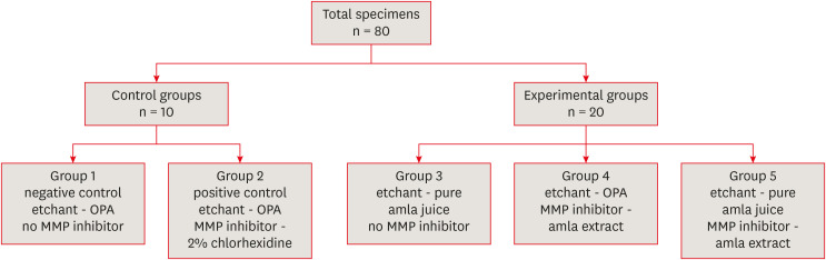
Figure 1 Study group distribution.OPA, 37% orthophosphoric acid; MMP, matrix metalloproteinase.

Figure 2 Scanning electron microscopic images evaluating and comparing the etching effect of 37% OPA with pure amla juice on dentin samples at ×5,000 magnification. (A) Etched with 37% OPA. (B) Etched with pure amla juice.OPA, orthophosphoric acid.
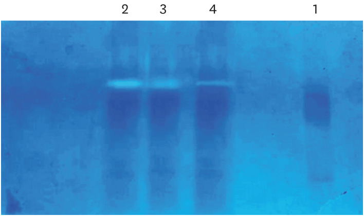
Figure 3 Effective concentration at which amla extract acts as an matrix metalloproteinase inhibitor as determined using gelatin zymography.Lane 1, untreated mineralized dentin powder (negative control); Lane 2, dentin powder demineralized with 1% OPA for 10 minutes at 4ºC (positive control); Lane 3, dentin powder demineralized with 1% OPA for 10 minutes at 4ºC and then incubated with 50% amla extract for 30 minutes; Lane 4, dentin powder demineralized with 1% OPA for 10 minutes at 4ºC and then incubated with 100% amla extract for 30 minutes.OPA, orthophosphoric acid.
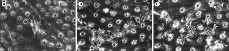
Figure 4 Scanning electron microscopic images evaluating and comparing different etchants and MMP inhibitors at ×5,000 magnification. (A) Etched with 37% OPA followed by 2% CHX as an MMP inhibitor. (B) Etched with 37% OPA followed by 100% amla extract as an MMP inhibitor. (C) Etched with pure amla juice followed by 100% amla extract as an MMP inhibitor.OPA, orthophosphoric acid; CHX, chlorhexidine; MMP, matrix metalloproteinase.
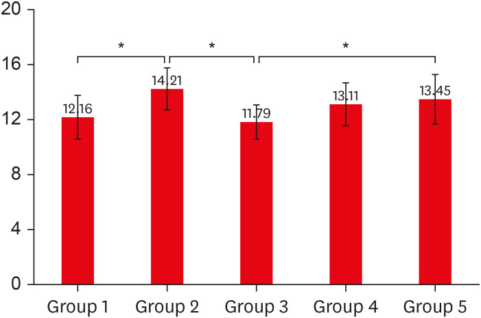
Figure 5 Comparison of mean microshear bond strength values between the control and experimental groups.Asterisk indicates statistically significant differences.
Reference
-
1. Mazzoni A, Angeloni V, Sartori N, Duarte S Jr, Maravic T, Tjäderhane L, Pashley DH, Tay FR, Breschi L. Substantivity of carbodiimide inhibition on dentinal enzyme activity over time. J Dent Res. 2017; 96:902–908. PMID: 28499097.
Article2. Mazzoni A, Nascimento FD, Carrilho M, Tersariol I, Papa V, Tjäderhane L, Di Lenarda R, Tay FR, Pashley DH, Breschi L. MMP activity in the hybrid layer detected with in situ zymography. J Dent Res. 2012; 91:467–472. PMID: 22354448.
Article3. Heshmat H, Jahangiri MH, Hoorizad Ganjkar M, Emami Arjomand M, Kharazifard MJ. Effect of chlorhexidine on micro-shear bond strength of two adhesive systems after aging. J Islam Dent Assoc Iran. 2018; 30:9–14.
Article4. Hass V, Luque-Martinez IV, Gutierrez MF, Moreira CG, Gotti VB, Feitosa VP, Koller G, Otuki MF, Loguercio AD, Reis A. Collagen cross-linkers on dentin bonding: Stability of the adhesive interfaces, degree of conversion of the adhesive, cytotoxicity and in situ MMP inhibition. Dent Mater. 2016; 32:732–741. PMID: 27087688.
Article5. Pashley DH, Tay FR, Yiu C, Hashimoto M, Breschi L, Carvalho RM, Ito S. Collagen degradation by host-derived enzymes during aging. J Dent Res. 2004; 83:216–221. PMID: 14981122.
Article6. Fawzy AS, Priyadarshini BM, Selvan ST, Lu TB, Neo J. Proanthocyanidins-Loaded Nanoparticles Enhance Dentin Degradation Resistance. J Dent Res. 2017; 96:780–789. PMID: 28182862.
Article7. de Macedo FA, Souza NO, Lemos MV, De-Paula DM, Santiago SL, Feitosa VP. Dentin bonding and physicochemical properties of adhesives incorporated with epigallocatechin-3-gallate. Odontology. 2019; 107:23–28. PMID: 29796959.
Article8. Carrilho MR, Geraldeli S, Tay F, de Goes MF, Carvalho RM, Tjäderhane L, Reis AF, Hebling J, Mazzoni A, Breschi L, Pashley D. In vivo preservation of the hybrid layer by chlorhexidine. J Dent Res. 2007; 86:529–533. PMID: 17525352.
Article9. Breschi L, Martin P, Mazzoni A, Nato F, Carrilho M, Tjäderhane L, Visintini E, Cadenaro M, Tay FR, De Stefano Dorigo E, Pashley DH. Use of a specific MMP-inhibitor (galardin) for preservation of hybrid layer. Dent Mater. 2010; 26:571–578. PMID: 20299089.
Article10. Jain PK, Das D. Pharmacognostic comparison of bacopa monnieri, cyperus rotundus and emblica officinalis. Innov J Ayruvedic Sci. 2016; 4:16–26.11. Fujii T, Wakaizumi M, Ikami T, Saito M. Amla (Emblica officinalis Gaertn.) extract promotes procollagen production and inhibits matrix metalloproteinase-1 in human skin fibroblasts. J Ethnopharmacol. 2008; 119:53–57. PMID: 18588964.
Article12. Abraham S, Kumar MS, Sehgal PK, Nitish S, Jayakumar ND. Evaluation of the inhibitory effect of triphala on PMN-type matrix metalloproteinase (MMP-9). J Periodontol. 2005; 76:497–502. PMID: 15857087.
Article13. Breschi L, Mazzoni A, Ruggeri A, Cadenaro M, Di Lenarda R, De Stefano Dorigo E. Dental adhesion review: aging and stability of the bonded interface. Dent Mater. 2008; 24:90–101. PMID: 17442386.
Article14. De Munck J, Mine A, Van den Steen PE, Van Landuyt KL, Poitevin A, Opdenakker G, Van Meerbeek B. Enzymatic degradation of adhesive-dentin interfaces produced by mild self-etch adhesives. Eur J Oral Sci. 2010; 118:494–501. PMID: 20831584.
Article15. Green B, Yao X, Ganguly A, Xu C, Dusevich V, Walker MP, Wang Y. Grape seed proanthocyanidins increase collagen biodegradation resistance in the dentin/adhesive interface when included in an adhesive. J Dent. 2010; 38:908–915. PMID: 20709136.
Article16. Susin AH, Alves LS, Melo GP, Lenzi TL. Comparative scanning electron microscopic study of the effect of different dental conditioners on dentin micromorphology. J Appl Oral Sci. 2008; 16:100–105. PMID: 19089199.
Article17. Mazzoni A, Mannello F, Tay FR, Tonti GA, Papa S, Mazzotti G, Di Lenarda R, Pashley DH, Breschi L. Zymographic analysis and characterization of MMP-2 and -9 forms in human sound dentin. J Dent Res. 2007; 86:436–440. PMID: 17452564.
Article18. Gupta P. Natural products as inhibitors of matrix metalloproteinases. Nat Prod Chem Res. 2016; 4:e114.
Article19. Breschi L, Mazzoni A, Nato F, Carrilho M, Visintini E, Tjäderhane L, Ruggeri A Jr, Tay FR, Dorigo ES, Pashley DH. Chlorhexidine stabilizes the adhesive interface: a 2-year in vitro study. Dent Mater. 2010; 26:320–325. PMID: 20045177.20. Breschi L, Cammelli F, Visintini E, Mazzoni A, Vita F, Carrilho M, Cadenaro M, Foulger S, Mazzoti G, Tay FR, Di Lenarda R, Pashley D. Influence of chlorhexidine concentration on the durability of etch-and-rinse dentin bonds: a 12-month in vitro study. J Adhes Dent. 2009; 11:191–198. PMID: 19603582.21. Kang HJ, Moon HJ, Shin DH; Hyun-Jung Kang. Effect of different chlorhexidine application times on microtensile bond strength to dentin in Class I cavities. Restor Dent Endod. 2012; 37:9–15.
Article22. Lapinska B, Klimek L, Sokolowski J, Lukomska-Szymanska M. Dentine surface morphology after chlorhexidine application-SEM study. Polymers (Basel). 2018; 10:905.
Article23. Ahmed AA, Hassan MM, Abdalla AI. Microshear bond strength of universal adhesives to dentin used in total-etch and self-etch modes. Tanta Dent J. 2018; 15:91–98.
Article
- Full Text Links
-
- Actions
-
Cited
- CITED
-
- Close
- Share
-
- Similar articles
-
- Bond strength of self-adhesive resin cements to composite submitted to different surface pretreatments
- The effect of various commercially available bleaching agents on the microshear bond strength of composite resin to enamel
- Microshear bond strength of a flowable resin to enamel according to the different adhesive systems
- The shear bond strength of two adhesives bonded to composite resin and glass ionomer cement restorations
- Effect of pre-heating on some physical properties of composite resin
