Restor Dent Endod.
2022 Feb;47(1):e6. 10.5395/rde.2022.47.e6.
Morphological characteristics of the mesiobuccal root in the presence of a second mesiobuccal canal: a micro-CT study
- Affiliations
-
- 1Department of Oral Diagnosis, Division of Oral Radiology, Piracicaba Dental School, University of Campinas (UNICAMP), Piracicaba, São Paulo, Brazil
- 2Department of Pathology and Dentistry Clinic, School of Dentistry, Federal University of Piauí (UFPI), Teresina, Piauí, Brazil
- 3Department of Propedeutics and Integrated Clinic, Division of Oral Radiology, School of Dentistry, Federal University of Bahia (UFBA), Salvador, Bahia, Brazil
- KMID: 2548115
- DOI: http://doi.org/10.5395/rde.2022.47.e6
Abstract
Objectives
This study investigated the internal morphology of mesiobuccal (MB) roots of maxillary molars with a second mesiobuccal (MB2) canal.
Materials and Methods
Forty-seven maxillary first or second molars from Brazilians were scanned using micro-computed tomography. The following measurements were obtained from the MB roots: root thickness, root width, and dentin thickness of the buccal aspect of the first mesiobuccal (MB1) canal, between the MB1 and MB2 canals, and the palatal aspect of the MB2 and MB1 canals at 3 mm from the root apex and in the furcation region. For statistical analysis, the Student’s t-test and analysis of variance with the post-hoc Tukey test were used (α = 0.05).
Results
In maxillary molars with an MB2 canal, MB roots were significantly thicker (p = 0.0014) and narrower (p = 0.0016) than in maxillary molars without an MB2 canal. The dentin thickness of the palatal aspect of the MB1 canal was also significantly greater than that of MB roots without an MB2 canal at 3 mm from the root apex (p = 0.0007) and in the furcation region (p < 0.0001). In the furcation region of maxillary molars with an MB2 canal, the dentin thickness between the MB1 and MB2 canals was significantly smaller than that in the buccal and palatal aspects (p < 0.0001).
Conclusions
The internal morphology of MB roots of maxillary molars with an MB2 canal revealed differences in dentin thickness, root diameter, and distance between the canals when compared with maxillary molars without an MB2 canal.
Figure
-
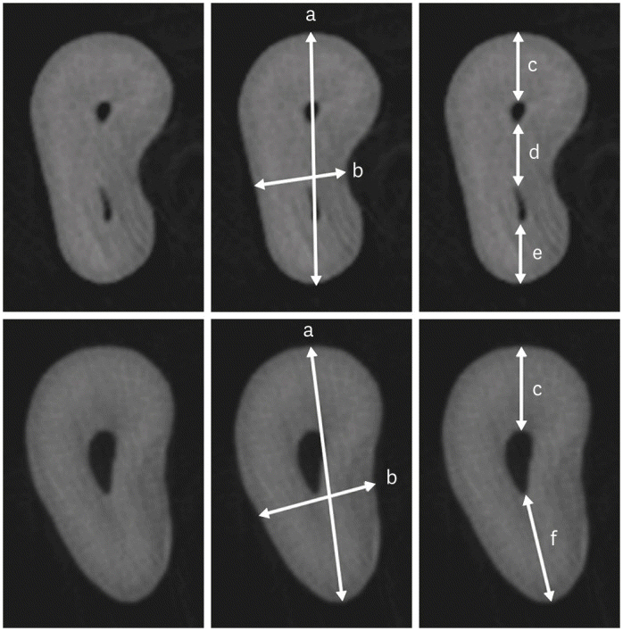
Figure 1 Cropped micro-CT axial sections at 3 mm from the apex of the mesiobuccal root with (upper row) and without an MB2 canal (lower row). For better visualization, the same root is shown in triplicate. (a) Root thickness, (b) Root width, (c) MB1 buccal segment, (d) MB1-MB2 segment, (e) MB2 palatal segment, (f) MB1 palatal segment.CT, computed tomography; MB, mesiobuccal.

Figure 2 Cropped axial sections of measurements taken from the furcation region of maxillary molars with an MB2 canal (A) and without an MB2 canal (B). (c) MB1 buccal segment, (d) MB1-MB2 segment, (e) MB2 palatal segment, (f) MB1 palatal segment.MB, mesiobuccal.
Reference
-
1. Karabucak B, Bunes A, Chehoud C, Kohli MR, Setzer F. Prevalence of apical periodontitis in endodontically treated premolars and molars with untreated canal: a cone-beam computed tomography study. J Endod. 2016; 42:538–541. PMID: 26873567.
Article2. Nascimento EHL, Gaêta-Araujo H, Andrade MFS, Freitas DQ. Prevalence of technical errors and periapical lesions in a sample of endodontically treated teeth: a CBCT analysis. Clin Oral Investig. 2018; 22:2495–2503.
Article3. do Carmo WD, Verner FS, Aguiar LM, Visconti MA, Ferreira MD, Lacerda MF, Junqueira RB. Missed canals in endodontically treated maxillary molars of a Brazilian subpopulation: prevalence and association with periapical lesion using cone-beam computed tomography. Clin Oral Investig. 2021; 25:2317–2323.
Article4. Tomaszewska IM, Jarzębska A, Skinningsrud B, Pękala PA, Wroński S, Iwanaga J. An original micro-CT study and meta-analysis of the internal and external anatomy of maxillary molars-implications for endodontic treatment. Clin Anat. 2018; 31:838–853. PMID: 29732629.
Article5. Martins JNR, Alkhawas MAM, Altaki Z, Bellardini G, Berti L, Boveda C, Chaniotis A, Flynn D, Gonzalez JA, Kottoor J, Marques MS, Monroe A, Ounsi HF, Parashos P, Plotino G, Ragnarsson MF, Aguilar RR, Santiago F, Seedat HC, Vargas W, von Zuben M, Zhang Y, Gu Y, Ginjeira A. Worldwide analyses of maxillary first molar second mesiobuccal prevalence: a multicenter cone-beam computed tomographic study. J Endod. 2018; 44:1641–1649.e1. PMID: 30243661.
Article6. Das S, Warhadpande MM, Redij SA, Jibhkate NG, Sabir H. Frequency of second mesiobuccal canal in permanent maxillary first molars using the operating microscope and selective dentin removal: a clinical study. Contemp Clin Dent. 2015; 6:74–78. PMID: 25684916.
Article7. Estrela C, Holland R, Estrela CR, Alencar AH, Sousa-Neto MD, Pécora JD. Characterization of successful root canal treatment. Braz Dent J. 2014; 25:3–11. PMID: 24789284.
Article8. Sipavičiūtė E, Manelienė R. Pain and flare-up after endodontic treatment procedures. Stomatologija. 2014; 16:25–30. PMID: 24824057.9. Somma F, Leoni D, Plotino G, Grande NM, Plasschaert A. Root canal morphology of the mesiobuccal root of maxillary first molars: a micro-computed tomographic analysis. Int Endod J. 2009; 42:165–174. PMID: 19134045.
Article10. Verma P, Love RM. A Micro CT study of the mesiobuccal root canal morphology of the maxillary first molar tooth. Int Endod J. 2011; 44:210–217. PMID: 20880136.
Article11. Degerness RA, Bowles WR. Dimension, anatomy and morphology of the mesiobuccal root canal system in maxillary molars. J Endod. 2010; 36:985–989. PMID: 20478451.
Article12. Kim Y, Chang SW, Lee JK, Chen IP, Kaufman B, Jiang J, Cha BY, Zhu Q, Safavi KE, Kum KY. A micro-computed tomography study of canal configuration of multiple-canalled mesiobuccal root of maxillary first molar. Clin Oral Investig. 2013; 17:1541–1546.
Article13. Wolf TG, Paqué F, Woop AC, Willershausen B, Briseño-Marroquín B. Root canal morphology and configuration of 123 maxillary second molars by means of micro-CT. Int J Oral Sci. 2017; 9:33–37. PMID: 28106044.
Article14. Ordinola-Zapata R, Martins JNR, Versiani MA, Bramante CM. Micro-CT analysis of danger zone thickness in the mesiobuccal roots of maxillary first molars. Int Endod J. 2019; 52:524–529. PMID: 30295947.
Article15. Harris SP, Bowles WR, Fok A, McClanahan SB. An anatomic investigation of the mandibular first molar using micro-computed tomography. J Endod. 2013; 39:1374–1378. PMID: 24139257.
Article16. Hiebert BM, Abramovitch K, Rice D, Torabinejad M. Prevalence of second mesiobuccal canals in maxillary first molars detected using cone-beam computed tomography, direct occlusal access, and coronal plane grinding. J Endod. 2017; 43:1711–1715. PMID: 28735796.
Article17. van der Vyver PJ, Paleker F, Vorster M, de Wet FA. Root canal shaping using nickel titanium, M-wire, and Gold wire: a micro-computed tomographic comparative study of one shape, ProTaper Next, and WaveOne gold instruments in maxillary first molars. J Endod. 2019; 45:62–67. PMID: 30446405.
Article
- Full Text Links
-
- Actions
-
Cited
- CITED
-
- Close
- Share
-
- Similar articles
-
- Dilemmas pertaining to three canals in the mesiobuccal root of a maxillary second molar: a case report
- In-depth morphological study of mesiobuccal root canal systems in maxillary first molars: review
- Apical periodontitis in mesiobuccal roots of maxillary molars: influence of anatomy and quality of root canal treatment, a CBCT study
- The canal system in the mesiobuccal root of the maxillary first molar
- A cone-beam computed tomography study of the prevalence and location of the second mesiobuccal root canal in maxillary molars
