Restor Dent Endod.
2022 Nov;47(4):e37. 10.5395/rde.2022.47.e37.
Apical periodontitis in mesiobuccal roots of maxillary molars: influence of anatomy and quality of root canal treatment, a CBCT study
- Affiliations
-
- 1Department of Endodontic, São Leopoldo Mandic School of Dentistry, Campinas, SP, Brazil
- KMID: 2548141
- DOI: http://doi.org/10.5395/rde.2022.47.e37
Abstract
Objectives
This study aimed to evaluate the prevalence of apical periodontitis (AP) in the mesiobuccal roots of root canal-treated maxillary molars.
Materials and Methods
One thousand cone-beam computed tomography images of the teeth were examined by 2 dental specialists in oral radiology and endodontics. The internal anatomy of the roots, Vertucci’s classification, quality of root canal treatment, and presence of missed canals were evaluated; additionally, the correlation between these variables and AP was ascertained.
Results
A total of 1,000 roots (692 first molars and 308 second molars) encompassing 1,549 canals were assessed, and the quality of the root canal filling in the majority (56.9%) of the canals was satisfactory. AP was observed in 54.4% of the teeth. A mesiolingual canal in the mesiobuccal root (MB2 canal) was observed in 54.9% of the images, and the majority (83.5%) of these canals were not filled. Significant associations were observed between the presence of an MB2 canal and the quality of the root canal filling and the presence of AP.
Conclusions
AP was detected in more than half of the images. The MB2 canals were frequently missed or poorly filled.
Figure
-
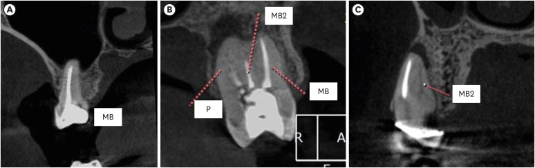
Figure 1 Representative images showing (A) satisfactory filling, (B) satisfactory filling (MB), unsatisfactory (MB2) and (C) Untreated (MB2).MB, mesiobuccal; MB2, second mesiobuccal; P, palatal.
Reference
-
1. American Association of Endodontists. Treatment standards. updated 2020. cited May 2, 2022. Available from: https://www.aae.org/specialty/wp-content/uploads/sites/2/2018/04/TreatmentStandards_Whitepaper.pdf.2. Yoo YJ, Perinpanayagam H, Oh S, Kim AR, Han SH, Kum KY. Endodontic biofilms: contemporary and future treatment options. Restor Dent Endod. 2019; 44:e7. PMID: 30834229.
Article3. Kulild JC, Peters DD. Incidence and configuration of canal systems in the mesiobuccal root of maxillary first and second molars. J Endod. 1990; 16:311–317. PMID: 2081944.
Article4. Keskin C, Keleş A, Versiani MA. Mesiobuccal and palatal interorifice distance may predict the presence of the second mesiobuccal canal in maxillary second molars with fused roots. J Endod. 2021; 47:585–591. PMID: 33497731.
Article5. Martins JN, Kishen A, Marques D, Nogueira Leal Silva EJ, Caramês J, Mata A, Versiani MA. Preferred reporting items for epidemiologic cross-sectional studies on root and root canal anatomy using cone-beam computed tomographic technology: a systematized assessment. J Endod. 2020; 46:915–935. PMID: 32387077.
Article6. Martins JN, Alkhawas MA, Altaki Z, Bellardini G, Berti L, Boveda C, Chaniotis A, Flynn D, Gonzalez JA, Kottoor J, Marques MS, Monroe A, Ounsi HF, Parashos P, Plotino G, Ragnarsson MF, Aguilar RR, Santiago F, Seedat HC, Vargas W, von Zuben M, Zhang Y, Gu Y, Ginjeira A. Worldwide analyses of maxillary first molar second mesiobuccal prevalence: a multicenter cone-beam computed tomographic study. J Endod. 2018; 44:1641–1649.e1. PMID: 30243661.
Article7. Sjogren U, Hagglund B, Sundqvist G, Wing K. Factors affecting the long-term results of endodontic treatment. J Endod. 1990; 16:498–504. PMID: 2084204.
Article8. Saini HR, Tewari S, Sangwan P, Duhan J, Gupta A. Effect of different apical preparation sizes on outcome of primary endodontic treatment: a randomized controlled trial. J Endod. 2012; 38:1309–1315. PMID: 22980168.
Article9. Song M, Kim HC, Lee W, Kim E. Analysis of the cause of failure in nonsurgical endodontic treatment by microscopic inspection during endodontic microsurgery. J Endod. 2011; 37:1516–1519. PMID: 22000454.
Article10. do Carmo WD, Verner FS, Aguiar LM, Visconti MA, Ferreira MD, Lacerda MF, Junqueira RB. Missed canals in endodontically treated maxillary molars of a Brazilian subpopulation: prevalence and association with periapical lesion using cone-beam computed tomography. Clin Oral Investig. 2021; 25:2317–2323.
Article11. Baruwa AO, Martins JN, Meirinhos J, Pereira B, Gouveia J, Quaresma SA, Monroe A, Ginjeira A. The influence of missed canals on the prevalence of periapical lesions in endodontically treated teeth: a cross-sectional study. J Endod. 2020; 46:34–39.e1. PMID: 31733814.
Article12. Ricucci D, Siqueira JF Jr. Biofilms and apical periodontitis: study of prevalence and association with clinical and histopathologic findings. J Endod. 2010; 36:1277–1288. PMID: 20647081.
Article13. Ricucci D, Siqueira JF Jr, Bate AL, Pitt Ford TR. Histologic investigation of root canal-treated teeth with apical periodontitis: a retrospective study from twenty-four patients. J Endod. 2009; 35:493–502. PMID: 19345793.
Article14. Jang YE, Kim BS, Kim Y. Clinical factors associated with apical periodontitis visible on cone-beam computed tomography but missed with periapical radiographs: a retrospective clinical study. J Endod. 2020; 46:832–838. PMID: 32295704.
Article15. Vertucci FJ. Root canal anatomy of the human permanent teeth. Oral Surg Oral Med Oral Pathol. 1984; 58:589–599. PMID: 6595621.
Article16. Estrela C, Bueno MR, Azevedo BC, Azevedo JR, Pécora JD. A new periapical index based on cone beam computed tomography. J Endod. 2008; 34:1325–1331. PMID: 18928840.
Article17. Tibúrcio-Machado CS, Michelon C, Zanatta FB, Gomes MS, Marin JA, Bier CA. The global prevalence of apical periodontitis: a systematic review and meta-analysis. Int Endod J. 2021; 54:712–735. PMID: 33378579.
Article18. Chauhan N, Mittal S, Tewari S, Sen J, Laller K. Association of apical periodontitis with cardiovascular disease via noninvasive assessment of endothelial function and subclinical atherosclerosis. J Endod. 2019; 45:681–690. PMID: 31030979.
Article19. Jakovljevic A, Sljivancanin Jakovljevic T, Duncan HF, Nagendrababu V, Jacimovic J, Aminoshariae A, Milasin J, Dummer PM. The association between apical periodontitis and adverse pregnancy outcomes: a systematic review. Int Endod J. 2021; 54:1527–1537. PMID: 33908039.
Article20. Poornima L, Ravishankar P, Abbott PV, Subbiya A, PradeepKumar AR. Impact of root canal treatment on high-sensitivity C-reactive protein levels in systemically healthy adults with apical periodontitis - a preliminary prospective, longitudinal interventional study. Int Endod J. 2021; 54:501–508. PMID: 33185278.
Article21. Libby P, Ridker PM, Maseri A. Inflammation and atherosclerosis. Circulation. 2002; 105:1135–1143. PMID: 11877368.
Article22. Mozzo P, Procacci C, Tacconi A, Martini PT, Andreis IA. A new volumetric CT machine for dental imaging based on the cone-beam technique: preliminary results. Eur Radiol. 1998; 8:1558–1564. PMID: 9866761.
Article23. Parker JM, Mol A, Rivera EM, Tawil PZ. Cone-beam computed tomography uses in clinical endodontics: observer variability in detecting periapical lesions. J Endod. 2017; 43:184–187. PMID: 28024758.
Article24. Beacham JT, Geist JR, Yu Q, Himel VT, Sabey KA. Accuracy of cone-beam computed tomographic image interpretation by endodontists and endodontic residents. J Endod. 2018; 44:571–575. PMID: 29397216.
Article25. Martins JN, Marques D, Silva EJ, Caramês J, Mata A, Versiani MA. Second mesiobuccal root canal in maxillary molars—a systematic review and meta-analysis of prevalence studies using cone beam computed tomography. Arch Oral Biol. 2020; 113:104589. PMID: 31735252.
Article26. Alaçam T, Tinaz AC, Genç O, Kayaoglu G. Second mesiobuccal canal detection in maxillary first molars using microscopy and ultrasonics. Aust Endod J. 2008; 34:106–109. PMID: 19032644.
Article27. Coelho MS, Parker JM, Tawil PZ. Second mesiobuccal canal treatment in a predoctoral dental clinic: a retrospective clinical study. J Dent Educ. 2016; 80:726–730. PMID: 27251355.
Article28. Parker J, Mol A, Rivera EM, Tawil P. CBCT uses in clinical endodontics: the effect of CBCT on the ability to locate MB2 canals in maxillary molars. Int Endod J. 2017; 50:1109–1115. PMID: 27977863.
Article29. Vizzotto MB, Silveira PF, Arús NA, Montagner F, Gomes BP, da Silveira HE. CBCT for the assessment of second mesiobuccal (MB2) canals in maxillary molar teeth: effect of voxel size and presence of root filling. Int Endod J. 2013; 46:870–876. PMID: 23442087.
Article30. Costa FF, Pacheco-Yanes J, Siqueira JF Jr, Oliveira AC, Gazzaneo I, Amorim CA, Santos PH, Alves FR. Association between missed canals and apical periodontitis. Int Endod J. 2019; 52:400–406. PMID: 30284719.
Article31. Burry JC, Stover S, Eichmiller F, Bhagavatula P. Outcomes of primary endodontic therapy provided by endodontic specialists compared with other providers. J Endod. 2016; 42:702–705. PMID: 27004720.
Article32. Kruse C, Spin-Neto R, Evar Kraft DC, Vaeth M, Kirkevang LL. Diagnostic accuracy of cone beam computed tomography used for assessment of apical periodontitis: an ex vivo histopathological study on human cadavers. Int Endod J. 2019; 52:439–450. PMID: 30267421.
Article
- Full Text Links
-
- Actions
-
Cited
- CITED
-
- Close
- Share
-
- Similar articles
-
- Assessment of Root and Root Canal Morphology of Human Primary Molars using CBCT
- Assessment of the relationship between the maxillary molars and adjacent structures using cone beam computed tomography
- Discrepancy in Root Apex Closure Timing of Maxillary First Molars: CBCT Study
- Morphological characteristics of the mesiobuccal root in the presence of a second mesiobuccal canal: a micro-CT study
- A Study of Root Canals Morphology in Primary Molars using Computerized Tomography
