Age-dependent root canal instrumentation techniques: a comprehensive narrative review
- Affiliations
-
- 1Department of Endodontics, Israel Defense Forces (IDF) Medical Corps, Tel Hashomer, Israel
- 2Department of Conservative Dentistry, School of Dentistry, Dental Research Institute, Pusan National University, Yangsan, Korea
- 3Private Practice, Tel Aviv, Israel
- KMID: 2503516
- DOI: http://doi.org/10.5395/rde.2020.45.e21
Abstract
- The aim of this article was to review age-dependent clinical recommendations for appropriate root canal instrumentation techniques. A comprehensive narrative review of canal morphology, the structural characteristics of dentin, and endodontic outcomes at different ages was undertaken instead of a systematic review. An electronic literature search was carried out, including the Medline (Ovid), PubMed, and Web of Science databases. The searches used controlled vocabulary and free-text terms, as follows: ‘age-related root canal treatment,’ ‘age-related instrumentation,’ ‘age-related chemo-mechanical preparation,’ ‘age-related endodontic clinical recommendations,’ ‘root canal instrumentation at different ages,’ ‘geriatric root canal treatment,’ and ‘pediatric root canal treatment.’ Due to the lack of literature with practical age-based clinical recommendations for an appropriate root canal instrumentation technique, a narrative review was conducted to suggest a clinical algorithm for choosing the most appropriate instrumentation technique during root canal treatment. Based on the evidence found through the narrative review, an age-related clinical algorithm for choosing appropriate instrumentation during root canal treatment was proposed. Age affects the morphology of the root canal system and the structural characteristics of dentin. The clinician’s awareness of root canal morphology and dentin characteristics can influence the choice of instruments for root canal treatment.
Keyword
Figure
-
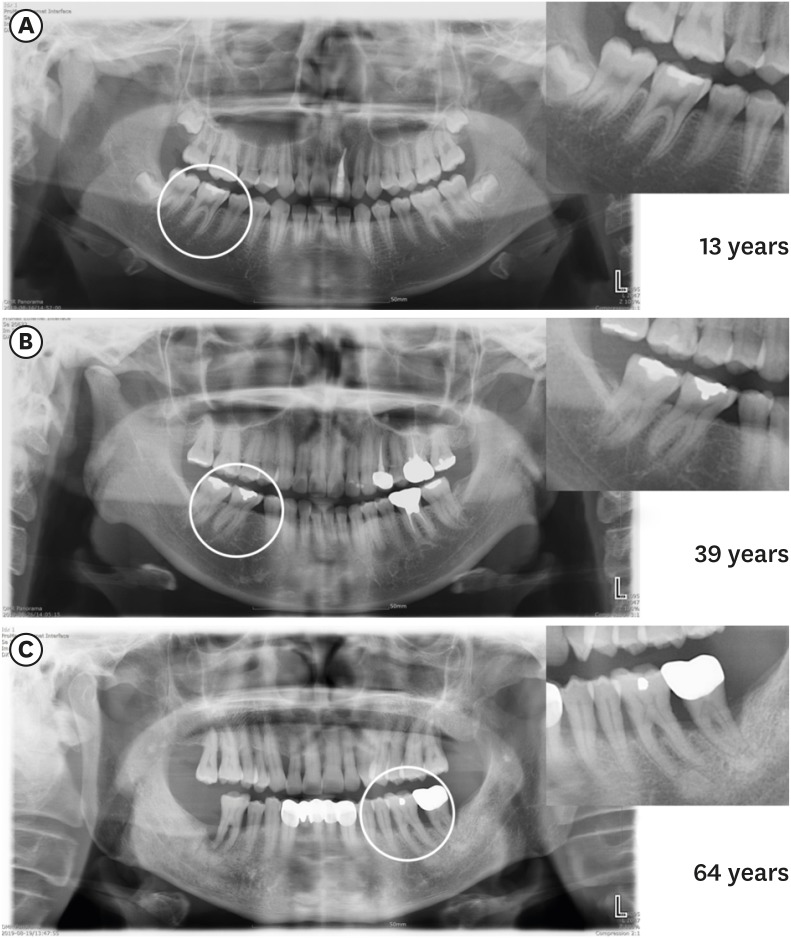
Figure 1 Panoramic radiography images showing the chamber and canal sizes according at different ages. (A) Individuals under 20 years old (13 years), (B) individuals between 20 and 40 years old (39 years), (C) individuals over 40 years old (64 years). The image in (A) shows a large chamber and a straight direction to the canal orifice. The image in (B) shows a dentin shelf area and an angulated direction to canal orifice. The image in (C) shows a thin chamber area and sclerotic canals.
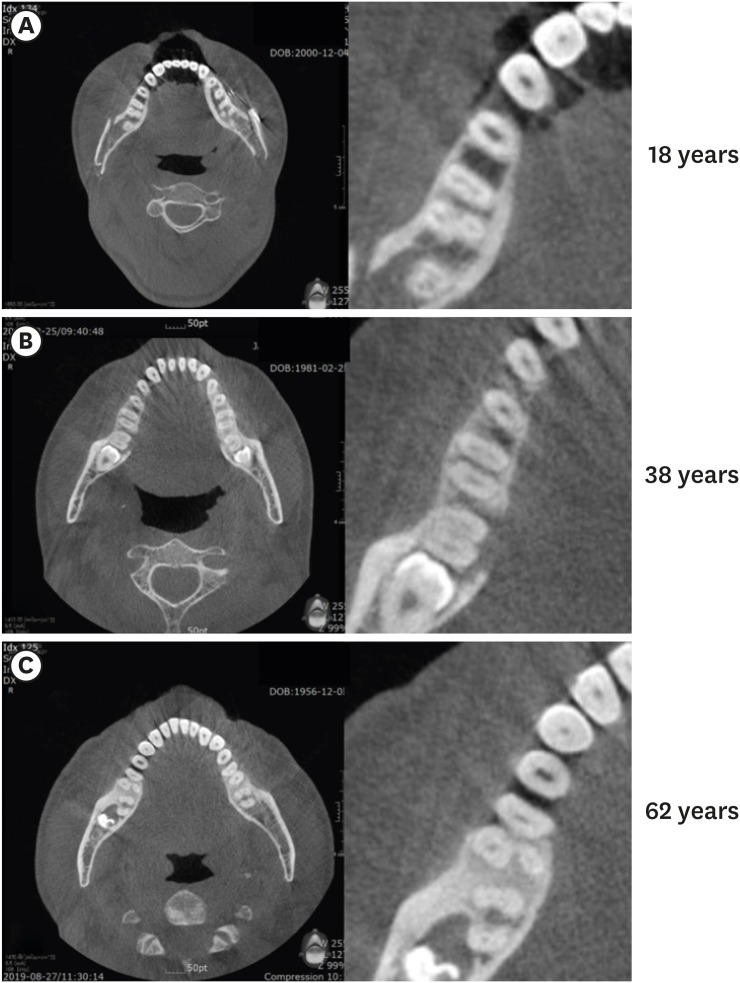
Figure 2 Axial cone-beam computed tomography images of 3 mandibular first molars, showing isthmus characteristics at different ages. (A) Individuals under 20 years old (18 years): a wide canal without an isthmus, (B) individuals between 20 and 40 years old (38 years): an isthmus in the mesial root, (C) individuals who are 40 years old or more (62 years): sclerotic mesial canals without an isthmus, resembling a single canal.
Reference
-
1. Kassebaum NJ, Bernabé E, Dahiya M, Bhandari B, Murray CJ, Marcenes W. Global burden of untreated caries: a systematic review and metaregression. J Dent Res. 2015; 94:650–658. PMID: 25740856.2. Gani O, Visvisian C. Apical canal diameter in the first upper molar at various ages. J Endod. 1999; 25:689–691. PMID: 10687530.
Article3. Yared G. Canal preparation using only one Ni-Ti rotary instrument: preliminary observations. Int Endod J. 2008; 41:339–344. PMID: 18081803.
Article4. Ahn SY, Kim HC, Kim E. Kinematic effects of nickel-titanium instruments with reciprocating or continuous rotation motion: a systematic review of in vitro studies. J Endod. 2016; 42:1009–1017. PMID: 27185740.5. Pérez-Higueras JJ, Arias A, de la Macorra JC. Cyclic fatigue resistance of K3, K3XF, and twisted file nickel-titanium files under continuous rotation or reciprocating motion. J Endod. 2013; 39:1585–1588. PMID: 24238452.
Article6. Martins JN, Alkhawas MA, Altaki Z, Bellardini G, Berti L, Boveda C, Chaniotis A, Flynn D, Gonzalez JA, Kottoor J, Marques MS, Monroe A, Ounsi HF, Parashos P, Plotino G, Ragnarsson MF, Aguilar RR, Santiago F, Seedat HC, Vargas W, von Zuben M, Zhang Y, Gu Y, Ginjeira A. Worldwide analyses of maxillary first molar second mesiobuccal prevalence: a multicenter cone-beam computed tomographic study. J Endod. 2018; 44:1641–1649.e1. PMID: 30243661.
Article7. Martins JN, Ordinola-Zapata R, Marques D, Francisco H, Caramês J. Differences in root canal system configuration in human permanent teeth within different age groups. Int Endod J. 2018; 51:931–941. PMID: 29363147.
Article8. Peiris HR, Pitakotuwage TN, Takahashi M, Sasaki K, Kanazawa E. Root canal morphology of mandibular permanent molars at different ages. Int Endod J. 2008; 41:828–835. PMID: 18822010.
Article9. Zheng QH, Wang Y, Zhou XD, Wang Q, Zheng GN, Huang DM. A cone-beam computed tomography study of maxillary first permanent molar root and canal morphology in a Chinese population. J Endod. 2010; 36:1480–1484. PMID: 20728713.
Article10. Guo J, Vahidnia A, Sedghizadeh P, Enciso R. Evaluation of root and canal morphology of maxillary permanent first molars in a North American population by cone-beam computed tomography. J Endod. 2014; 40:635–639. PMID: 24767556.11. Hess W, Zurcher E. The anatomy of root canals of the teeth of the permanent and deciduous dentitions. New York, NY: William Wood & Co;1925.12. Ørstavik D, Qvist V, Stoltze K. A multivariate analysis of the outcome of endodontic treatment. Eur J Oral Sci. 2004; 112:224–230. PMID: 15154919.
Article13. Vertucci FJ. Root canal morphology and its relationship to endodontic procedures. Endod Topics. 2005; 10:3–29.
Article14. Manning SA. Root canal anatomy of mandibular second molars. Part I. Int Endod J. 1990; 23:34–39. PMID: 2391179.
Article15. Thomas RP, Moule AJ, Bryant R. Root canal morphology of maxillary permanent first molar teeth at various ages. Int Endod J. 1993; 26:257–267. PMID: 8300257.
Article16. Nosrat A, Deschenes RJ, Tordik PA, Hicks ML, Fouad AF. Middle mesial canals in mandibular molars: incidence and related factors. J Endod. 2015; 41:28–32. PMID: 25266468.17. Pattanshetti N, Gaidhane M, Al Kandari AM. Root and canal morphology of the mesiobuccal and distal roots of permanent first molars in a Kuwait population--a clinical study. Int Endod J. 2008; 41:755–762. PMID: 18637850.18. Liu J, Luo J, Dou L, Yang D. CBCT study of root and canal morphology of permanent mandibular incisors in a Chinese population. Acta Odontol Scand. 2014; 72:26–30. PMID: 24255962.
Article19. Al-Fouzan KS. C-shaped root canals in mandibular second molars in a Saudi Arabian population. Int Endod J. 2002; 35:499–504. PMID: 12190906.
Article20. Akbarzadeh N, Aminoshariae A, Khalighinejad N, Palomo JM, Syed A, Kulild JC, Sadeghi G, Mickel A. The association between the anatomic landmarks of the pulp chamber floor and the prevalence of middle mesial canals in mandibular first molars: an in vivo analysis. J Endod. 2017; 43:1797–1801. PMID: 28864218.21. Kaya S, Adiguzel O, Yavuz I, Tumen EC, Akkus Z. Cone-beam dental computerized tomography for evaluating changes of aging in the dimensions central superior incisor root canals. Med Oral Patol Oral Cir Bucal. 2011; 16:e463–e466. PMID: 20711137.22. Reis AG, Grazziotin-Soares R, Barletta FB, Fontanella VR, Mahl CR. Second canal in mesiobuccal root of maxillary molars is correlated with root third and patient age: a cone-beam computed tomographic study. J Endod. 2013; 39:588–592. PMID: 23611373.
Article23. Neaverth EJ, Kotler LM, Kaltenbach RF. Clinical investigation. Clinical investigation (in vivo) of endodontically treated maxillary first molars. J Endod. 1987; 13:506–512. PMID: 3482228.24. Gilles J, Reader A. An SEM investigation of the mesiolingual canal in human maxillary first and second molars. Oral Surg Oral Med Oral Pathol. 1990; 70:638–643. PMID: 2234885.
Article25. Fogel HM, Peikoff MD, Christie WH. Canal configuration in the mesiobuccal root of the maxillary first molar: a clinical study. J Endod. 1994; 20:135–137. PMID: 7996086.
Article26. Yoshioka T, Kikuchi I, Fukumoto Y, Kobayashi C, Suda H. Detection of the second mesiobuccal canal in mesiobuccal roots of maxillary molar teeth ex vivo . Int Endod J. 2005; 38:124–128. PMID: 15667634.27. Lee JH, Kim KD, Lee JK, Park W, Jeong JS, Lee Y, Gu Y, Chang SW, Son WJ, Lee WC, Baek SH, Bae KS, Kum KY. Mesiobuccal root canal anatomy of Korean maxillary first and second molars by cone-beam computed tomography. Oral Surg Oral Med Oral Pathol Oral Radiol Endod. 2011; 111:785–791. PMID: 21439860.
Article28. al Shalabi RM, Omer OE, Glennon J, Jennings M, Claffey NM. Root canal anatomy of maxillary first and second permanent molars. Int Endod J. 2000; 33:405–414. PMID: 11307458.29. Gu L, Wei X, Ling J, Huang X. A microcomputed tomographic study of canal isthmuses in the mesial root of mandibular first molars in a Chinese population. J Endod. 2009; 35:353–356. PMID: 19249594.
Article30. Villas-Bôas MH, Bernardineli N, Cavenago BC, Marciano M, Del Carpio-Perochena A, de Moraes IG, Duarte MH, Bramante CM, Ordinola-Zapata R. Micro-computed tomography study of the internal anatomy of mesial root canals of mandibular molars. J Endod. 2011; 37:1682–1686. PMID: 22099905.
Article31. Srivastava S, Alrogaibah NA, Aljarbou G. Cone-beam computed tomographic analysis of middle mesial canals and isthmus in mesial roots of mandibular first molars-prevalence and related factors. J Conserv Dent. 2018; 21:526–530. PMID: 30294115.
Article32. Oi T, Saka H, Ide Y. Three-dimensional observation of pulp cavities in the maxillary first premolar tooth using micro-CT. Int Endod J. 2004; 37:46–51. PMID: 14718057.
Article33. Miller WA, Eick JD, Neiders ME. Inorganic components of the peritubular dentin in young human permanent teeth. Caries Res. 1971; 5:264–278. PMID: 5284352.
Article34. Kishen A. Biomechanics of fractures in endodontically treated teeth. Endod Topics. 2015; 33:3–13.
Article35. Tronstad L. Ultrastructural observations on human coronal dentin. Scand J Dent Res. 1973; 81:101–111. PMID: 4513437.
Article36. Carrigan PJ, Morse DR, Furst ML, Sinai IH. A scanning electron microscopic evaluation of human dentinal tubules according to age and location. J Endod. 1984; 10:359–363. PMID: 6590745.
Article37. Ozdemir HO, Buzoglu HD, Calt S, Stabholz A, Steinberg D. Effect of ethylenediaminetetraacetic acid and sodium hypochlorite irrigation on Enterococcus faecalis biofilm colonization in young and old human root canal dentin: in vitro study. J Endod. 2010; 36:842–846. PMID: 20416430.38. Stanley HR, Pereira JC, Spiegel E, Broom C, Schultz M. The detection and prevalence of reactive and physiologic sclerotic dentin, reparative dentin and dead tracts beneath various types of dental lesions according to tooth surface and age. J Oral Pathol. 1983; 12:257–289. PMID: 6193259.
Article39. Paqué F, Luder HU, Sener B, Zehnder M. Tubular sclerosis rather than the smear layer impedes dye penetration into the dentine of endodontically instrumented root canals. Int Endod J. 2006; 39:18–25. PMID: 16409324.
Article40. Kakoli P, Nandakumar R, Romberg E, Arola D, Fouad AF. The effect of age on bacterial penetration of radicular dentin. J Endod. 2009; 35:78–81. PMID: 19084130.
Article41. Sánchez-Sanhueza G, Bello-Toledo H, González-Rocha G, Gonçalves AT, Valenzuela V, Gallardo-Escárate C. Metagenomic study of bacterial microbiota in persistent endodontic infections using next-generation sequencing. Int Endod J. 2018; 51:1336–1348. PMID: 29786880.
Article42. Ketterl W. Age-induced changes in the teeth and their attachment apparatus. Int Dent J. 1983; 33:262–271. PMID: 6579031.43. Morse DR. Age-related changes of the dental pulp complex and their relationship to systemic aging. Oral Surg Oral Med Oral Pathol. 1991; 72:721–745. PMID: 1812456.
Article44. Love RM, Jenkinson HF. Invasion of dentinal tubules by oral bacteria. Crit Rev Oral Biol Med. 2002; 13:171–183. PMID: 12097359.45. Kinney JH, Nalla RK, Pople JA, Breunig TM, Ritchie RO. Age-related transparent root dentin: mineral concentration, crystallite size, and mechanical properties. Biomaterials. 2005; 26:3363–3376. PMID: 15603832.
Article46. Rigden MD, Baier C, Ramirez-Arcos S, Liao M, Wang M, Dillon JA. Identification of the coiled-coil domains of Enterococcus faecalis DivIVA that mediate oligomerization and their importance for biological function. J Biochem. 2008; 144:63–76. PMID: 18388125.47. Kishen A. Mechanisms and risk factors for fracture predilection in endodontically treated teeth. Endod Topics. 2006; 13:57–83.
Article48. Arola D, Reprogel RK. Effects of aging on the mechanical behavior of human dentin. Biomaterials. 2005; 26:4051–4061. PMID: 15626451.
Article49. Mireku AS, Romberg E, Fouad AF, Arola D. Vertical fracture of root filled teeth restored with posts: the effects of patient age and dentine thickness. Int Endod J. 2010; 43:218–225. PMID: 20158533.
Article50. Yan W, Montoya C, Øilo M, Ossa A, Paranjpe A, Zhang H, Arola D. Reduction in fracture resistance of the root with aging. J Endod. 2017; 43:1494–1498. PMID: 28712639.51. Koester KJ, Ager JW 3rd, Ritchie RO. The effect of aging on crack-growth resistance and toughening mechanisms in human dentin. Biomaterials. 2008; 29:1318–1328. PMID: 18164757.
Article52. Nazari A, Bajaj D, Zhang D, Romberg E, Arola D. Aging and the reduction in fracture toughness of human dentin. J Mech Behav Biomed Mater. 2009; 2:550–559. PMID: 19627862.
Article53. Tamse A. Iatrogenic vertical root fractures in endodontically treated teeth. Endod Dent Traumatol. 1988; 4:190–196. PMID: 3073952.
Article54. Testori T, Badino M, Castagnola M. Vertical root fractures in endodontically treated teeth: a clinical survey of 36 cases. J Endod. 1993; 19:87–91. PMID: 8509743.
Article55. Bajaj D, Sundaram N, Nazari A, Arola D. Age, dehydration and fatigue crack growth in dentin. Biomaterials. 2006; 27:2507–2517. PMID: 16338002.
Article56. PradeepKumar AR, Shemesh H, Chang JW, Bhowmik A, Sibi S, Gopikrishna V, Lakshmi-Narayanan L, Kishen A. Preexisting dentinal microcracks in nonendodontically treated teeth: an ex vivo micro-computed tomographic analysis. J Endod. 2017; 43:896–900. PMID: 28457637.57. Sathorn C, Palamara JE, Palamara D, Messer HH. Effect of root canal size and external root surface morphology on fracture susceptibility and pattern: a finite element analysis. J Endod. 2005; 31:288–292. PMID: 15793386.
Article58. Tang W, Wu Y, Smales RJ. Identifying and reducing risks for potential fractures in endodontically treated teeth. J Endod. 2010; 36:609–617. PMID: 20307732.
Article59. Swartz DB, Skidmore AE, Griffin JA Jr. Twenty years of endodontic success and failure. J Endod. 1983; 9:198–202. PMID: 6574207.
Article60. Sjogren U, Hagglund B, Sundqvist G, Wing K. Factors affecting the long-term results of endodontic treatment. J Endod. 1990; 16:498–504. PMID: 2084204.61. Dammaschke T, Steven D, Kaup M, Ott KH. Long-term survival of root-canal-treated teeth: a retrospective study over 10 years. J Endod. 2003; 29:638–643. PMID: 14606785.
Article62. Kojima K, Inamoto K, Nagamatsu K, Hara A, Nakata K, Morita I, Nakagaki H, Nakamura H. Success rate of endodontic treatment of teeth with vital and nonvital pulps. A meta-analysis. Oral Surg Oral Med Oral Pathol Oral Radiol Endod. 2004; 97:95–99. PMID: 14716263.
Article63. Grossman LI, Shepard LI, Pearson LA. Roentgenologic and clinical evaluation of endodontically treated teeth. Oral Surg Oral Med Oral Pathol. 1964; 17:368–374. PMID: 14130241.
Article64. Harty FJ, Parkins BJ, Wengraf AM. Success rate in root canal therapy. A retrospective study of conventional cases. Br Dent J. 1970; 128:65–70. PMID: 5270224.
Article65. Smith CS, Setchell DJ, Harty FJ. Factors influencing the success of conventional root canal therapy--a five-year retrospective study. Int Endod J. 1993; 26:321–333. PMID: 8144241.
Article66. Imura N, Pinheiro ET, Gomes BP, Zaia AA, Ferraz CC, Souza-Filho FJ. The outcome of endodontic treatment: a retrospective study of 2000 cases performed by a specialist. J Endod. 2007; 33:1278–1282. PMID: 17963946.
Article67. Seltzer S, Bender IB, Turkenkopf S. Factors affecting successful repairs after root canal therapy. J Am Dent Assoc. 1963; 67:651–662. PMID: 14054365.68. Caplan DJ, Weintraub JA. Factors related to loss of root canal filled teeth. J Public Health Dent. 1997; 57:31–39. PMID: 9150061.
Article69. Lazarski MP, Walker WA 3rd, Flores CM, Schindler WG, Hargreaves KM. Epidemiological evaluation of the outcomes of nonsurgical root canal treatment in a large cohort of insured dental patients. J Endod. 2001; 27:791–796. PMID: 11771594.
Article70. Eriksen HM. Epidemiology of apical periodontitis. In : Orstavik D, Pitt Ford TR, editors. Essential endodontology. Prevention and treatment of apical periodontitis. London: Blackwell Science Ltd;1998. p. 179–191.71. Azim AA, Griggs JA, Huang GT. The Tennessee study: factors affecting treatment outcome and healing time following nonsurgical root canal treatment. Int Endod J. 2016; 49:6–16. PMID: 25582870.
Article72. Lloberas J, Celada A. Effect of aging on macrophage function. Exp Gerontol. 2002; 37:1325–1331. PMID: 12559402.
Article73. Renshaw M, Rockwell J, Engleman C, Gewirtz A, Katz J, Sambhara S. Cutting edge: impaired Toll-like receptor expression and function in aging. J Immunol. 2002; 169:4697–4701. PMID: 12391175.
Article74. Okiji T, Kosaka T, Kamal AM, Kawashima N, Suda H. Age-related changes in the immunoreactivity of the monocyte/macrophage system in rat molar pulp. Arch Oral Biol. 1996; 41:453–460. PMID: 8809308.
Article75. Tranasi M, Sberna MT, Zizzari V, D'Apolito G, Mastrangelo F, Salini L, Stuppia L, Tetè S. Microarray evaluation of age-related changes in human dental pulp. J Endod. 2009; 35:1211–1217. PMID: 19720218.
Article76. Prince MJ, Wu F, Guo Y, Gutierrez Robledo LM, O'Donnell M, Sullivan R, Yusuf S. The burden of disease in older people and implications for health policy and practice. Lancet. 2015; 385:549–562. PMID: 25468153.
Article77. Aminoshariae A, Kulild JC, Mickel A, Fouad AF. Association between systemic diseases and endodontic outcome: a systematic review. J Endod. 2017; 43:514–519. PMID: 28190585.
Article78. Fouad AF, Burleson J. The effect of diabetes mellitus on endodontic treatment outcome: data from an electronic patient record. J Am Dent Assoc. 2003; 134:43–51. PMID: 12555956.79. Lima SM, Grisi DC, Kogawa EM, Franco OL, Peixoto VC, Gonçalves-Júnior JF, Arruda MP, Rezende TM. Diabetes mellitus and inflammatory pulpal and periapical disease: a review. Int Endod J. 2013; 46:700–709. PMID: 23442003.
Article80. Siqueira JF Jr, Alves FR, Almeida BM, de Oliveira JC, Rôças IN. Ability of chemomechanical preparation with either rotary instruments or self-adjusting file to disinfect oval-shaped root canals. J Endod. 2010; 36:1860–1865. PMID: 20951301.
Article81. Lin J, Shen Y, Haapasalo M. A comparative study of biofilm removal with hand, rotary nickel-titanium, and self-adjusting file instrumentation using a novel in vitro biofilm model. J Endod. 2013; 39:658–663. PMID: 23611386.82. Metzger Z, Solomonov M, Kfir A. The role of mechanical instrumentation in the cleaning of root canals. Endod Topics. 2013; 29:87–109.
Article83. Alves FR, Marceliano-Alves MF, Sousa JC, Silveira SB, Provenzano JC, Siqueira JF Jr. Removal of root canal fillings in curved canals using either reciprocating single- or rotary multi-instrument systems and a supplementary step with the XP-Endo Finisher. J Endod. 2016; 42:1114–1119. PMID: 27215810.
Article84. Arvaniti IS, Khabbaz MG. Influence of root canal taper on its cleanliness: a scanning electron microscopic study. J Endod. 2011; 37:871–874. PMID: 21787508.
Article85. Kim HC, Lee MH, Yum J, Versluis A, Lee CJ, Kim BM. Potential relationship between design of nickel-titanium rotary instruments and vertical root fracture. J Endod. 2010; 36:1195–1199. PMID: 20630298.
Article86. Philippas GG, Applebaum E. Age factor in secondary dentin formation. J Dent Res. 1966; 45:778–789. PMID: 5222482.
Article87. Tziafas D. Mechanisms controlling secondary initiation of dentinogenesis: a review. Int Endod J. 1994; 27:61–74. PMID: 8034367.
Article88. Bender IB, Seltzer S. The effect of periodontal disease on the pulp. Oral Surg Oral Med Oral Pathol. 1972; 33:458–474. PMID: 4621862.
Article
- Full Text Links
-
- Actions
-
Cited
- CITED
-
- Close
- Share
-
- Similar articles
-
- Review of root canal irrigant delivery techniques and devices
- Selection of Nickel-Titanium Files according to the Clinical Procedure and Factors of File Fracture: A Narrative Review
- Evaluation of time-dependent antimicrobial effect of sodium dichloroisocyanurate (NaDCC) on Enterococcus faecalis in the root canal
- Comparison of canal transportation in simulated curved canals prepared with ProTaper Universal and ProTaper Gold systems
- A comparison of master apical file size according to instrumentation in type II root canal
