J Pathol Transl Med.
2018 Jan;52(1):61-63. 10.4132/jptm.2017.06.13.
Liquid-Based Cytology of the Cerebrospinal Fluid in a Case of Cryptococcal Meningitis
- Affiliations
-
- 1Department of Pathology, Yonsei University Wonju College of Medicine, Wonju, Korea.
- 2Department of Pathology, Yonsei University College of Medicine, Seoul, Korea. paxco@yuhs.ac
- KMID: 2403260
- DOI: http://doi.org/10.4132/jptm.2017.06.13
Abstract
- Cryptococcus neoformans is the most common microorganism found in cerebrospinal fluid (CSF) cytology and causes life-threatening infections in immunocompromised hosts. Although its cytomorphologic features in conventional smear cytology have been well described, those in liquid-based cytology have rarely been. A 73-year-old woman with diffuse large B-cell lymphoma presented with mental confusion and a spiking fever. To rule out infectious conditions, CSF examination was performed. A cytology slide that was prepared using the ThinPrep method showed numerous spherical yeast-form organisms with diameters of 4-11 μm and thick capsules. Occasional asymmetrical, narrow-based budding but no true hyphae or pseudohyphae were observed. Gomori methenamine silver staining was positive. Cryptococcosis was confirmed in blood and CSF through the cryptococcal antigen test and culture. Liquid-based cytology allows for a clean background and additional slides for ancillary testing, facilitating the detection of microorganisms in CSF specimens, particularly when the number of organisms is small.
MeSH Terms
Figure
-
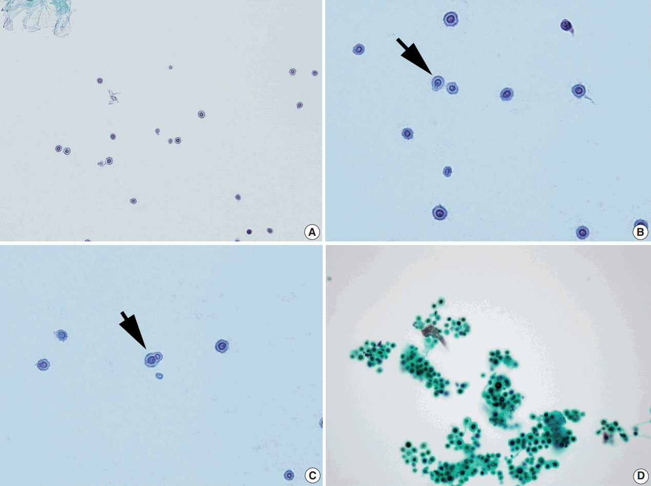
Fig. 1. Representative images of ThinPrep slide preparations of the cerebrospinal fluid. (A) Liquid-based cytology shows numerous spherical yeast cells against a clean background (Papanicolau stain). (B, C) The cells have varying sizes, clear halos, and show occasional narrow-based single budding (arrows) (Papanicolau stain). (D) Black staining is observed with the Gomori methenamine silver stain.
Reference
-
1. Prayson RA, Fischler DF. Cerebrospinal fluid cytology: an 11-year experience with 5951 specimens. Arch Pathol Lab Med. 1998; 122:47–51.2. Sharma S, Gupta N, Behera D, Rajwanshi A. Detection of cryptococcosis in liquid-based sputum cytology. Cytopathology. 2017; 28:177–8.
Article3. Levitz SM. The ecology of Cryptococcus neoformans and the epidemiology of cryptococcosis. Rev Infect Dis. 1991; 13:1163–9.4. Saigo P, Rosen PP, Kaplan MH, Solan G, Melamed MR. Identification of Cryptococcus neoformans in cytologic preparations of cerebrospinal fluid. Am J Clin Pathol. 1977; 67:141–5.5. Powers CN. Diagnosis of infectious diseases: a cytopathologist’s perspective. Clin Microbiol Rev. 1998; 11:341–65.
Article6. Walts AE. Cerebrospinal fluid cytology: selected issues. Diagn Cytopathol. 1992; 8:394–408.
Article7. Dixit A, Carroll SF, Qureshi ST. Cryptococcus gattii: an emerging cause of fungal disease in North America. Interdiscip Perspect Infect Dis. 2009; 2009:840452.8. Kwon-Chung KJ, Polacheck I, Bennett JE. Improved diagnostic medium for separation of Cryptococcus neoformans var. neoformans (serotypes A and D) and Cryptococcus neoformans var. gattii (serotypes B and C). J Clin Microbiol. 1982; 15:535–7.9. Kobayashi TK, Ueda M, Nishino T, Moritani S, Higaki T, Bamba M. Cytologic detection of cryptococcosis coexisting with herpes simplex virus infection in sputum: use of liquid-based, thin-layer preparations. Acta Cytol. 2003; 47:103–6.10. Williamson JD, Silverman JF, Mallak CT, Christie JD. Atypical cytomorphologic appearance of Cryptococcus neoformans: a report of five cases. Acta Cytol. 1996; 40:363–70.11. Kanazawa M, Ishii M, Sato Y, Kitamura K, Oshiro H, Inayama Y. Capsule-deficient meningeal cryptococcosis. Acta Cytol. 2008; 52:266–8.
Article
- Full Text Links
-
- Actions
-
Cited
- CITED
-
- Close
- Share
-
- Similar articles
-
- Complete Binocular Blindness as the First Manifestation of HIV-Related Cryptococcal Meningitis
- A Case of Cryptococcal Meningitis with Bilateral Internuclear Ophthalmoplegia and Ptosis
- A Case of Cryptococcal Meningitis in a Patient with Systemic Lupus Erythematosus
- A Case of Cryptococcal Meningitis Presenting as Bilateral Sensorineural Hearing Loss
- A Case of Cryptococcal Meningitis Presenting as Multiple Cerebral Infarctions
