Cone-beam computed tomography based evaluation of rotational patterns of dentofacial structures in skeletal Class III deformity with mandibular asymmetry
- Affiliations
-
- 1Department of Orthodontics, Wonkwang Dental Research Institute, College of Dentistry, Wonkwang University, Iksan, Korea. pigtail@wku.ac.kr
- 2Department of Orthodontics, College of Dentistry, Gangneung-Wonju National University, Gangneung, Korea.
- KMID: 2400573
- DOI: http://doi.org/10.4041/kjod.2015.45.4.153
Abstract
OBJECTIVE
The purpose of this study was to assess rotational patterns of dentofacial structures according to different vertical skeletal patterns by cone-beam computed tomography (CBCT) and analyze their influence on menton deviation in skeletal Class III deformity with mandibular asymmetry.
METHODS
The control group consisted of 30 young adults (15 men, 15 women) without any severe skeletal deformity. The asymmetry group included 55 adults (28 men, 27 women) with skeletal Class III deformity and at least 3-mm menton deviation from the midsagittal plane; it was divided into the hyperdivergent and hypodivergent subgroups using a mandibular plane angle cutoff of 35degrees. Fourteen rotational variables of the dental arches and mandible were measured and compared among the groups. Correlations between menton deviation and the other variables were evaluated.
RESULTS
The asymmetry group showed significantly larger measurements of roll and yaw in the mandible than the control group. The hypodivergent subgroup showed significant differences in maxillary posterior measurements of yaw (p < 0.01) and maxillary anterior shift (p < 0.05) compared with the hyperdivergent subgroup. All the mandibular measurements had significant correlations with menton deviation (p < 0.01). Most measurements of roll were positively correlated with one another (p < 0.01). Measurements of yaw and roll in the posterior regions were also positively correlated (p < 0.05).
CONCLUSIONS
Menton deviation in skeletal Class III deformity with mandibular asymmetry is influenced by rotation of mandibular posterior dentofacial structures. The rotational patterns vary slightly according to the vertical skeletal pattern.
MeSH Terms
Figure
-

Figure 1 Landmarks used in this study. A, Facial skeleton. B, Maxillary dental arch. C, Mandibular dental arch. FZP, Or, and Go are bilateral landmarks. See Table 2 for definitions.
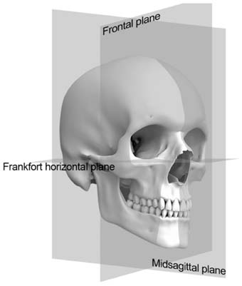
Figure 2 Reference planes used in this study. See Table 3 for definitions.

Figure 3 Positive upper canine roll relative to the direction of menton deviation.
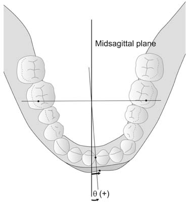
Figure 4 Positive sign of lower anterior yaw relative to the direction of menton deviation.
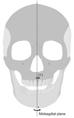
Figure 5 Positive upper anterior shift relative to the direction of menton deviation.
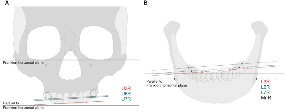
Figure 6 Measurements of roll in this study. A, Maxillary dental arch. B, Mandibular dental arch. See Table 4 for definitions.
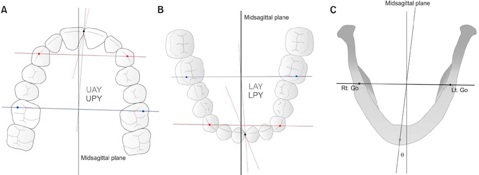
Figure 7 Measurements of yaw in this study. A, Maxillary dental arch. B, Mandibular dental arch. C, Mandible. LAY, Lower anterior yaw; LPY, Lower posterior yaw.

Figure 8 Measurements of shift in this study. See Table 4 for definitions.
Cited by 2 articles
-
Comparison of changes in the transverse dental axis between patients with skeletal Class III malocclusion and facial asymmetry treated by orthognathic surgery with and without presurgical orthodontic treatment
Han-Sol Song, Sung-Hwan Choi, Jung-Yul Cha, Kee-Joon Lee, Hyung-Seog Yu
Korean J Orthod. 2017;47(4):256-267. doi: 10.4041/kjod.2017.47.4.256.Cone-beam computed tomography analysis of transverse dental compensation in patients with skeletal Class III malocclusion and facial asymmetry
Ji-Yea Lee, Sung-Hoon Han, Hyeong-Seok Ryu, Hee-Min Lee, Sang-Cheol Kim
Korean J Orthod. 2018;48(6):357-366. doi: 10.4041/kjod.2018.48.6.357.
Reference
-
1. Enlow DH. Facial growth. 3rd ed. Philadelphia: Saunders;1990.2. Shah SM, Joshi MR. An assessment of asymmetry in the normal craniofacial complex. Angle Orthod. 1978; 48:141–148.3. Pirttiniemi P, Miettinen J, Kantomaa T. Combined effects of errors in frontal-view asymmetry diagnosis. Eur J Orthod. 1996; 18:629–636.
Article4. Padwa BL, Kaiser MO, Kaban LB. Occlusal cant in the frontal plane as a reflection of facial asymmetry. J Oral Maxillofac Surg. 1997; 55:811–816.
Article5. Trpkova B, Prasad NG, Lam EW, Raboud D, Glover KE, Major PW. Assessment of facial asymmetries from posteroanterior cephalograms: validity of reference lines. Am J Orthod Dentofacial Orthop. 2003; 123:512–520.
Article6. Vannier MW, Marsh JL, Warren JO. Three dimensional CT reconstruction images for craniofacial surgical planning and evaluation. Radiology. 1984; 150:179–184.
Article7. Fuhrmann RA, Schnappauf A, Diedrich PR. Three-dimensional imaging of craniomaxillofacial structures with a standard personal computer. Dentomaxillofac Radiol. 1995; 24:260–263.
Article8. Vannier MW, Hildebolt CF, Conover G, Knapp RH, Yokoyama-Crothers N, Wang G. Three-dimensional dental imaging by spiral CT. A progress report. Oral Surg Oral Med Oral Pathol Oral Radiol Endod. 1997; 84:561–570.9. Kim MG, Lee JW, Cha KS, Chung DH, Lee SM. Three-dimensional symmetry and parallelism of the skeletal and soft-tissue poria in patients with facial asymmetry. Korean J Orthod. 2014; 44:62–68.
Article10. Ackerman JL, Proffit WR, Sarver DM, Ackerman MB, Kean MR. Pitch, roll, and yaw: describing the spatial orientation of dentofacial traits. Am J Orthod Dentofacial Orthop. 2007; 131:305–310.
Article11. Kim JM. 3D rotational pattern of the dental arch and mandible in class III patients with facial asymmetry [PhD. dissertation]. Seoul: Dankook University;2009.12. Severt TR, Proffit WR. The prevalence of facial asymmetry in the dentofacial deformities population at the University of North Carolina. Int J Adult Orthodon Orthognath Surg. 1997; 12:171–176.13. Baek SH, Cho IS, Chang YI, Kim MJ. Skeletodental factors affecting chin point deviation in female patients with class III malocclusion and facial asymmetry: a three-dimensional analysis using computed tomography. Oral Surg Oral Med Oral Pathol Oral Radiol Endod. 2007; 104:628–639.
Article14. Hwang HS, Hwang CH, Lee KH, Kang BC. Maxillofacial 3-dimensional image analysis for the diagnosis of facial asymmetry. Am J Orthod Dentofacial Orthop. 2006; 130:779–785.
Article15. Baek SH, Yang WS. A soft tissue analysis on facial esthetics of Korean young adults. Korean J Orthod. 1991; 21:131–170.16. Baek C, Paeng JY, Lee JS, Hong J. Morphologic evaluation and classification of facial asymmetry using 3-dimensional computed tomography. J Oral Maxillofac Surg. 2012; 70:1161–1169.
Article17. Maeda M, Katsumata A, Ariji Y, Muramatsu A, Yoshida K, Goto S, et al. 3D-CT evaluation of facial asymmetry in patients with maxillofacial deformities. Oral Surg Oral Med Oral Pathol Oral Radiol Endod. 2006; 102:382–390.
Article18. Haraguchi S, Takada K, Yasuda Y. Facial asymmetry in subjects with skeletal Class III deformity. Angle Orthod. 2002; 72:28–35.19. Chebib FS, Chamma AM. Indices of craniofacial asymmetry. Angle Orthod. 1981; 51:214–226.20. Adams GL, Gansky SA, Miller AJ, Harrell WE Jr, Hatcher DC. Comparison between traditional 2-dimensional cephalometry and a 3-dimensional approach on human dry skulls. Am J Orthod Dentofacial Orthop. 2004; 126:397–409.
Article21. Türp JC, Alt KW, Vach W, Harbich K. Mandibular condyles and rami are asymmetric structures. Cranio. 1998; 16:51–56.
Article22. El-Beialy AR, Fayed MS, El-Bialy AM, Mostafa YA. Accuracy and reliability of cone-beam computed tomography measurements: Influence of head orientation. Am J Orthod Dentofacial Orthop. 2011; 140:157–165.
Article23. Jacobson A. Radiographic cephalometry from basics to videoimaging. Carol Stream, IL: Quintessence Publishing;1995.24. Lascala CA, Panella J, Marques MM. Analysis of the accuracy of linear measurements obtained by cone beam computed tomography (CBCT-NewTom). Dentomaxillofac Radiol. 2004; 33:291–294.
Article25. Park JU, Kook YA, Kim Y. Assessment of asymmetry in a normal occlusion sample and asymmetric patients with three-dimensional cone beam computed tomography: a study for a transverse reference plane. Angle Orthod. 2012; 82:860–867.
Article26. Cho HJ. A three-dimensional cephalometric analysis. J Clin Orthod. 2009; 43:235–252.27. Kim SJ, Lee KJ, Lee SH, Baik HS. Morphologic relationship between the cranial base and the mandible in patients with facial asymmetry and mandibular prognathism. Am J Orthod Dentofacial Orthop. 2013; 144:330–340.
Article28. Hwang HS, Min YS, Lee SC, Sun MK, Lim HS. Change of lip-line cant after 1-jaw orthognathic surgery in patients with mandibular asymmetry. Am J Orthod Dentofacial Orthop. 2009; 136:564–569.
Article29. Lee MS, Chung DH, Lee JW, Cha KS. Assessing soft-tissue characteristics of facial asymmetry with photographs. Am J Orthod Dentofacial Orthop. 2010; 138:23–31.
Article
- Full Text Links
-
- Actions
-
Cited
- CITED
-
- Close
- Share
-
- Similar articles
-
- Cone-beam computed tomography analysis of transverse dental compensation in patients with skeletal Class III malocclusion and facial asymmetry
- Location and shape of the mandibular lingula: Comparison of skeletal class I and class III patients using panoramic radiography and cone-beam computed tomography
- Differences in positions of cone-beam computed tomography landmarks in patients with skeletal Class III facial asymmetry according to midsagittal planes
- Differences in facial soft tissue deviations in Class III patients with different types of mandibular asymmetry: A cone-beam computed tomography study
- Cone-beam computed tomographic evaluation of the temporomandibular joint and dental characteristics of patients with Class II subdivision malocclusion and asymmetry
