Cone-beam computed tomographic evaluation of the temporomandibular joint and dental characteristics of patients with Class II subdivision malocclusion and asymmetry
- Affiliations
-
- 1The Affiliated Stomatology Hospital, Chongqing Medical University, Chongqing, China. zhengleileicqmu@hospital.cqmu.edu.cn
- 2Chongqing Key Laboratory of Oral Diseases and Biomedical Sciences, Chongqing, China.
- 3Chongqing Municipal Key Laboratory of Oral Biomedical Engineering of Higher Education, Chongqing, China.
- KMID: 2389143
- DOI: http://doi.org/10.4041/kjod.2017.47.5.277
Abstract
OBJECTIVE
Treating Class II subdivision malocclusion with asymmetry has been a challenge for orthodontists because of the complicated characteristics of asymmetry. This study aimed to explore the characteristics of dental and skeletal asymmetry in Class II subdivision malocclusion, and to assess the relationship between the condyle-glenoid fossa and first molar.
METHODS
Cone-beam computed tomographic images of 32 patients with Class II subdivision malocclusion were three-dimensionally reconstructed using the Mimics software. Forty-five anatomic landmarks on the reconstructed structures were selected and 27 linear and angular measurements were performed. Paired-samples t-tests were used to compare the average differences between the Class I and Class II sides; Pearson correlation coefficient (r) was used for analyzing the linear association.
RESULTS
The faciolingual crown angulation of the mandibular first molar (p < 0.05), sagittal position of the maxillary and mandibular first molars (p < 0.01), condylar head height (p < 0.01), condylar process height (p < 0.05), and angle of the posterior wall of the articular tubercle and coronal position of the glenoid fossa (p < 0.01) were significantly different between the two sides. The morphology and position of the condyle-glenoid fossa significantly correlated with the three-dimensional changes in the first molar.
CONCLUSIONS
Asymmetry in the sagittal position of the maxillary and mandibular first molars between the two sides and significant lingual inclination of the mandibular first molar on the Class II side were the dental characteristics of Class II subdivision malocclusion. Condylar morphology and glenoid fossa position asymmetries were the major components of skeletal asymmetry and were well correlated with the three-dimensional position of the first molar.
MeSH Terms
Figure
-
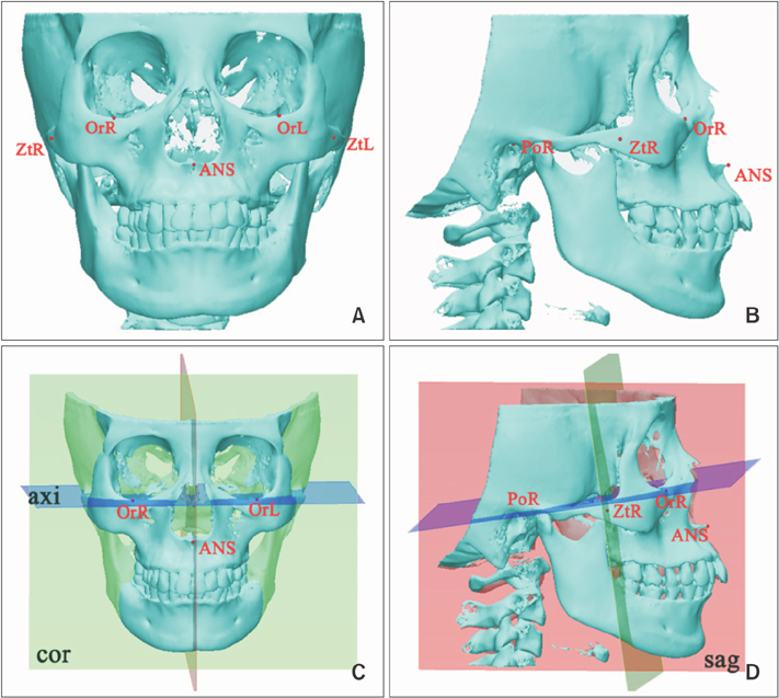
Figure 1 Reference planes and landmarks used in the study. A, Frontal view of the craniofacial bone. B, Lateral view of the craniofacial bone. C, D, Setting of the three perpendicular reference planes. The red plane represents the sagittal plane (sag), the blue one represents the axial plane (axi), and the green one represents the coronal plane (cor). Terms and definitions are listed in Table 1.
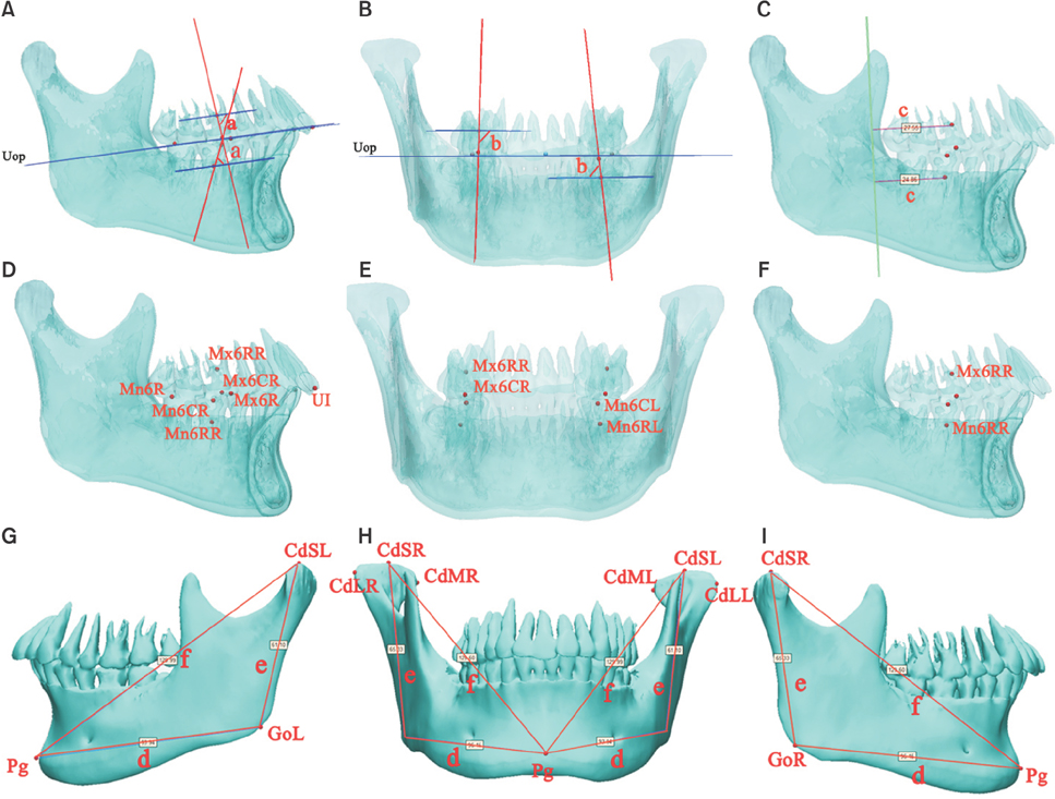
Figure 2 The measurements of the teeth and mandibular bone (Table 1 and Table 2). A and D, Right perspective view of the mandibular bone: mesiodistal crown angulation of the first molar (a); B and E, frontal perspective view of the mandibular bone: faciolingual crown angulation of the first molar (b); C and F, right perspective view of the mandibular bone: sagittal position of the first molar (c); G, left view of the mandibular bone; H, frontal view of the mandibular bone; I, right view of the mandibular bone: the length of the mandibular body (d), height of the ramus (e), and length of the entire mandibular bone (f).
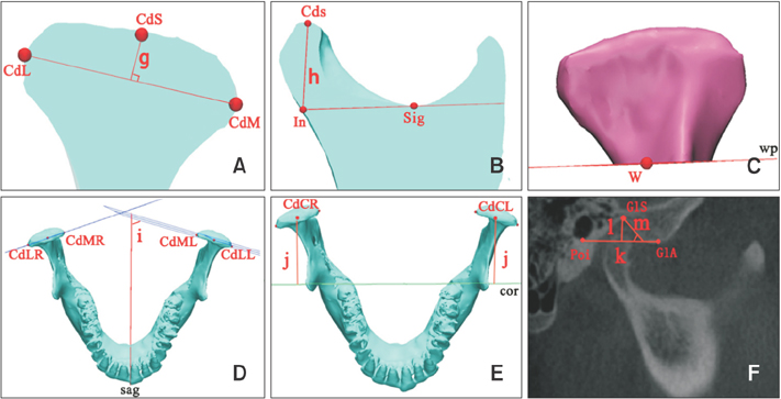
Figure 3 The measurements of the condyle and glenoid fossa (Table 1 and Table 2). A, Mediolateral cut surface of the condyle: the height of the condylar head (g); B, anteroposterior cut surface of the condyle: the height of the condylar process (h); C, condylar process reconstructed by cutting the condyle along the wp plane: condylar area and volume; D and E, top view of the mandible: the angle between the mediolateral plane of the condyle and the sagittal plane (i); the vertical distance from the geometric centers of the condyles to the coronal plane (j); F, the condyle and glenoid fossa in a cone-beam computed tomography image: the width of the glenoid fossa (k), depth of the glenoid fossa (l), and angle of the posterior wall of the articular tubercle (m).
Reference
-
1. Janson G, de Lima KJ, Woodside DG, Metaxas A, de Freitas MR, Henriques JF. Class II subdivision malocclusion types and evaluation of their asymmetries. Am J Orthod Dentofacial Orthop. 2007; 131:57–66.
Article2. Azevedo AR, Janson G, Henriques JF, Freitas MR. Evaluation of asymmetries between subjects with Class II subdivision and apparent facial asymmetry and those with normal occlusion. Am J Orthod Dentofacial Orthop. 2006; 129:376–383.
Article3. Janson GR, Metaxas A, Woodside DG, de Freitas MR, Pinzan A. Three-dimensional evaluation of skeletal and dental asymmetries in Class II subdivision malocclusions. Am J Orthod Dentofacial Orthop. 2001; 119:406–418.
Article4. Meloti AF, Gonçalves Rde C, Silva E, Martins LP, dos Santos-Pinto A. Lateral cephalometric diagnosis of asymmetry in Angle Class II subdivision compared to Class I and II. Dental Press J Orthod. 2014; 19:80–88.
Article5. Sanders DA, Rigali PH, Neace WP, Uribe F, Nanda R. Skeletal and dental asymmetries in Class II subdivision malocclusions using cone-beam computed tomography. Am J Orthod Dentofacial Orthop. 2010; 138:542–543.
Article6. Minich CM, Araújo EA, Behrents RG, Buschang PH, Tanaka OM, Kim KB. Evaluation of skeletal and dental asymmetries in Angle Class II subdivision malocclusions with cone-beam computed tomography. Am J Orthod Dentofacial Orthop. 2013; 144:57–66.
Article7. Li J, He Y, Wang Y, Chen T, Xu Y, Xu X, et al. Dental, skeletal asymmetries and functional characteristics in Class II subdivision malocclusions. J Oral Rehabil. 2015; 42:588–599.
Article8. Kurusu A, Horiuchi M, Soma K. Relationship between occlusal force and mandibular condyle morphology. Evaluated by limited cone-beam computed tomography. Angle Orthod. 2009; 79:1063–1069.9. Vitral RW, Telles Cde S, Fraga MR, de Oliveira RS, Tanaka OM. Computed tomography evaluation of temporomandibular joint alterations in patients with class II division 1 subdivision malocclusions: condyle-fossa relationship. Am J Orthod Dentofacial Orthop. 2004; 126:48–52.
Article10. Rodrigues AF, Fraga MR, Vitral RW. Computed tomography evaluation of the temporomandibular joint in Class II Division 1 and Class III malocclusion patients: condylar symmetry and condyle-fossa relationship. Am J Orthod Dentofacial Orthop. 2009; 136:199–206.
Article11. Fraga MR, Rodrigues AF, Ribeiro LC, Campos MJ, Vitral RW. Anteroposterior condylar position: a comparative study between subjects with normal occlusion and patients with Class I, Class II Division 1, and Class III malocclusions. Med Sci Monit. 2013; 19:903–907.
Article12. Krisjane Z, Urtane I, Krumina G, Zepa K. Three-dimensional evaluation of TMJ parameters in Class II and Class III patients. Stomatologija. 2009; 11:32–36.13. Saccucci M, D'Attilio M, Rodolfino D, Festa F, Polimeni A, Tecco S. Condylar volume and condylar area in class I, class II and class III young adult subjects. Head Face Med. 2012; 8:34.
Article14. Schulze D, Heiland M, Thurmann H, Adam G. Radiation exposure during midfacial imaging using 4- and 16-slice computed tomography, cone beam computed tomography systems and conventional radiography. Dentomaxillofac Radiol. 2004; 33:83–86.
Article15. Cevidanes LH, Styner MA, Proffit WR. Image analysis and superimposition of 3-dimensional cone-beam computed tomography models. Am J Orthod Dentofacial Orthop. 2006; 129:611–618.
Article16. Salemi F, Shokri A, Mortazavi H, Baharvand M. Diagnosis of simulated condylar bone defects using panoramic radiography, spiral tomography and cone-beam computed tomography: a comparison study. J Clin Exp Dent. 2015; 7:e34–e39.
Article17. Ludlow JB, Laster WS, See M, Bailey LJ, Hershey HG. Accuracy of measurements of mandibular anatomy in cone beam computed tomography images. Oral Surg Oral Med Oral Pathol Oral Radiol Endod. 2007; 103:534–542.
Article18. Neiva MB, Soares ÁC, Lisboa Cde O, Vilella Ode V, Motta AT. Evaluation of cephalometric landmark identification on CBCT multiplanar and 3D reconstructions. Angle Orthod. 2015; 85:11–17.
Article19. Pinho T. Treatment of a Class II subdivision based on occlusal plane control: a clinical case. Orthodontics (Chic.). 2012; 13:128–137.20. Kurt G, Uysal T, Sisman Y, Ramoglu SI. Mandibular asymmetry in Class II subdivision malocclusion. Angle Orthod. 2008; 78:32–37.
Article21. Mohlin BO, Derweduwen K, Pilley R, Kingdon A, Shaw WC, Kenealy P. Malocclusion and temporomandibular disorder: a comparison of adolescents with moderate to severe dysfunction with those without signs and symptoms of temporomandibular disorder and their further development to 30 years of age. Angle Orthod. 2004; 74:319–327.22. Egermark I, Magnusson T, Carlsson GE. A 20-year follow-up of signs and symptoms of temporomandibular disorders and malocclusions in subjects with and without orthodontic treatment in childhood. Angle Orthod. 2003; 73:109–115.23. Paknahad M, Shahidi S. Association between mandibular condylar position and clinical dysfunction index. J Craniomaxillofac Surg. 2015; 43:432–436.
Article24. Talaat W, Al Bayatti S, Al Kawas S. CBCT analysis of bony changes associated with temporomandibular disorders. Cranio. 2016; 34:88–94.
Article25. Slavicek R. Relationship between occlusion and temporomandibular disorders: implications for the gnathologist. Am J Orthod Dentofacial Orthop. 2011; 139:10. 12. 14 passim.
Article26. Tanne K, Okamoto T, Su SC, Mitsuyoshi T, Asakawa-Tanne Y, Tanimoto K. Current status of temporomandibular joint disorders and the therapeutic system derived from a series of biomechanical, histological, and biochemical studies. APOS Trends Orthod. 2015; 5:4–21.
Article27. Burstone CJ. Diagnosis and treatment planning of patients with asymmetries. Semin Orthod. 1998; 4:153–164.28. Chung KR, Kim SH, Chaffee MP, Nelson G. Molar distalization with a partially integrated mini-implant to correct unilateral Class II malocclusion. Am J Orthod Dentofacial Orthop. 2010; 138:810–819.
Article29. Bock NC, Reiser B, Ruf S. Class II subdivision treatment with the Herbst appliance. Angle Orthod. 2013; 83:327–333.
Article
- Full Text Links
-
- Actions
-
Cited
- CITED
-
- Close
- Share
-
- Similar articles
-
- Three-dimensional assessment of the temporomandibular joint and mandibular dimensions after early correction of the maxillary arch form in patients with Class II division 1 or division 2 malocclusion
- Cone-beam computed tomography analysis of transverse dental compensation in patients with skeletal Class III malocclusion and facial asymmetry
- Comparison of the condyle-fossa relationship between skeletal class III malocclusion patients with and without asymmetry: a retrospective three-dimensional cone-beam computed tomograpy study
- Three-dimensional finite element analysis of unilateral mastication in malocclusion cases using cone-beam computed tomography and a motion capture system
- A Study on the Nasal Index of Malocclusion Patients Using Cone-Beam Computed Tomography 3D Program
