Comparison of the condyle-fossa relationship between skeletal class III malocclusion patients with and without asymmetry: a retrospective three-dimensional cone-beam computed tomograpy study
- Affiliations
-
- 1Department of Orthodontics, Graduate School of Clinical Dental Science, The Catholic University of Korea, Seoul, Korea. juice@catholic.ac.kr
- 2Department of Oral and Maxillofacial Surgery, Dental Clinic, Uijeongbu St. Mary's Hospital, The Catholic University of Korea, Uijeongbu, Korea.
- 3Department of Orthodontics, Seoul St. Mary's Hospital, The Catholic University of Korea, Seoul, Korea.
- KMID: 2273412
- DOI: http://doi.org/10.4041/kjod.2013.43.5.209
Abstract
OBJECTIVE
This study investigated whether temporomandibular joint (TMJ) condyle-fossa relationships are bilaterally symmetric in class III malocclusion patients with and without asymmetry and compared to those with normal occlusion. The hypothesis was a difference in condyle-fossa relationships exists in asymmetric patients.
METHODS
Group 1 comprised 40 Korean normal occlusion subjects. Groups 2 and 3 comprised patients diagnosed with skeletal class III malocclusion, who were grouped according to the presence of mandibular asymmetry: Group 2 included symmetric mandibles, while group 3 included asymmetric mandibles. Pretreatment three-dimensional cone-beam computed tomography (3D CBCT) images were obtained. Right- and left-sided TMJ spaces in groups 1 and 2 or deviated and non-deviated sides in group 3 were evaluated, and the axial condylar angle was compared.
RESULTS
The TMJ spaces demonstrated no significant bilateral differences in any group. Only group 3 had slightly narrower superior spaces (p < 0.001). The axial condylar angles between group 1 and 2 were not significant. However, group 3 showed a statistically significant bilateral difference (p < 0.001); toward the deviated side, the axial condylar angle was steeper.
CONCLUSIONS
Even in the asymmetric group, the TMJ spaces were similar between deviated and non-deviated sides, indicating a bilateral condyle-fossa relationship in patients with asymmetry that may be as symmetrical as that in patients with symmetry. However, the axial condylar angle had bilateral differences only in asymmetric groups. The mean TMJ space value and the bilateral difference may be used for evaluating condyle-fossa relationships with CBCT.
Keyword
MeSH Terms
Figure
-

Figure 1 The 3 views of the condyle in the cone-beam computed tomography (CBCT) image: A, axial view; B, coronal view; C, sagittal view. The CBCT images were reoriented with the horizontal reference plane connecting the bilateral orbitales and Frankfurt horizontal plane,17,18 and the vertical midline and horizontal reference planes were set accordingly. The sagittal slice (C) was evaluated at the point where the mediolateral diameter of the right or left condyles was greatest (A) in the axial view.

Figure 2 Measurement of the joint space in the sagittal view. Anterior joint space (AS), superior joint space (SS), and posterior joint space (PS) were measured from the most prominent anterior, posterior, and superior condylar points to that of the glenoid fossa with the methods previously reported.7 The plane parallel to the Frankfurt horizontal (FH) plane was used as the reference plane.
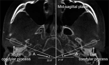
Figure 3 Measurement of the axial condylar angle of the condylar process (A). It was defined as the angle between the long axis of the mandibular condylar process and a perpendicular line to the midsagittal plane.7

Figure 4 Cone-beam computed tomography image of a sample case showing the largest axial condylar angle difference between the deviated and non-deviated sides.
Cited by 1 articles
-
Comparison of the three-dimensional structures of mandibular condyles between adults with and without facial asymmetry: A retrospective study
Min-Hee Oh, Sung-Ja Kang, Jin-Hyoung Cho
Korean J Orthod. 2018;48(2):73-80. doi: 10.4041/kjod.2018.48.2.73.
Reference
-
1. Katsavrias EG, Halazonetis DJ. Condyle and fossa shape in Class II and Class III skeletal patterns: a morphometric tomographic study. Am J Orthod Dentofacial Orthop. 2005; 128:337–346.
Article2. Ahn SJ, Lee SP, Nahm DS. Relationship between temporomandibular joint internal derangement and facial asymmetry in women. Am J Orthod Dentofacial Orthop. 2005; 128:583–591.
Article3. Byun ES, Ahn SJ, Kim TW. Relationship between internal derangement of the temporomandibular joint and dentofacial morphology in women with anterior open bite. Am J Orthod Dentofacial Orthop. 2005; 128:87–95.
Article4. Vitral RW, da Silva Campos MJ, Rodrigues AF, Fraga MR. Temporomandibular joint and normal occlusion: Is there anything singular about it? A computed tomographic evaluation. Am J Orthod Dentofacial Orthop. 2011; 140:18–24.
Article5. Rodrigues AF, Fraga MR, Vitral RW. Computed tomography evaluation of the temporomandibular joint in Class I malocclusion patients: condylar symmetry and condyle-fossa relationship. Am J Orthod Dentofacial Orthop. 2009; 136:192–198.
Article6. Pullinger AG, Solberg WK, Hollender L, Petersson A. Relationship of mandibular condylar position to dental occlusion factors in an asymptomatic population. Am J Orthod Dentofacial Orthop. 1987; 91:200–206.
Article7. Cohlmia JT, Ghosh J, Sinha PK, Nanda RS, Currier GF. Tomographic assessment of temporomandibular joints in patients with malocclusion. Angle Orthod. 1996; 66:27–35.8. Vitral RW, Telles Cde S, Fraga MR, de Oliveira RS, Tanaka OM. Computed tomography evaluation of temporomandibular joint alterations in patients with class II division 1 subdivision malocclusions: condyle-fossa relationship. Am J Orthod Dentofacial Orthop. 2004; 126:48–52.
Article9. Zuckerberg Al, Yaster M. Chapter 26. Anesthesia for orthopedic surgery. In : Davis PJ, Cladis FP, Motoyama EK, Smith RM, editors. Smith's anesthesia for infants and children. 8th ed. Philadelphia: Elsevier Mosby;2011. p. 842–869.10. Rodrigues AF, Fraga MR, Vitral RW. Computed tomography evaluation of the temporomandibular joint in Class II Division 1 and Class III malocclusion patients: condylar symmetry and condyle-fossa relationship. Am J Orthod Dentofacial Orthop. 2009; 136:199–206.
Article11. Pandis N, Karpac J, Trevino R, Williams B. A radiographic study of condyle position at various depths of cut in dry skulls with axially corrected lateral tomograms. Am J Orthod Dentofacial Orthop. 1991; 100:116–122.
Article12. Burke G, Major P, Glover K, Prasad N. Correlations between condylar characteristics and facial morphology in Class II preadolescent patients. Am J Orthod Dentofacial Orthop. 1998; 114:328–336.
Article13. Lee W, Park JU. Three-dimensional evaluation of positional change of the condyle after mandibular setback by means of bilateral sagittal split ramus osteotomy. Oral Surg Oral Med Oral Pathol Oral Radiol Endod. 2002; 94:305–309.
Article14. Hilgers ML, Scarfe WC, Scheetz JP, Farman AG. Accuracy of linear temporomandibular joint measurements with cone beam computed tomography and digital cephalometric radiography. Am J Orthod Dentofacial Orthop. 2005; 128:803–811.
Article15. Librizzi ZT, Tadinada AS, Valiyaparambil JV, Lurie AG, Mallya SM. Cone-beam computed tomography to detect erosions of the temporomandibular joint: Effect of field of view and voxel size on diagnostic efficacy and effective dose. Am J Orthod Dentofacial Orthop. 2011; 140:e25–e30.
Article16. Kim YI, Cho BH, Jung YH, Son WS, Park SB. Cone-beam computerized tomography evaluation of condylar changes and stability following two-jaw surgery: Le Fort I osteotomy and mandibular setback surgery with rigid fixation. Oral Surg Oral Med Oral Pathol Oral Radiol Endod. 2011; 111:681–687.
Article17. Kook YA, Kim Y. Evaluation of facial asymmetry with three-dimensional cone-beam computed tomography. J Clin Orthod. 2011; 45:112–115. quiz 92.18. Park JU, Kook YA, Kim Y. Assessment of asymmetry in a normal occlusion sample and asymmetric patients with three-dimensional cone beam computed tomography: a study for a transverse reference plane. Angle Orthod. 2012; 82:860–867.
Article19. Blackwood HJ. Pathology of the temporomandibular joint. J Am Dent Assoc. 1969; 79:118–124.
Article20. Westesson PL, Bifano JA, Tallents RH, Hatala MP. Increased horizontal angle of the mandibular condyle in abnormal temporomandibular joints. A magnetic resonance imaging study. Oral Surg Oral Med Oral Pathol. 1991; 72:359–363.
Article21. Ueki K, Nakagawa K, Takatsuka S, Shimada M, Marukawa K, Takazakura D, et al. Temporomandibular joint morphology and disc position in skeletal class III patients. J Craniomaxillofac Surg. 2000; 28:362–368.
Article22. Ahn SJ, Lee SJ, Kim TW. Orthodontic effects on dentofacial morphology in women with bilateral TMJ disk displacement. Angle Orthod. 2007; 77:288–295.
Article
- Full Text Links
-
- Actions
-
Cited
- CITED
-
- Close
- Share
-
- Similar articles
-
- Cone-beam computed tomographic evaluation of the temporomandibular joint and dental characteristics of patients with Class II subdivision malocclusion and asymmetry
- Cone-beam computed tomography analysis of transverse dental compensation in patients with skeletal Class III malocclusion and facial asymmetry
- Three-dimensional evaluation of the mandibular condyle in adults with various skeletal patterns
- Positional change in mandibular condyle in facial asymmetric patients after orthognathic surgery: cone-beam computed tomography study
- The relationship between condyle position, morphology and chin deviation in skeletal Class III patients with facial asymmetry using cone-beam CT
