Korean J Orthod.
2023 Mar;53(2):67-76. 10.4041/kjod22.076.
Three-dimensional evaluation of the mandibular condyle in adults with various skeletal patterns
- Affiliations
-
- 1Jiangsu Key Laboratory of Oral Disease, Nanjing Medical University, Nanjing, China
- 2Department of Orthodontics, Affiliated Hospital of Stomatology, Nanjing Medical University, Nanjing, China
- 3Department of Stomatology, Dushu Lake Hospital Affiliated to Soochow University, Suzhou, China
- 4Department of Stomatology, Medical Center of Soochow University, Suzhou, China
- KMID: 2540946
- DOI: http://doi.org/10.4041/kjod22.076
Abstract
Objective
Morphometric and morphological evaluation of the mandibular condyle in adults and to identify its correlation with skeletal malocclusion patterns.
Methods
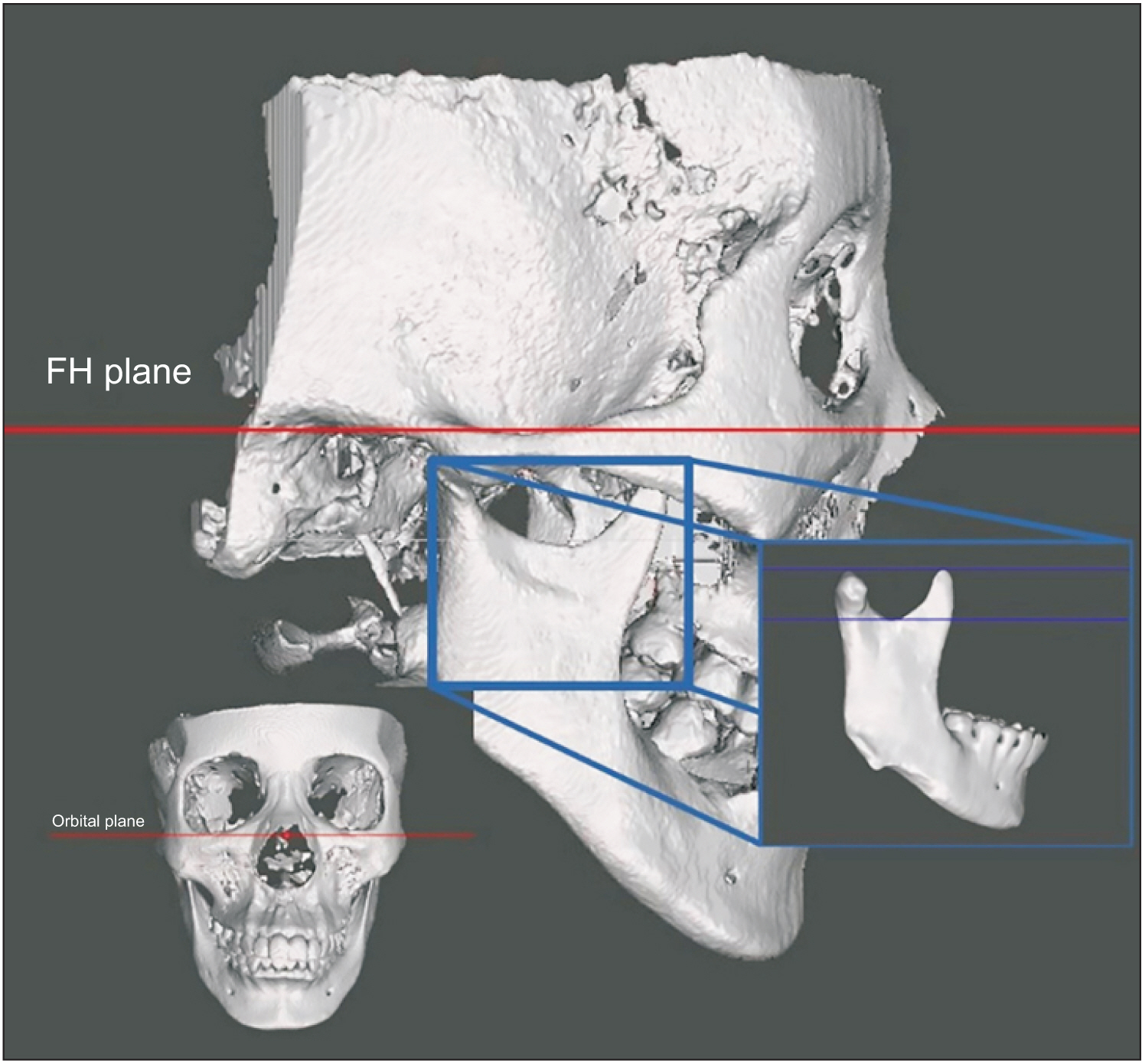
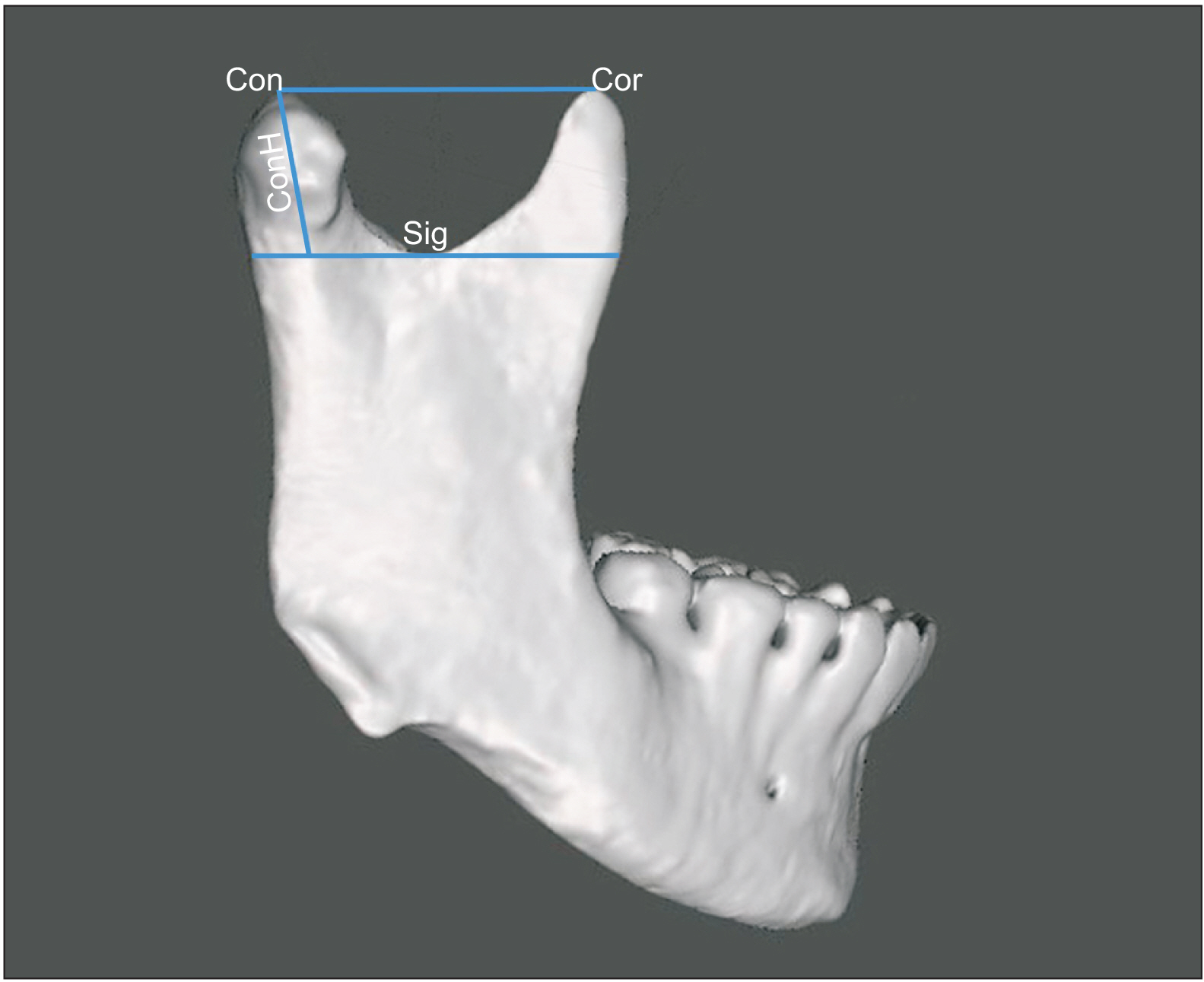
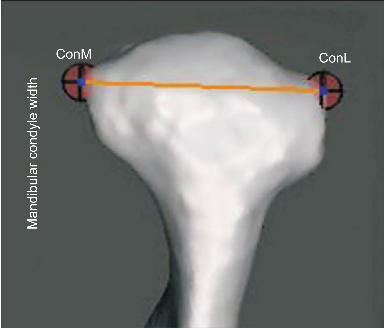
Cone-beam computed tomography scans of 135 adult patients were used in this study and classified into groups according to four criteria: (1) sex (male and female); (2) sagittal skeletal discrepancy (Class I, Class II, and Class III); (3) vertical skeletal discrepancy (hyperdivergent, normodivergent, and hypodivergent); and age (group 1 ≤ 20 years, 21 ≤ group 2 < 30, and group 3 ≥ 30 years). The morphometrical variables were mandibular condyle height and width, and the morphological variable was the mandibular condyle shape in coronal and sagittal sections. Three-dimensional standard tessellation language files were created using itk-snap (open-source software), and measurements were performed using Meshmixer (open-source software).
Results
The mandibular condyle height was significantly greater (p < 0.05) in patients with class III malocclusion than in those with class I or II malocclusion; the mandibular condyle width was not significantly different among different sexes, age groups, and sagittal and vertical malocclusions. There were no statistical associations between various mandibular condyle shapes and the sexes, age groups, and skeletal malocclusions.
Conclusions
The condylar height was greatest in patients with class III malocclusion. The condylar height and width were greater among males than in females. The mandibular condyle shapes observed in sagittal and coronal sections did not affect the skeletal malocclusion patterns.
Keyword
Figure
Reference
-
1. Jablonski S. 1982. Illustrated dictionary of dentistry. W.B Saunders Co.;Philadelphia: DOI: 10.1016/0002-9416(82)90326-8.2. Mohl N, Zarb G, Carlsson G, Rugh J. 1988. Textbook of occlusion. Quintessence Publishing Co.;Chicago: p. 139–40.3. Kandasamy S, Greene CS, Rinchuse DJ, Stockstill JW. 2015. TMD and orthodontics: a clinical guide for the orthodontist. Springer;Cham: DOI: 10.1007/978-3-319-19782-1.4. Snell RS. 1995. Clinical anatomy for medical students. Little Brown & Co.;Boston: DOI: 10.7326/0003-4819-123-5-199509010-00049.5. Williams PL BL, Berry MM, Collins P, Dyson M, Dussek JE, et al. Williams PL BL, Berry MM, Collins P, Dyson M, Dussek JE, editors. 1995. Skeletal system-individual cranial bones. Gray's anatomy: The anatomical basis of medicine and surgery. 38th ed. Churchill Livingstone;Great Britain: p. 577.6. Ardakani FE, Niafar N. 2004; Evaluation of changes in the mandibular angular cortex using panoramic images. J Contemp Dent Pract. 5:1–15. DOI: 10.5005/jcdp-5-3-1. PMID: 15318252.7. Solberg WK, Bibb CA, Nordström BB, Hansson TL. 1986; Malocclusion associated with temporomandibular joint changes in young adults at autopsy. Am J Orthod. 89:326–30. DOI: 10.1016/0002-9416(86)90055-2. PMID: 3457531.8. Gray H, Standring S. 2005. Gray's anatomy: the anatomical basis of clinical practice. Churchill Livingstone;Edinburgh: DOI: 10.5860/choice.43-1300.9. Alomar X, Medrano J, Cabratosa J, Clavero JA, Lorente M, Serra I, et al. 2007; Anatomy of the temporomandibular joint. Semin Ultrasound CT MR. 28:170–83. DOI: 10.1053/j.sult.2007.02.002. PMID: 17571700.10. Hegde S, Praveen BN, Shetty SR. 2013; Morphological and radiological variations of mandibular condyles in health and diseases: a systematic review. Dentistry. 3:154. DOI: 10.4172/2161-1122.1000154.11. Krisjane Z, Urtane I, Krumina G, Bieza A, Zepa K, Rogovska I. 2007; Condylar and mandibular morphological criteria in the 2D and 3D MSCT imaging for patients with Class II division 1 subdivision malocclusion. Stomatologija. 9:67–71. PMID: 17993738.12. Al-koshab M, Nambiar P, John J. 2015; Assessment of condyle and glenoid fossa morphology using CBCT in South-East Asians. PLoS One. 10:e0121682. DOI: 10.1371/journal.pone.0121682. PMID: 25803868. PMCID: PMC4372412. PMID: d2bf1af7723d46099cc547a255c99578.13. Chae JM, Park JH, Tai K, Mizutani K, Uzuka S, Miyashita W, et al. 2020; Evaluation of condyle-fossa relationships in adolescents with various skeletal patterns using cone-beam computed tomography. Angle Orthod. 90:224–32. DOI: 10.2319/052919-369.1. PMID: 31638857. PMCID: PMC8051241.14. Song J, Cheng M, Qian Y, Chu F. 2020; Cone-beam CT evaluation of temporomandibular joint in permanent dentition according to Angle's classification. Oral Radiol. 36:261–6. DOI: 10.1007/s11282-019-00403-3. PMID: 31385140.15. Zhang Y, Xu X, Liu Z. 2017; Comparison of morphologic parameters of temporomandibular joint for asymptomatic subjects using the two-dimensional and three-dimensional measuring methods. J Healthc Eng. 2017:5680708. DOI: 10.1155/2017/5680708. PMID: 29065621. PMCID: PMC5434231.16. Krisjane Z, Urtane I, Krumina G, Zepa K. 2009; Three-dimensional evaluation of TMJ parameters in Class II and Class III patients. Stomatologija. 11:32–6. PMID: 19423969.17. Noh KJ, Baik HS, Han SS, Jang W, Choi YJ. 2021; Differences in mandibular condyle and glenoid fossa morphology in relation to vertical and sagittal skeletal patterns: a cone-beam computed tomography study. Korean J Orthod. 51:126–34. DOI: 10.4041/kjod.2021.51.2.126. PMID: 33678628. PMCID: PMC7940806.18. Tassoker M, Kabakci ADA, Akin D, Sener S. 2017; Evaluation of mandibular notch, coronoid process, and mandibular condyle configurations with cone beam computed tomography. Biomed Res. 28:8327–35.19. Isaac B, Holla SJ. 2001; Variations in the shape of the coronoid process in the adult human mandible. J Anat Soc India. 50:137–9.20. Ishwarkumar S, Pillay P, De-Gama B, Satyapal K. 2019; Osteometric and radiological study of the mandibular notch. Int J Morphol. 37:491–7. DOI: 10.4067/S0717-95022019000200491.21. Oliveira-Santos C, Bernardo RT, Capelozza A. 2009; Mandibular condyle morphology on panoramic radiographs of asymptomatic temporomandibular joints. Int J Dent. 8.22. Sahithi D, Reddy S, Teja DD, Koneru J, Praveen KNS, Sruthi R. 2016; Reveal the concealed - morphological variations of the coronoid process, condyle and sigmoid notch in personal identification. Egypt J Forensic Sci. 6:108–13. DOI: 10.1016/j.ejfs.2015.11.003. PMID: 531bda3ca9b24f43b360e5ae8a40339d.23. Nagaraj T, Nigam H, Santosh H, Gogula S, Sumana C, Sahu P. 2017; Morphological variations of the coronoid process, condyle and sigmoid notch as an adjunct in personal identification. J Med Radiol Pathol Surg. 4:1–5. DOI: 10.15713/ins.jmrps.86.24. Hwang HS, Hwang CH, Lee KH, Kang BC. 2006; Maxillofacial 3-dimensional image analysis for the diagnosis of facial asymmetry. Am J Orthod Dentofacial Orthop. 130:779–85. DOI: 10.1016/j.ajodo.2005.02.021. PMID: 17169741.25. Lopez TT, Michel-Crosato E, Benedicto EN, Paiva LA, Silva DC, Biazevic MG. 2017; Accuracy of mandibular measurements of sexual dimorphism using stabilizer equipment. Braz Oral Res. 31:e1. DOI: 10.1590/1807-3107bor-2017.vol31.0001. PMID: 28076494.26. Saccucci M, D'Attilio M, Rodolfino D, Festa F, Polimeni A, Tecco S. 2012; Condylar volume and condylar area in class I, class II and class III young adult subjects. Head Face Med. 8:34. DOI: 10.1186/1746-160X-8-34. PMID: 23241136. PMCID: PMC3558396. PMID: 3df2f567c37b40cd83ffc473945335b2.27. Wolff J. 2012. The law of bone remodelling. Springer Science & Business Media;Berlin: DOI: 10.1007/978-3-642-71031-5.28. Charalampidou M, Kjellberg H, Georgiakaki I, Kiliaridis S. 2008; Masseter muscle thickness and mechanical advantage in relation to vertical craniofacial morphology in children. Acta Odontol Scand. 66:23–30. DOI: 10.1080/00016350701884604. PMID: 18320415.29. Proffit WR, Fields HW, Nixon WL. 1983; Occlusal forces in normal- and long-face adults. J Dent Res. 62:566–70. DOI: 10.1177/00220345830620051201. PMID: 6573373.30. Gomes SGF. 2010. [Effect of facial vertical pattern on mastication and its parameters] [PhD dissertation]. UNICAMP Universidade Estadual de Campinas;Piracicaba:31. Yale SH, Allison BD, Hauptfuehrer JD. 1966; An epidemiological assessment of mandibular condyle morphology. Oral Surg Oral Med Oral Pathol. 21:169–77. DOI: 10.1016/0030-4220(66)90238-6. PMID: 5215976.32. Ribeiro EC, Sanches ML, Alonso LG, Smith RL. 2015; Shape and symmetry of human condyle and mandibular fossa. Int J Odontostomat. 9:65–72. DOI: 10.4067/S0718-381X2015000100010.33. Chaudhary S, Srivastava D, Jaetli V, Tirth A. 2015; Evaluation of condylar morphology using panoramic radiography in normal adult population. Int J Sci Stud. 2:164–8.34. Shubhasini AR, Praveen BN, Shubha G, Keerthi G, Darshana SN. 2016; Study of three dimensional morphology of mandibular condyle using cone beam computed tomography. MJDS. 1:7–12.35. Vahanwala S, Pagare S, Gavand K, Roy C. 2016; Evaluation of condylar morphology using panoramic radiography. J Adv Clin Res Insights. 3:5–8. DOI: 10.15713/ins.jcri.94.36. Anisuzzaman MM, Khan SR, Khan MTI, Abdullah MK, Afrin A. 2019; Evaluation of mandibular condylar morphology by orthopantomogram in Bangladeshi population. Update Dent Coll J. 9:29–31. DOI: 10.3329/updcj.v9i1.41203.
- Full Text Links
- Actions
-
Cited
- CITED
-
- Close
- Share
- Similar articles
-
- The Evaluation of the Positional Change of the Mandibular Condyle after Bilateral Sagittal Split Ramus Osteotomy using Three Dimensional Computed Tomography in Skeletal Class Iii Patients
- Overview of Mandibular Condyle Fracture
- Novel three-dimensional position analysis of the mandibular foramen in patients with skeletal class III mandibular prognathism
- Biomechanics in various mandibular widening procedures
- Three-dimensional cone-beam computed tomography based comparison of condylar position and morphology according to the vertical skeletal pattern


