Three-dimensional cone-beam computed tomography based comparison of condylar position and morphology according to the vertical skeletal pattern
- Affiliations
-
- 1Department of Orthodontics, Hallym University Sacred Heart Hospital, Anyang, Korea.
- 2Department of Orthodontics, Hallym University Kangdong Sacred Heart Hospital, Seoul, Korea. dentpark64@hanmail.net
- KMID: 1787607
- DOI: http://doi.org/10.4041/kjod.2015.45.2.66
Abstract
OBJECTIVE
To compare condylar position and morphology among different vertical skeletal patterns.
METHODS
Diagnostic cone-beam computed tomography images of 60 adult patients (120 temporomandibular joints) who visited the orthodontic clinic of Hallym University Sacred Heart Hospital were reviewed. The subjects were divided into three equal groups according to the mandibular plane angle: hypodivergent, normodivergent, and hyperdivergent groups. Morphology of the condyle and mandibular fossa and condylar position were compared among the groups.
RESULTS
The hypodivergent and hyperdivergent groups showed significant differences in superior joint spaces, antero-posterior condyle width, medio-lateral condyle width, condyle head angle, and condylar shapes.
CONCLUSIONS
Condylar position and morphology vary according to vertical facial morphology. This relationship should be considered for predicting and establishing a proper treatment plan for temporomandibular diseases during orthodontic treatment.
Figure
-

Figure 1 Sagittal measurements. 1, Anterior joint space; 2, superior joint space; 3, posterior joint spaces, 4, angulation of the posterior wall of articular tubercle; 5, depth of the mandibular fossa.

Figure 2 Measurements from the axial view. 1, Antero-posterior width; 2, medio-lateral width of the condyle; 3, the angle between the condylar process and the midsagittal plane.
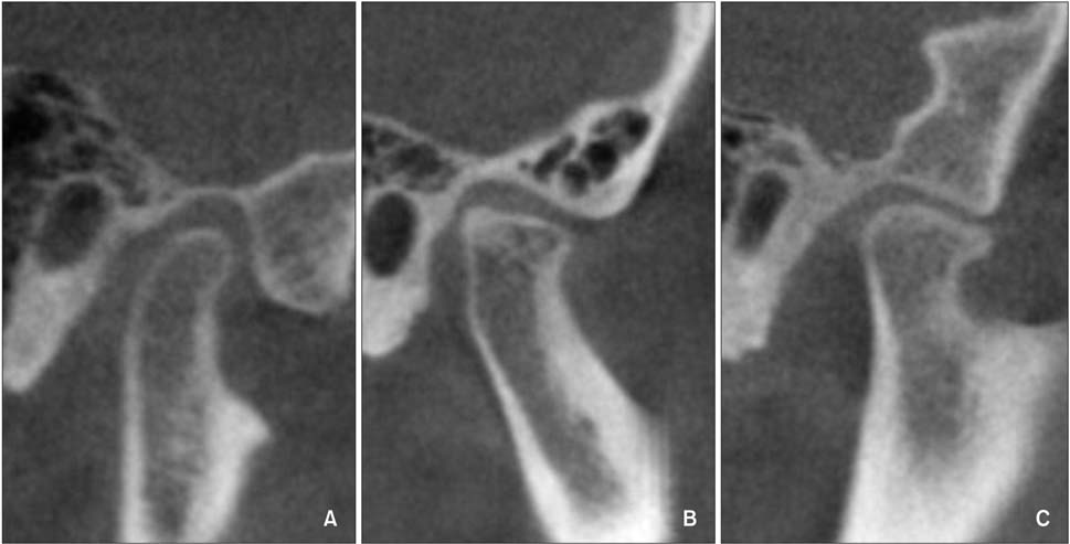
Figure 3 Different shapes of condyles. A, Normal. B, Flattened. C, Osteophyte.
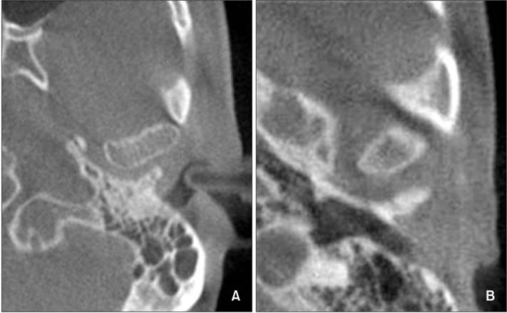
Figure 4 Condylar shape difference within the normal group. A, Oval. B, Round.
Cited by 2 articles
-
Reliability of cone-beam computed tomography for temporomandibular joint analysis
Hande Gorucu-Coskuner, Ezgi Atik, Hakan El
Korean J Orthod. 2019;49(2):81-88. doi: 10.4041/kjod.2019.49.2.81.Differences in mandibular condyle and glenoid fossa morphology in relation to vertical and sagittal skeletal patterns: A cone-beam computed tomography study
Kyoung Jin Noh, Hyoung-Seon Baik, Sang-Sun Han, Woowon Jang, Yoon Jeong Choi
Korean J Orthod. 2021;51(2):126-134. doi: 10.4041/kjod.2021.51.2.126.
Reference
-
1. Girardot RA Jr. Comparison of condylar position in hyperdivergent and hypodivergent facial skeletal types. Angle Orthod. 2001; 71:240–246.2. Ponces MJ, Tavares JP, Lopes JD, Ferreira AP. Comparison of condylar displacement between three biotypological facial groups by using mounted models and a mandibular position indicator. Korean J Orthod. 2014; 44:312–319.
Article3. Vitral RW, Telles Cde S, Fraga MR, de Oliveira RS, Tanaka OM. Computed tomography evaluation of temporomandibular joint alterations in patients with class II division 1 subdivision malocclusions: condyle-fossa relationship. Am J Orthod Dentofacial Orthop. 2004; 126:48–52.
Article4. Rodrigues AF, Fraga MR, Vitral RW. Computed tomography evaluation of the temporomandibular joint in Class I malocclusion patients: condylar symmetry and condyle-fossa relationship. Am J Orthod Dentofacial Orthop. 2009; 136:192–198.
Article5. Schudy FF. Treatment of adult midline deviation by condylar repositioning. J Clin Orthod. 1996; 30:343–347.6. Matsumoto MA, Bolognese AM. Radiographic morphology of the temporomandibular joint related to occlusal characteristics. Braz Dent J. 1994; 5:115–120.7. Burley M. An examination of the relation between the radiographic appearance of the temporomandibular joint and some features of the occlusion. Br Dent J. 1961; 110:195–200.8. Custodio W, Gomes SG, Faot F, Garcia RC, Del Bel Cury AA. Occlusal force, electromyographic activity of masticatory muscles and mandibular flexure of subjects with different facial types. J Appl Oral Sci. 2011; 19:343–349.
Article9. Stringert HG, Worms FW. Variations in skeletal and dental patterns in patients with structural and functional alterations of the temporomandibular joint: a preliminary report. Am J Orthod. 1986; 89:285–297.
Article10. Burke G, Major P, Glover K, Prasad N. Correlations between condylar characteristics and facial morphology in Class II preadolescent patients. Am J Orthod Dentofacial Orthop. 1998; 114:328–336.
Article11. Cohlmia JT, Ghosh J, Sinha PK, Nanda RS, Currier GF. Tomographic assessment of temporomandibular joints in patients with malocclusion. Angle Orthod. 1996; 66:27–35.12. Gianelly AA, Petras JC, Boffa J. Condylar position and Class II deep-bite, no-overjet malocclusions. Am J Orthod Dentofacial Orthop. 1989; 96:428–432.13. Naeije M, Te Veldhuis AH, Te Veldhuis EC, Visscher CM, Lobbezoo F. Disc displacement within the human temporomandibular joint: a systematic review of a 'noisy annoyance'. J Oral Rehabil. 2013; 40:139–158.
Article14. Dalili Z, Khaki N, Kia SJ, Salamat F. Assessing joint space and condylar position in the people with normal function of temporomandibular joint with cone-beam computed tomography. Dent Res J (Isfahan). 2012; 9:607–612.
Article15. Tsiklakis K, Syriopoulos K, Stamatakis HC. Radiographic examination of the temporomandibular joint using cone beam computed tomography. Dentomaxillofac Radiol. 2004; 33:196–201.
Article16. Hayashi T, Ito J, Koyama J, Hinoki A, Kobayashi F, Torikai Y, et al. Detectability of anterior displacement of the articular disk in the temporomandibular joint on helical computed tomography: the value of open mouth position. Oral Surg Oral Med Oral Pathol Oral Radiol Endod. 1999; 88:106–111.
Article17. Ikeda K, Kawamura A. Assessment of optimal condylar position with limited cone-beam computed tomography. Am J Orthod Dentofacial Orthop. 2009; 135:495–501.
Article18. Seren E, Akan H, Toller MO, Akyar S. An evaluation of the condylar position of the temporomandibular joint by computerized tomography in Class III malocclusions: a preliminary study. Am J Orthod Dentofacial Orthop. 1994; 105:483–488.
Article19. Vitral RW, Telles Cde S. Computed tomography evaluation of temporomandibular joint alterations in class II Division 1 subdivision patients: condylar symmetry. Am J Orthod Dentofacial Orthop. 2002; 121:369–375.
Article20. Hilgers ML, Scarfe WC, Scheetz JP, Farman AG. Accuracy of linear temporomandibular joint measurements with cone beam computed tomography and digital cephalometric radiography. Am J Orthod Dentofacial Orthop. 2005; 128:803–811.
Article21. Saccucci M, Polimeni A, Festa F, Tecco S. Do skeletal cephalometric characteristics correlate with condylar volume, surface and shape? A 3D analysis. Head Face Med. 2012; 8:15.
Article22. Katsavrias EG. Morphology of the temporomandibular joint in subjects with Class II Division 2 malocclusions. Am J Orthod Dentofacial Orthop. 2006; 129:470–478.
Article23. Rodrigues AF, Fraga MR, Vitral RW. Computed tomography evaluation of the temporomandibular joint in Class II Division 1 and Class III malocclusion patients: condylar symmetry and condyle-fossa relationship. Am J Orthod Dentofacial Orthop. 2009; 136:199–206.
Article24. Shahidi S, Vojdani M, Paknahad M. Correlation between articular eminence steepness measured with cone-beam computed tomography and clinical dysfunction index in patients with temporomandibular joint dysfunction. Oral Surg Oral Med Oral Pathol Oral Radiol. 2013; 116:91–97.
Article25. Ahn SJ, Kim TW, Lee DY, Nahm DS. Evaluation of internal derangement of the temporomandibular joint by panoramic radiographs compared with magnetic resonance imaging. Am J Orthod Dentofacial Orthop. 2006; 129:479–485.
Article26. Zhang ZL, Cheng JG, Li G, Zhang JZ, Zhang ZY, Ma XC. Measurement accuracy of temporomandibular joint space in Promax 3-dimensional cone-beam computerized tomography images. Oral Surg Oral Med Oral Pathol Oral Radiol. 2012; 114:112–117.
Article27. Yang IH, Moon BS, Lee SP, Ahn SJ. Skeletal differences in patients with temporomandibular joint disc displacement according to sagittal jaw relationship. J Oral Maxillofac Surg. 2012; 70:e349–e360.
Article28. Ahmad M, Hollender L, Anderson Q, Kartha K, Ohrbach R, Truelove EL, et al. Research diagnostic criteria for temporomandibular disorders (RDC/TMD): development of image analysis criteria and examiner reliability for image analysis. Oral Surg Oral Med Oral Pathol Oral Radiol Endod. 2009; 107:844–860.
Article29. Nah KS. Condylar bony changes in patients with temporomandibular disorders: a CBCT study. Imaging Sci Dent. 2012; 42:249–253.
Article30. Ahn SJ, Baek SH, Kim TW, Nahm DS. Discrimination of internal derangement of temporomandibular joint by lateral cephalometric analysis. Am J Orthod Dentofacial Orthop. 2006; 130:331–339.
Article31. Katsavrias EG, Halazonetis DJ. Condyle and fossa shape in Class II and Class III skeletal patterns: a morphometric tomographic study. Am J Orthod Dentofacial Orthop. 2005; 128:337–346.
Article
- Full Text Links
-
- Actions
-
Cited
- CITED
-
- Close
- Share
-
- Similar articles
-
- Differences in mandibular condyle and glenoid fossa morphology in relation to vertical and sagittal skeletal patterns: A cone-beam computed tomography study
- Cone-beam computed tomographic evaluation of the temporomandibular joint and dental characteristics of patients with Class II subdivision malocclusion and asymmetry
- Three-dimensional imaging modalities in endodontics
- Positional change in mandibular condyle in facial asymmetric patients after orthognathic surgery: cone-beam computed tomography study
- Evaluation of condylar dimension and position following rapid maxillary expansion with toothor tooth-bone-borne appliances
