Korean J Orthod.
2021 Mar;51(2):126-134. 10.4041/kjod.2021.51.2.126.
Differences in mandibular condyle and glenoid fossa morphology in relation to vertical and sagittal skeletal patterns: A cone-beam computed tomography study
- Affiliations
-
- 1Private Practice, Seoul, Korea
- 2Department of Orthodontics, The Institute of Craniofacial Deformity, Yonsei University College of Dentistry, Seoul, Korea
- 3Department of Oral and Maxillofacial Radiology, Yonsei University College of Dentistry, Seoul, Korea
- KMID: 2513581
- DOI: http://doi.org/10.4041/kjod.2021.51.2.126
Abstract
Objective
This study aimed to evaluate the following null hypothesis: there are no differences in the morphology of the temporomandibular joint (TMJ) structures in relation to vertical and sagittal cephalometric patterns.
Methods
This retrospective study was performed with 131 participants showing no TMJ symptoms. The participants were divided into Class I, II, and III groups on the basis of their sagittal cephalometric relationships and into hyperdivergent, normodivergent, and hypodivergent groups on the basis of their vertical cephalometric relationships. The following measurements were performed using cone-beam computed tomography images and compared among the groups: condylar volume, condylar size (width, length, and height), fossa size (length and height), and condyle-to-fossa joint spaces at the anterior, superior, and posterior condylar poles.
Results
The null hypothesis was rejected. The Class III group showed larger values for condylar width, condylar height, and fossa height than the Class II group (p < 0.05). Condylar volume and superior joint space in the hyperdivergent group were significantly smaller than those in the other two vertical groups (p < 0.001), whereas fossa length and height were significantly larger in the hyperdivergent group than in the other groups (p < 0.01). The hypodivergent group showed a greater condylar width than the hyperdivergent group (p < 0.01). The sagittal and vertical cephalometric patterns showed statistically significant interactions for fossa length and height.
Conclusions
TMJ morphology differed across diverse skeletal cephalometric patterns. The fossa length and height were affected by the interactions of the vertical and sagittal skeletal patterns.
Figure
-
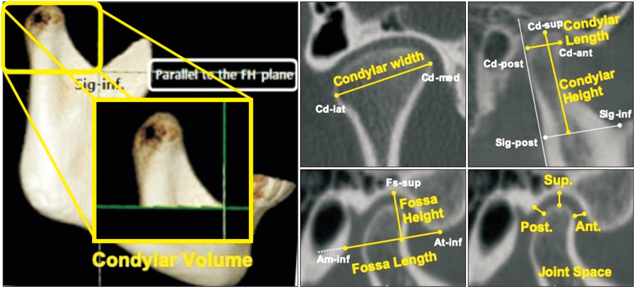
Figure 1 Measurements for the temporomandibular joint structure. Condylar volume was measured from a three-dimensional reconstructed image; condylar width was measured on the coronal section; and other measurements, including condylar length and height, fossa length and height, and superior, anterior, and posterior joint spaces, were measured on the same sagittal section. Please refer to Tables 2 and 3 for definitions of the abbreviations and measurements.
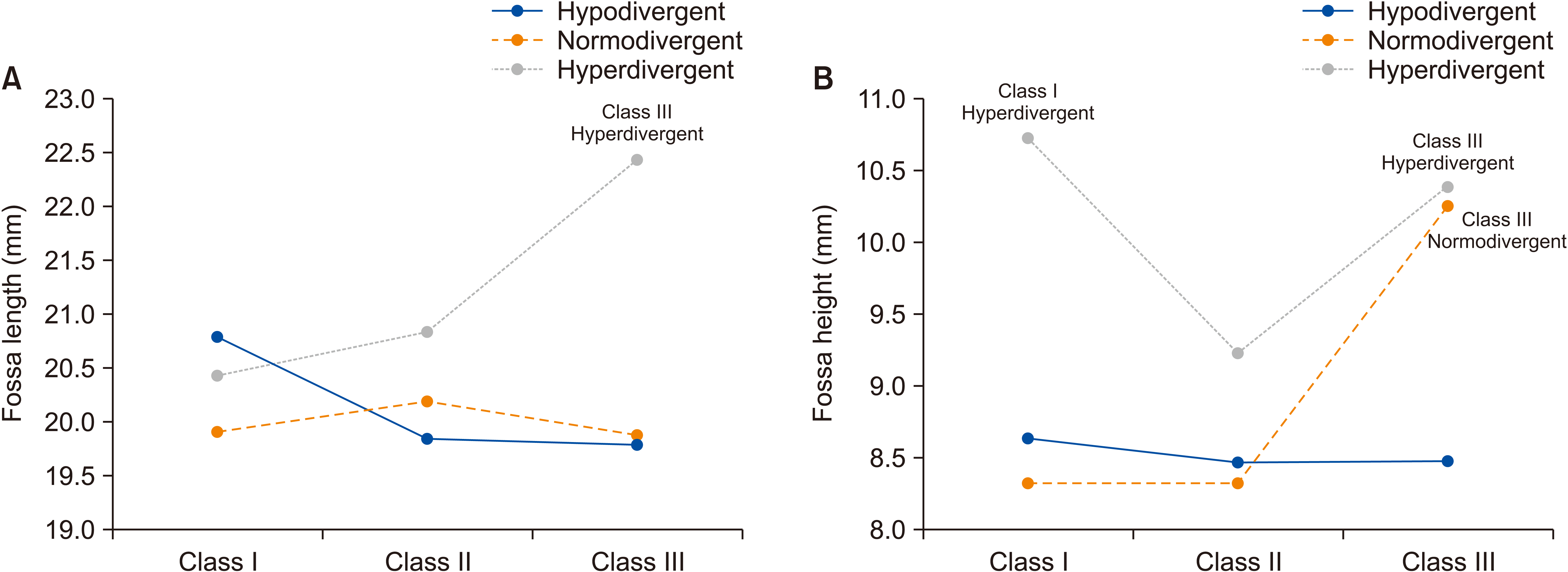
Figure 2 Interaction between vertical and sagittal skeletal patterns and fossa length (A) and height (B).
Reference
-
1. Krisjane Z, Urtane I, Krumina G, Bieza A, Zepa K, Rogovska I. 2007; Condylar and mandibular morphological criteria in the 2D and 3D MSCT imaging for patients with Class II division 1 subdivision malocclusion. Stomatologija. 9:67–71. PMID: 17993738.2. Tecco S, Saccucci M, Nucera R, Polimeni A, Pagnoni M, Cordasco G, et al. 2010; Condylar volume and surface in Caucasian young adult subjects. BMC Med Imaging. 10:28. DOI: 10.1186/1471-2342-10-28. PMID: 21194477. PMCID: PMC3019198.
Article3. Katsavrias EG. 2006; Morphology of the temporomandibular joint in subjects with Class II Division 2 malocclusions. Am J Orthod Dentofacial Orthop. 129:470–8. DOI: 10.1016/j.ajodo.2005.01.018. PMID: 16627172.
Article4. Katsavrias EG, Halazonetis DJ. 2005; Condyle and fossa shape in Class II and Class III skeletal patterns: a morphometric tomographic study. Am J Orthod Dentofacial Orthop. 128:337–46. DOI: 10.1016/j.ajodo.2004.05.024. PMID: 16168330.
Article5. Krisjane Z, Urtane I, Krumina G, Zepa K. 2009; Three-dimensional evaluation of TMJ parameters in Class II and Class III patients. Stomatologija. 11:32–6. PMID: 19423969.6. Park IY, Kim JH, Park YH. 2015; Three-dimensional cone-beam computed tomography based comparison of condylar position and morphology according to the vertical skeletal pattern. Korean J Orthod. 45:66–73. DOI: 10.4041/kjod.2015.45.2.66. PMID: 25798412. PMCID: PMC4367133.
Article7. Saccucci M, D'Attilio M, Rodolfino D, Festa F, Polimeni A, Tecco S. 2012; Condylar volume and condylar area in class I, class II and class III young adult subjects. Head Face Med. 8:34. DOI: 10.1186/1746-160X-8-34. PMID: 23241136. PMCID: PMC3558396.
Article8. Saccucci M, Polimeni A, Festa F, Tecco S. 2012; Do skeletal cephalometric characteristics correlate with condylar volume, surface and shape? A 3D analysis. Head Face Med. 8:15. DOI: 10.1186/1746-160X-8-15. PMID: 22587445. PMCID: PMC3484012.
Article9. Vitral RW, Telles Cde S, Fraga MR, de Oliveira RS, Tanaka OM. 2004; Computed tomography evaluation of temporomandibular joint alterations in patients with class II division 1 subdivision malocclusions: condyle-fossa relationship. Am J Orthod Dentofacial Orthop. 126:48–52. DOI: 10.1016/j.ajodo.2003.06.012. PMID: 15224058.
Article10. Kurita H, Uehara S, Yokochi M, Nakatsuka A, Kobayashi H, Kurashina K. 2006; A long-term follow-up study of radiographically evident degenerative changes in the temporomandibular joint with different conditions of disk displacement. Int J Oral Maxillofac Surg. 35:49–54. DOI: 10.1016/j.ijom.2005.04.004. PMID: 15961278.
Article11. Sümbüllü MA, Cağlayan F, Akgül HM, Yilmaz AB. 2012; Radiological examination of the articular eminence morphology using cone beam CT. Dentomaxillofac Radiol. 41:234–40. DOI: 10.1259/dmfr/24780643. PMID: 22074873. PMCID: PMC3520296.
Article12. Yamada K, Tsuruta A, Hanada K, Hayashi T. 2004; Morphology of the articular eminence in temporomandibular joints and condylar bone change. J Oral Rehabil. 31:438–44. DOI: 10.1111/j.1365-2842.2004.01255.x. PMID: 15140169.
Article13. Hasegawa H, Saitoh I, Nakakura-Ohshima K, Shigeta K, Yoshihara T, Suenaga S, et al. 2011; Condylar shape in relation to anterior disk displacement in juvenile females. Cranio. 29:100–10. DOI: 10.1179/crn.2011.018. PMID: 21661584.
Article14. Paknahad M, Shahidi S, Akhlaghian M, Abolvardi M. 2016; Is mandibular fossa morphology and articular eminence inclination associated with temporomandibular dysfunction? J Dent (Shiraz). 17:134–41. PMID: 27284559. PMCID: PMC4885671.15. Weinmann JP, Sicher H. 1947. Bone and bones: fundamentals of bone biology. Mosby;St. Louis:16. Mupparapu M, Chow I, Uppal A. 2011; Hard tissue structural changes in TMJ morphology prior to orthodontic therapy: a complex motion tomographic study. Quintessence Int. 42:427–34. PMID: 21519564.17. Al-Riyami S, Moles DR, Cunningham SJ. 2009; Orthognathic treatment and temporomandibular disorders: a systematic review. Part 1. A new quality-assessment technique and analysis of study characteristics and classifications. Am J Orthod Dentofacial Orthop. 136:624.e1–15. discussion 624–5. DOI: 10.1016/j.ajodo.2009.02.021. PMID: 19892268.18. Chae JM, Park JH, Tai K, Mizutani K, Uzuka S, Miyashita W, et al. 2020; Evaluation of condyle-fossa relationships in adolescents with various skeletal patterns using cone-beam computed tomography. Angle Orthod. 90:224–32. DOI: 10.2319/052919-369.1. PMID: 31638857.
Article19. Kim JY, Lee SJ, Kim TW, Nahm DS, Chang YI. 2005; Classification of the skeletal variation in normal occlusion. Angle Orthod. 75:311–9. DOI: 10.1043/0003-3219(2005)75[311:COTSVI]2.0.CO;2. PMID: 15898366.20. Petersson A. 2010; What you can and cannot see in TMJ imaging--an overview related to the RDC/TMD diagnostic system. J Oral Rehabil. 37:771–8. DOI: 10.1111/j.1365-2842.2010.02108.x. PMID: 20492436.21. Nur RB, Çakan DG, Arun T. 2016; Evaluation of facial hard and soft tissue asymmetry using cone-beam computed tomography. Am J Orthod Dentofacial Orthop. 149:225–37. DOI: 10.1016/j.ajodo.2015.07.038. PMID: 26827979.
Article22. Kim SJ, Kim KH, Yu HS, Baik HS. 2014; Dentoalveolar compensation according to skeletal discrepancy and overjet in skeletal Class III patients. Am J Orthod Dentofacial Orthop. 145:317–24. DOI: 10.1016/j.ajodo.2013.11.014. PMID: 24582023.
Article23. Kim JH, Gansukh O, Amarsaikhan B, Lee SJ, Kim TW. 2011; Comparison of cephalometric norms between Mongolian and Korean adults with normal occlusions and well-balanced profiles. Korean J Orthod. 41:42–50. DOI: 10.4041/kjod.2011.41.1.42.
Article24. Al-koshab M, Nambiar P, John J. 2015; Assessment of condyle and glenoid fossa morphology using CBCT in South-East Asians. PLoS One. 10:e0121682. DOI: 10.1371/journal.pone.0121682. PMID: 25803868. PMCID: PMC4372412.
Article25. Hilgers ML, Scarfe WC, Scheetz JP, Farman AG. 2005; Accuracy of linear temporomandibular joint measurements with cone beam computed tomography and digital cephalometric radiography. Am J Orthod Dentofacial Orthop. 128:803–11. DOI: 10.1016/j.ajodo.2005.08.034. PMID: 16360924.
Article26. Bjork A. 1963; Variations in the growth pattern of the human mandible: longitudinal radiographic study by the implant method. J Dent Res. 42Pt 2:400–11. DOI: 10.1177/00220345630420014701. PMID: 13971295.
Article27. Björk A. 1969; Prediction of mandibular growth rotation. Am J Orthod. 55:585–99. DOI: 10.1016/0002-9416(69)90036-0. PMID: 6594058.
Article28. Burke G, Major P, Glover K, Prasad N. 1998; Correlations between condylar characteristics and facial morphology in Class II preadolescent patients. Am J Orthod Dentofacial Orthop. 114:328–36. DOI: 10.1016/S0889-5406(98)70216-1. PMID: 9743139.
Article29. Cohlmia JT, Ghosh J, Sinha PK, Nanda RS, Currier GF. 1996; Tomographic assessment of temporomandibular joints in patients with malocclusion. Angle Orthod. 66:27–35. DOI: 10.1043/0003-3219(1996)066<0027:TAOTJI>2.3.CO;2. PMID: 8678342.30. Arnett GW, Gunson MJ. 2004; Facial planning for orthodontists and oral surgeons. Am J Orthod Dentofacial Orthop. 126:290–5. DOI: 10.1016/j.ajodo.2004.06.006. PMID: 15356488.
Article
- Full Text Links
-
- Actions
-
Cited
- CITED
-
- Close
- Share
-
- Similar articles
-
- Mandibular condyle position in cone beam computed tomography
- Three-dimensional cone-beam computed tomography based comparison of condylar position and morphology according to the vertical skeletal pattern
- Cone-beam computed tomographic evaluation of the temporomandibular joint and dental characteristics of patients with Class II subdivision malocclusion and asymmetry
- Temporomandibular joint morphology in Korean using cone-beam computed tomography: influence of age and gender
- Cone beam computed tomography findings of ectopic mandibular third molar in the mandibular condyle: report of a case
