Imaging Sci Dent.
2016 Jun;46(2):77-85. 10.5624/isd.2016.46.2.77.
Novel three-dimensional position analysis of the mandibular foramen in patients with skeletal class III mandibular prognathism
- Affiliations
-
- 1Department of Oral and Maxillofacial Surgery, National Health Insurance Service Ilsan Hospital, Goyang, Korea. omfs1ksh@daum.net
- 2Department of Oral and Maxillofacial Surgery, College of Dentistry, Yonsei University, Seoul, Korea.
- KMID: 2308870
- DOI: http://doi.org/10.5624/isd.2016.46.2.77
Abstract
- PURPOSE
To analyze the relative position of the mandibular foramina (MnFs) in patients diagnosed with skeletal class III malocclusion.
MATERIALS AND METHODS
Computed tomography (CT) images were collected from 85 patients. The vertical lengths of each anatomic point from the five horizontal planes passing through the MnF were measured at the coronoid process, sigmoid notch, condyle, and the gonion. The distance from the anterior ramus point to the posterior ramus point on the five horizontal planes was designated the anteroposterior horizontal distance of the ramus for each plane. The perpendicular distance from each anterior ramus point to each vertical plane through the MnF was designated the horizontal distance from the anterior ramus to the MnF. The horizontal and vertical positions were examined by regression analysis.
RESULTS
Regression analysis showed the heights of the coronoid process, sigmoid notch, and condyle for the five horizontal planes were significantly related to the height of the MnF, with the highest significance associated with the MnF-mandibular plane (coefficients of determination (R2): 0.424, 0.597, and 0.604, respectively). The horizontal anteroposterior length of the ramus and the distance from the anterior ramus point to the MnF were significant by regression analysis.
CONCLUSION
The relative position of the MnF was significantly related to the vertical heights of the sigmoid notch, coronoid process, and condyle as well as to the horizontal anteroposterior length of the ascending ramus. These findings should be clinically useful for patients with skeletal class III mandibular prognathism.
Keyword
MeSH Terms
Figure
-
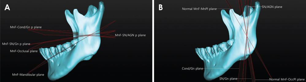
Fig. 1 Reference planes. A. Five horizontal planes are defined that pass through the mandibular foramen (MnF). B. Five vertical planes are defined that pass through the MnF.
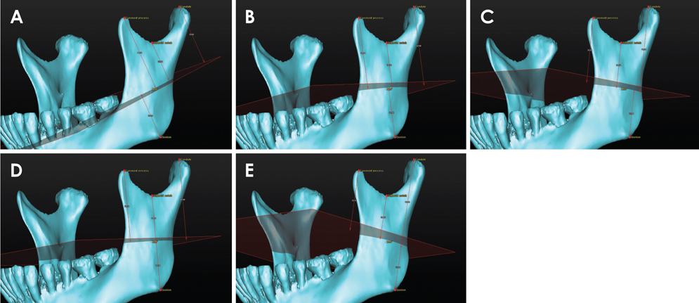
Fig. 2 Five horizontal planes are used as reference planes for measuring the vertical height of the mandible: A. the MnF-mandibular plane; B. the MnF-occlusal plane; C. the MnF-SN/AGN p plane; D. the MnF-SN/Gn p plane; and E. the MnF-Cond/Gn p plane.

Fig. 3 Five vertical planes are used as reference planes for measuring the horizontal distance from the anterior ramus to the mandibular foramen (MnF). A. the normal MnF-MnPl plane; B. the normal MnF-OccPl plane; C. the SN/AGN plane; D. the SN/Gn plane; and E. the Cond/Gn plane.
Reference
-
1. Sekerci AE, Cantekin K, Aydinbelge M. Cone beam computed tomographic analysis of the shape, height, and location of the mandibular lingula in a population of children. Biomed Res Int. 2013; 2013:825453.
Article2. Findik Y, Yildirim D, Baykul T. Three-dimensional anatomic analysis of the lingula and mandibular foramen: a cone beam computed tomography study. J Craniofac Surg. 2014; 25:607–610.3. Park KR, Kim SY, Kim GJ, Park HS, Jung YS. Anatomic study to determine a safe surgical reference point for mandibular ramus osteotomy. J Craniomaxillofac Surg. 2014; 42:22–27.
Article4. Sekerci AE, Sisman Y. Cone-beam computed tomography analysis of the shape, height, and location of the mandibular lingula. Surg Radiol Anat. 2014; 36:155–162.
Article5. Alves N, Deana NF. Morphometric study of mandibular foramen in macerated skulls to contribute to the development of sagittal split ramus osteotomy (SSRO) technique. Surg Radiol Anat. 2014; 36:839–845.
Article6. Yu IH, Wong YK. Evaluation of mandibular anatomy related to sagittal split ramus osteotomy using 3-dimensional computed tomography scan images. Int J Oral Maxillofac Surg. 2008; 37:521–528.
Article7. Kim MS, Lee EJ, Song IJ, Lee JS, Kang BC, Yoon SJ. The location of midfacial landmarks according to the method of establishing the midsagittal reference plane in three-dimensional computed tomography analysis of facial asymmetry. Imaging Sci Dent. 2015; 45:227–232.
Article8. Kang SH, Byun IY, Kim JH, Park HK, Kim MK. Three-dimensional anatomic analysis of mandibular foramen with mandibular anatomic landmarks for inferior alveolar nerve block anesthesia. Oral Surg Oral Med Oral Pathol Oral Radiol. 2013; 115:e17–e23.
Article9. Kositbowornchai S, Siritapetawee M, Damrongrungruang T, Khongkankong W, Chatrchaiwiwatana S, Khamanarong K, et al. Shape of the lingula and its localization by panoramic radiograph versus dry mandibular measurement. Surg Radiol Anat. 2007; 29:689–694.
Article10. Monnazzi MS, Passeri LA, Gabrielli MF, Bolini PD, de Carvalho WR, da Costa Machado H. Anatomic study of the mandibular foramen, lingula and antilingula in dry mandibles, and its statistical relationship between the true lingula and the antilingula. Int J Oral Maxillofac Surg. 2012; 41:74–78.
Article
- Full Text Links
-
- Actions
-
Cited
- CITED
-
- Close
- Share
-
- Similar articles
-
- Radiologic study of mandibular foramen of mandibular prognathism by three-dimensional computed tomography
- Comparison of cranial base morphology between the mandibular prognathism and maxillary retrognathism in skeletal class III patients
- A Comparative Study On The Location Of The Mandibular Foramen In Panoramic Radiographs Of Normal Occlusion and Mandibular Prognathism
- Evaluation of craniofacial growth prediction method on Class III malocclusion patients
- A comparative study on the location of the mandibular foramen in CBCT of normal occlusion and skeletal class II and III malocclusion
