The relationship between condyle position, morphology and chin deviation in skeletal Class III patients with facial asymmetry using cone-beam CT
- Affiliations
-
- 1Department of Orthodontics, School of Dentistry, Pusan National University, Korea. wsson@pusan.ac.kr
- KMID: 2274245
- DOI: http://doi.org/10.4041/kjod.2011.41.2.87
Abstract
OBJECTIVE
Facial asymmetry is usually evaluated from the difference in length and angulation of the maxilla and mandible. However, asymmetric position or shape of the condyle can also affect the expression of asymmetry. The purpose of this study was to evaluate the correlation between condylar asymmetry and chin point deviation in facial asymmetry.
METHODS
Cone-beam CT images of fifty adult skeletal Class III patients were studied. Thirty patients who had more than 4 mm menton deviation were categorized in the asymmetric group. Twenty patients with less than 4 mm menton deviation were assigned to the symmetric group. Anteroposterior and transverse condyle positions were evaluated from the cranial base. The greatest mediolateral diameter (GMD) of the condyle in the axial plane and angulation to the coronal plane were measured. The height and volume of the condyles were evaluated.
RESULTS
The symmetric group had no statistical difference between both condyles in position, angulation, GMD, height and volume. In the asymmetric group, the non-deviated side condyle was larger in GMD, height and volume than the deviated side. There was no statistical difference in condyle position and angulation. The GMD, height difference and condylar volume ratio (non-deviated/deviated) were positively correlated with chin deviation. From the linear regression analysis, condylar volume ratio was a significant factor affecting chin deviation.
CONCLUSIONS
These findings suggests that the non-deviated side condyle is larger than the deviated side. In addition, condylar asymmetry can affect the expression of facial asymmetry.
MeSH Terms
Figure
-
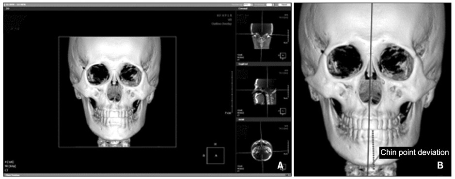
Fig. 1 Reference planes and chin point deviation. A, Constructed reference planes; B, chin point deviation measured from MSR plane to menton point (inferior).
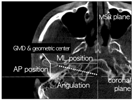
Fig. 2 Measurement of GMD, angulation to coronal plane. The geometric center defined as the center of GMD. The anteroposterior position to coronal plane and mediolateral position to midsagittal reference plane of geometric center were measured. GMD, Greateast mediolateral diameter; AP, anteroposterior; ML, mediolateral; MSR plane, midsagittal reference plane.
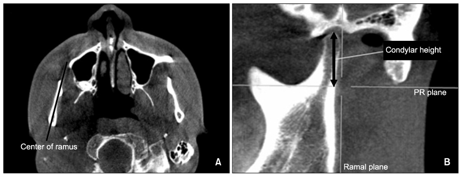
Fig. 3 Measurement of condylar height. A, Center of ramus. This line bisects the axial plane of ramus; B, ramal plane was constructed by a line perpendicular to the center of ramus and tangent to the posterior ramus. PR (Perpendicular to ramal) plane, a line passing through the deepest point of sigmoid notch and tagent to the ramal plane. Condylar height was measured from the PR plane to the highest point of the condyle (parallel to ramal plane).
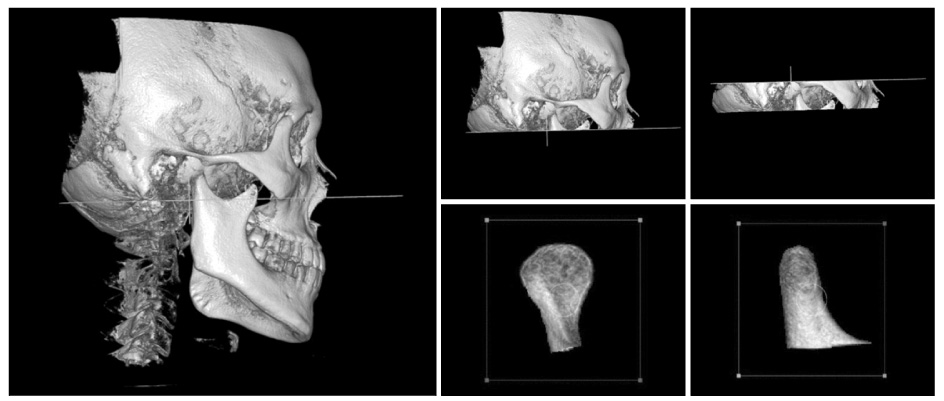
Fig. 4 Condyle segmentation was done for condylar volume measurement.
Cited by 2 articles
-
Correction of dental Class III with posterior open bite by simple biomechanics using an anterior C-tube miniplate
Hyo-Won Ahn, Kyu-Rhim Chung, Suk-Man Kang, Lu Lin, Gerald Nelson, Seong-Hun Kim
Korean J Orthod. 2012;42(5):270-278. doi: 10.4041/kjod.2012.42.5.270.The genial tubercle: A prospective novel landmark for the diagnosis of mandibular asymmetry
Seung-Youp Lee, Dong-Soon Choi, Insan Jang, Geun-Su Song, Bong-Kuen Cha
Korean J Orthod. 2017;47(1):50-58. doi: 10.4041/kjod.2017.47.1.50.
Reference
-
1. Grayson BH, McCarthy JG, Bookstein F. Analysis of craniofacial asymmetry by multiplane cephalometry. Am J Orthod. 1983. 84:217–224.
Article2. Jolley L, Huang JC, Maki K, Gansky SA, Miller AJ, Hatcher D. Development of three dimensional cone beam CT data into reconstructed lateral cephalometric images. Inf Orthod Kieferorthop. 2007. 39:173–186.
Article3. Hwang HS, Hwang CH, Lee KH, Kang BC. Maxillofacial 3-dimensional image analysis for the diagnosis of facial asymmetry. Am J Orthod Dentofacial Orthop. 2006. 130:779–785.
Article4. Park SH, Yu HS, Kim KD, Lee KJ, Baik HS. A proposal for a new analysis of craniofacial morphology by 3-dimensional computed tomography. Am J Orthod Dentofacial Orthop. 2006. 129:600.
Article5. Baek SH, Cho IS, Chang YI, Kim MJ. Skeletodental factorsaffecting chin point deviation in female patients with class III malocclusion and facial asymmetry: a three-dimensional analysis using computed tomography. Oral Surg Oral Med Oral Pathol Oral Radiol Endod. 2007. 104:628–639.
Article6. Kwon TG, Park HS, Ryoo HM, Lee SH. A comparison of craniofacial morphology in patients with and without facial asymmetry--a three-dimensional analysis with computed tomography. Int J Oral Maxillofac Surg. 2006. 35:43–48.
Article7. Ahn JS, Hwang HS. Relationship between perception of facial asymmetry and posteroanterior cephalometric measurements. Korean J Orthod. 2001. 31:489–498.8. Lee GH, Cho HK, Hwang HS, Kim JC. Studies of relationship between P-A cephalometric measurements and vidual facial asymmetry. Korean J Phys Anthropol. 1998. 11:41–48.
Article9. Haraguchi S, Takada K, Yasuda Y. Facial asymmetry in subjects with skeletal Class III deformity. Angle Orthod. 2002. 72:28–35.10. Rodrigues AF, Fraga MR, Vitral RW. Computed tomography evaluation of the temporomandibular joint in Class I malocclusion patients: condylar symmetry and condyle-fossa relationship. Am J Orthod Dentofacial Orthop. 2009. 136:192–198.
Article11. Rodrigues AF, Fraga MR, Vitral RW. Computed tomography evaluation of the temporomandibular joint in Class II Division 1 and Class III malocclusion patients: condylar symmetry and condyle-fossa relationship. Am J Orthod Dentofacial Orthop. 2009. 136:199–206.
Article12. Schlueter B, Kim KB, Oliver D, Sortiropoulos G. Cone beam computed tomography 3D reconstruction of the mandibular condyle. Angle Orthod. 2008. 78:880–888.
Article13. Hilgers ML, Scarfe WC, Scheetz JP, Farman AG. Accuracy of linear temporomandibular joint measurements with cone beam computed tomography and digital cephalometric radiography. Am J Orthod Dentofacial Orthop. 2005. 128:803–811.
Article14. El-Mangoury NH, Shaheen SI, Mostafa YA. Landmark identification in computerized posteroanterior cephalometrics. Am J Orthod Dentofacial Orthop. 1987. 91:57–61.
Article15. Cheon OJ, Suhr CH. A posteroanterior roentgenocephalometric study of skeletal craniofacial asymmetric patients. Korean J Orthod. 1990. 20:615–631.16. Lascala CA, Panella J, Marques MM. Analysis of the accuracy of linear measurements obtained by cone beam computed tomography (CBCT-NewTom). Dentomaxillofac Radiol. 2004. 33:291–294.
Article17. Marmulla R, Wörtche R, Mühling J, Hassfeld S. Geometric accuracy of the NewTom 9000 Cone Beam CT. Dentomaxillofac Radiol. 2005. 34:28–31.
Article18. Breiman RS, Beck JW, Korobkin M, Glenny R, Akwari OE, Heaston DK, et al. Volume determinations using computed tomography. AJR Am J Roentgenol. 1982. 138:329–333.
Article19. Shapurian T, Damoulis PD, Reiser GM, Griffin TJ, Rand WM. Quantitative evaluation of bone density using the Hounsfield index. Int J Oral Maxillofac Implants. 2006. 21:290–297.20. Rho JY, Hobatho MC, Ashman RB. Relations of mechanical properties to density and CT numbers in human bone. Med Eng Phys. 1995. 17:347–355.
Article21. Sgouros S, Natarajan K, Hockley AD, Goldin JH, Wake M. Skull base growth in childhood. Pediatr Neurosurg. 1999. 31:259–268.
Article22. St John D, Mulliken JB, Kaban LB, Padwa BL. Anthropometric analysis of mandibular asymmetry in infants with deformational posterior plagiocephaly. J Oral Maxillofac Surg. 2002. 60:873–877.
Article23. Obwegeser HL, Makek MS. Hemimandibular hyperplasia--hemimandibular elongation. J Maxillofac Surg. 1986. 14:183–208.
Article24. Pirttiniemi PM. Associations of mandibular and facial asymmetries--a review. Am J Orthod Dentofacial Orthop. 1994. 106:191–200.
Article25. Poikela A, Kantomaa T, Pirttiniemi P. Craniofacial growth after a period of unilateral masticatory function in young rabbits. Eur J Oral Sci. 1997. 105:331–337.
Article26. Bjoerk A, Bjoerk L. Artificial deformation and cranio-facial asymmetry in ancient Peruvians. J Dent Res. 1964. 43:353–362.
Article27. Persing J, James H, Swanson J, Kattwinkel J. American Academy of Pediatrics Committee on Practice and Ambulatory Medicine, Section on Plastic Surgery and Section on Neurological Surgery. Prevention and management of positional skull deformities in infants. American Academy of Pediatrics Committee on Practice and Ambulatory Medicine Section on Plastic Surgery and Section on Neurological Surgery. Pediatrics. 2003. 112(1 Pt 1):199–202.28. Sakurai A, Hirabayashi S, Sugawara Y, Harii K. Skeletal analysis of craniofacial asymmetries in plagiocephaly (unilateral coronal synostosis). Scand J Plast Reconstr Surg Hand Surg. 1998. 32:81–89.
Article
- Full Text Links
-
- Actions
-
Cited
- CITED
-
- Close
- Share
-
- Similar articles
-
- Cone-beam computed tomography analysis of transverse dental compensation in patients with skeletal Class III malocclusion and facial asymmetry
- Differences in facial soft tissue deviations in Class III patients with different types of mandibular asymmetry: A cone-beam computed tomography study
- Positional change in mandibular condyle in facial asymmetric patients after orthognathic surgery: cone-beam computed tomography study
- Differences in positions of cone-beam computed tomography landmarks in patients with skeletal Class III facial asymmetry according to midsagittal planes
- Cone-beam computed tomographic evaluation of the temporomandibular joint and dental characteristics of patients with Class II subdivision malocclusion and asymmetry
