Three-dimensional finite element analysis of unilateral mastication in malocclusion cases using cone-beam computed tomography and a motion capture system
- Affiliations
-
- 1Department of Anatomy, Dankook University College of Medicine, Cheonan, Korea.
- 2Department of Orthodontics, Yonsei University College of Dentistry, Seoul, Korea.
- 3Perio-Implant Research Center, Dankook University College of Dentistry, Cheonan, Korea.
- 4Department of Oral Anatomy, Dankook University College of Dentistry, Cheonan, Korea. jongtadent@gmail.com
- KMID: 2161986
- DOI: http://doi.org/10.5051/jpis.2016.46.2.96
Abstract
- PURPOSE
Stress distribution and mandible distortion during lateral movements are known to be closely linked to bruxism, dental implant placement, and temporomandibular joint disorder. The present study was performed to determine stress distribution and distortion patterns of the mandible during lateral movements in Class I, II, and III relationships.
METHODS
Five Korean volunteers (one normal, two Class II, and two Class III occlusion cases) were selected. Finite element (FE) modeling was performed using information from cone-beam computed tomographic (CBCT) scans of the subjects' skulls, scanned images of dental casts, and incisor movement captured by an optical motion-capture system.
RESULTS
In the Class I and II cases, maximum stress load occurred at the condyle of the balancing side, but, in the Class III cases, the maximum stress was loaded on the condyle of the working side. Maximum distortion was observed on the menton at the midline in every case, regardless of loading force. The distortion was greatest in Class III cases and smallest in Class II cases.
CONCLUSIONS
The stress distribution along and accompanying distortion of a mandible seems to be affected by the anteroposterior position of the mandible. Additionally, 3-D modeling of the craniofacial skeleton using CBCT and an optical laser scanner and reproduction of mandibular movement by way of the optical motion-capture technique used in this study are reliable techniques for investigating the masticatory system.
MeSH Terms
Figure
-
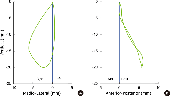
Figure 1 The trajectory of an incisor during lateral movement. (A) The motion of an incisor was captured from the anterior side. (B) The motion of an incisor was captured from the lateral side.

Figure 2 The reproduction of occlusion contacts (red colored area) at the time of initial contact on both sides during the closing phase of lateral movement. (A) The 3D simulation of lateral movement was performed based on CBCT images and captured motion information. (B) Occlusal contacts were examined on the occlusal aspects of the reconstructed image.
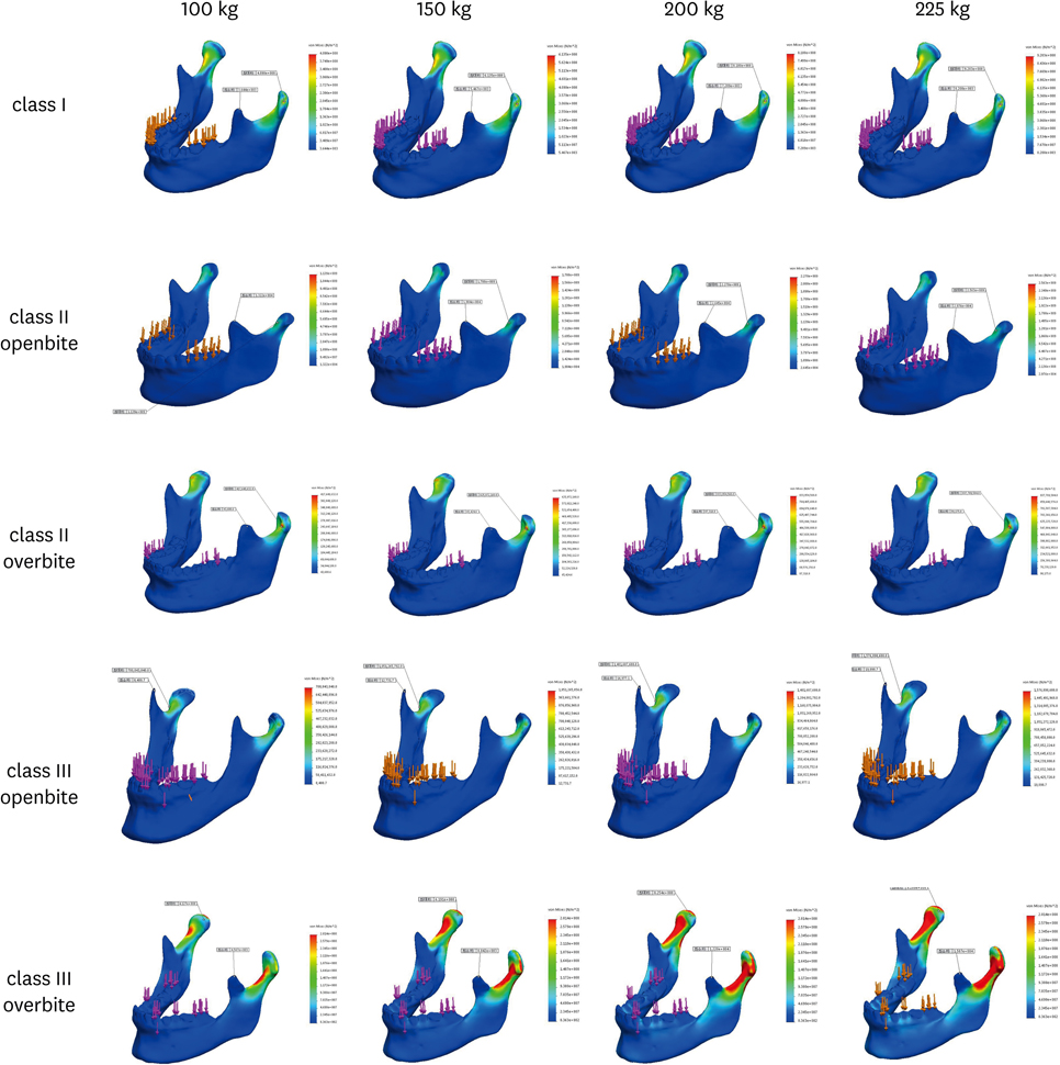
Figure 3 Stress distribution with simulation of lateral movement with 100, 150, 200 and 225kgf loading force in cases I~V.

Figure 4 Distortion of mandible with simulation of lateral movement with 100, 150, 200 and 225kgf loading force in cases I~V.

Figure 5 The maximum distortion of mandible according to the loading force in each case.
Reference
-
1. Commisso MS, Martínez-Reina J, Mayo J. A study of the temporomandibular joint during bruxism. Int J Oral Sci. 2014; 6:116–123.
Article2. Conti PC, Pertes RA, Heir GM, Nasri C, Cohen HV, Araújo CR. Orofacial pain: basic mechanisms and implication for successful management. J Appl Oral Sci. 2003; 11:1–7.
Article3. Koyano K, Esaki D. Occlusion on oral implants: current clinical guidelines. J Oral Rehabil. 2015; 42:153–161.
Article4. Choi AH, Conway RC, Taraschi V, Ben-Nissan B. Biomechanics and functional distortion of the human mandible. J Investig Clin Dent. 2015; 6:241–251.
Article5. Park JT, Lee JG, Won SY, Lee SH, Cha JY, Kim HJ. Realization of masticatory movement by 3-dimensional simulation of the temporomandibular joint and the masticatory muscles. J Craniofac Surg. 2013; 24:e347–51.
Article6. Akita K, Shimokawa T, Sato T. Positional relationships between the masticatory muscles and their innervating nerves with special reference to the lateral pterygoid and the midmedial and discotemporal muscle bundles of temporalis. J Anat. 2000; 197:291–302.
Article7. de Abreu RA, Pereira MD, Furtado F, Prado GP, Mestriner W Jr, Ferreira LM. Masticatory efficiency and bite force in individuals with normal occlusion. Arch Oral Biol. 2014; 59:1065–1074.
Article8. Davies JC, Charles M, Cantelmi D, Liebgott B, Ravichandiran M, Ravichandiran K, et al. Lateral pterygoid muscle: a three-dimensional analysis of neuromuscular partitioning. Clin Anat. 2012; 25:576–583.
Article9. Commisso MS, Martínez-Reina J, Ojeda J, Mayo J. Finite element analysis of the human mastication cycle. J Mech Behav Biomed Mater. 2015; 41:23–35.
Article10. Yamaguchi S, Itoh S, Watanabe Y, Tsuboi A, Watanabe M. Quantitative analysis of masticatory activity during unilateral mastication using muscle fMRI. Oral Dis. 2011; 17:407–413.
Article11. Koolstra JH, van Eijden TM. Consequences of viscoelastic behavior in the human temporomandibular joint disc. J Dent Res. 2007; 86:1198–1202.
Article12. Throckmorton GS, Finn RA, Bell WH. Biomechanics of differences in lower facial height. Am J Orthod. 1980; 77:410–420.
Article13. Ogami S, Yamada M, Kanazawa M, Takeda K, Kimura N, Mizutani H, et al. The effectiveness of a mouth guard to protect against strong occlusion caused by modified electroconvulsive therapy. Dent Traumatol. 2014; 30:368–373.
Article14. Murray GM, Orfanos T, Chan JY, Wanigaratne K, Klineberg IJ. Electromyographic activity of the human lateral pterygoid muscle during contralateral and protrusive jaw movements. Arch Oral Biol. 1999; 44:269–285.
Article15. Yamaguchi S, Rikimaru H, Yamaguchi K, Itoh M, Watanabe M. Overall activity of all masticatory muscles during lateral excursion. J Dent Res. 2006; 85:69–73.
Article16. Sessle BJ, Gurza SC. Jaw movement-related activity and reflexly induced changes in the lateral pterygoid muscle of the monkey Macaca fascicularis. Arch Oral Biol. 1982; 27:167–173.
Article17. Wood WW. A review of masticatory muscle function. J Prosthet Dent. 1987; 57:222–232.
Article18. Agur AM, Ng-Thow-Hing V, Ball KA, Fiume E, McKee NH. Documentation and three-dimensional modelling of human soleus muscle architecture. Clin Anat. 2003; 16:285–293.
Article19. Lemos RR, Epstein M, Herzog W, Wyvill B. A framework for structured modeling of skeletal muscle. Comput Methods Biomech Biomed Engin. 2004; 7:305–317.
Article20. Röhrle O, Waddell JN, Foster KD, Saini H, Pullan AJ. Using a motion-capture system to record dynamic articulation for application in CAD/CAM software. J Prosthodont. 2009; 18:703–710.
Article
- Full Text Links
-
- Actions
-
Cited
- CITED
-
- Close
- Share
-
- Similar articles
-
- A three-dimensional finite element analysis of the relationship between masticatory performance and skeletal malocclusion
- Three-dimensional imaging modalities in endodontics
- Finite element analysis of anterior maxillary segmental distraction osteogenesis using asymmetric distractors in patients with unilateral cleft lip and palate
- Axisymmetric Contact Stress analysis of an Artificial Hip Joint of the Conical Fitting Type
- A rare case of dilated invaginated odontome with talon cusp in a permanent maxillary central incisor diagnosed by cone beam computed tomography
