Imaging Sci Dent.
2013 Sep;43(3):209-213. 10.5624/isd.2013.43.3.209.
A rare case of dilated invaginated odontome with talon cusp in a permanent maxillary central incisor diagnosed by cone beam computed tomography
- Affiliations
-
- 1Department of Conservative Dentistry and Endodontics, Priyadarshini Dental College and Hospital, Chennai, India. endojaya@gmail.com
- KMID: 2167460
- DOI: http://doi.org/10.5624/isd.2013.43.3.209
Abstract
- It has been a challenge to establish the accurate diagnosis of developmental tooth anomalies based on periapical radiographs. Recently, three-dimensional imaging by cone beam computed tomography has provided useful information to investigate the complex anatomy of and establish the proper management for tooth anomalies. The most severe variant of dens invaginatus, known as dilated odontome, is a rare occurrence, and the cone beam computed tomographic findings of this anomaly have never been reported for an erupted permanent maxillary central incisor. The occurrence of talon cusp occurring along with dens invaginatus is also unusual. The aim of this report was to show the importance of cone beam computed tomography in contributing to the accurate diagnosis and evaluation of the complex anatomy of this rare anomaly.
Keyword
MeSH Terms
Figure
-

Fig. 1 A. Labial view clinically. B. Palatal view clinically showing the presence of talon cusp.
Fig. 2 A reformatted panoramic CBCT image shows the appearance of an immature root.
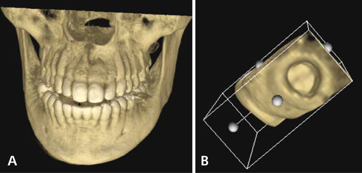
Fig. 3 A. CBCT three-dimensional frontal view. B. CBCT image shows the ring-like formation of the root.
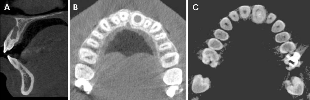
Fig. 4 A. CBCT parasagittal section shows the invagination and dilation in the root portion. B. CBCT axial image at the cervical level. C. CBCT cross section at the midroot level.

Fig. 5 A. Proximal view of the extracted tooth. B. The view from the root end of the extracted tooth showing the wide ring-like invagination.

Fig. 6 Histological examination of the contents of the inva-gination (H&E stain, 200×).
Reference
-
1. Hülsmann M. Dens invaginatus: aetiology, classification, prevalence, diagnosis, and treatment considerations. Int Endod J. 1997; 30:79–90.2. Alani A, Bishop K. Dens invaginatus. Part 1: classification, prevalence and aetiology. Int Endod J. 2008; 41:1123–1136.
Article3. Neville BW, Damm DD, Allen CM, Bouquot JE. Oral & maxillofacial pathology. 2nd Ed. Philadelphia: Saunders;2002. p. 80–81.4. Matsusue Y, Yamamoto K, Inagake K, Kirita T. A dilated odontoma in the second molar region of the mandible. Open Dent J. 2011; 5:150–153.
Article5. Cuković-Bagić I, Macan D, Dumancić J, Manojlović S, Hat J. Dilated odontome in the mandibular third molar region. Oral Surg Oral Med Oral Pathol Oral Radiol Endod. 2010; 109:e109–e113.6. Dumančić J, Kaić Z, Tolj M, Janković B. Talon cusp: a literature review and case report. Acta Stomatol Croat. 2006; 40:169–174.7. Mellor JK, Ripa LW. Talon cusp: a clinically significant anomaly. Oral Surg Oral Med Oral Pathol. 1970; 29:225–228.
Article8. Hattab FN, Yassin OM, al-Nimri KS. Talon cusp - clinical significance and management: case reports. Quintessence Int. 1995; 26:115–120.9. Segura JJ, Jiménez-Rubio A. Talon cusp affecting permanent maxillary lateral incisor in 2 family members. Oral Surg Oral Med Oral Pathol Oral Radiol Endod. 1999; 88:90–92.10. Durack C, Patel S. Cone beam computed tomography in endodontics. Braz Dent J. 2012; 23:179–191.
Article11. Patel S. New dimensions in endodontic imaging: Part 2. Cone beam computed tomography. Int Endod J. 2009; 42:463–475.
Article12. Oehlers FA. Dens invaginatus (dilated composite odontome). I. Variations of the invagination process and associated anterior crown forms. Oral Surg Oral Med Oral Pathol. 1957; 10:1204–1218.13. Ridell K, Mejàre I, Matsson L. Dens invaginatus: a retrospective study of prophylactic invagination treatment. Int J Paediatr Dent. 2001; 11:92–97.
Article14. Balcioğlu HA, Keklikoğlu N, Kökten G. Talon cusp: a morphological dental anomaly. Rom J Morphol Embryol. 2011; 52:179–181.15. al-Omari MA, Hattab FN, Darwazeh AM, Dummer PM. Clinical problems associated with unusual case of talon cusp. Int Endod J. 1999; 32:183–190.
- Full Text Links
-
- Actions
-
Cited
- CITED
-
- Close
- Share
-
- Similar articles
-
- Multidisciplinary management of a fused maxillary central incisor moved through the midpalatal suture: A case report
- Detection of maxillary second molar with two palatal roots using cone beam computed tomography: a case report
- Retrospective Analysis of Incisor Root Resorption Associated with Impacted Maxillary Canines
- Retreatment of failed regenerative endodontic of orthodontically treated immature permanent maxillary central incisor: a case report
- Crown-root angulations of the maxillary anterior teeth according to malocclusions: A cone-beam computed tomography study in Korean population
