Multidisciplinary management of a fused maxillary central incisor moved through the midpalatal suture: A case report
- Affiliations
-
- 1Department of Orthodontics, Faculty of Dentistry, Ege University, Izmir, Turkey.
- 2Private Practice, Istanbul, Turkey. pasaylin@hotmail.com
- KMID: 2392185
- DOI: http://doi.org/10.4041/kjod.2017.47.6.384
Abstract
- Fusion of teeth is a developmental anomaly. It occurs at the stage of tooth formation, which determines the shape and size of the tooth crown, when one or more teeth fuse at the dentin level during the morphodifferentiation of the dental germs. Such teeth show macrodontia and may cause crowding, as well as esthetic and endodontic problems. In this article, we report a rare case of a maxillary central incisor fused to a supernumerary tooth showing labial and palatal talon cusps, which was orthodontically moved across the midpalatal suture. A 13-year-old Caucasian boy sought treatment for the unesthetic appearance of his maxillary central incisor and anterior crowding. He was rehabilitated successfully via a multidisciplinary approach involving orthodontic, nonsurgical endodontic, periodontal, and prosthodontic treatments. After a 26-month treatment period, the patient's macroesthetics and microesthetics were improved. The overall improvement of this macrodontic tooth and its surrounding tissues through multidisciplinary treatment was documented using cone-beam computed tomography.
Keyword
MeSH Terms
Figure
-

Figure 1 Pretreatment photographs.
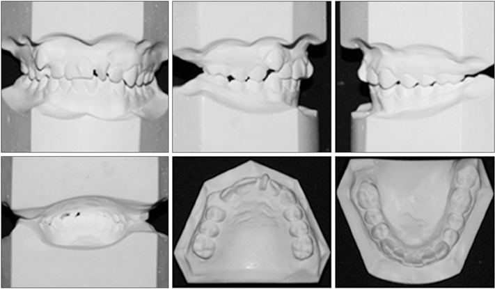
Figure 2 Pretreatment dental casts.
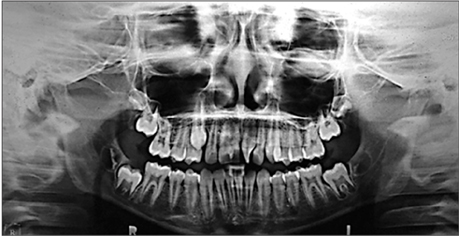
Figure 3 Pretreatment panoramic radiograph.
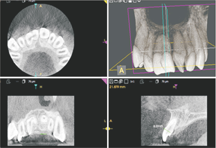
Figure 4 Pretreatment cone-beam computed tomography image.
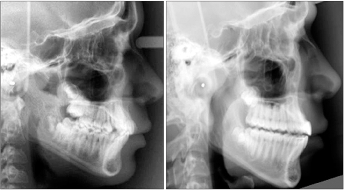
Figure 5 Pretreatment and post-treatment lateral cephalometric radiographs.
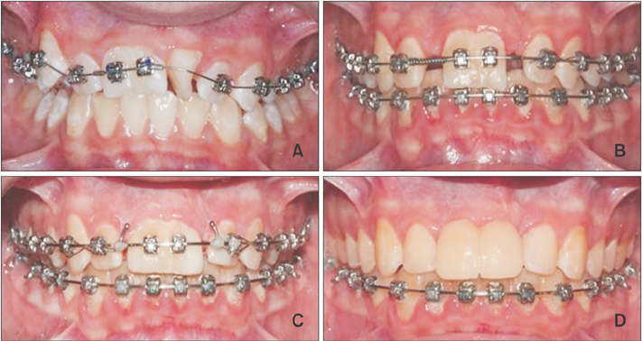
Figure 6 Treatment progress. A, Leveling and aligning phase of treatment with 0.014-inch preformed Damon Copper NiTi archwire. B, Movement of the fused teeth through the midpalatal suture by using a 0.019 × 0.025-inch stainless steel wire and open coil springs. C, Class II intermaxillary elastics (5/16-inch, 6 oz) are used for correcting the dental Class II relationship. D, Prosthetic restoration application after bracket removal.
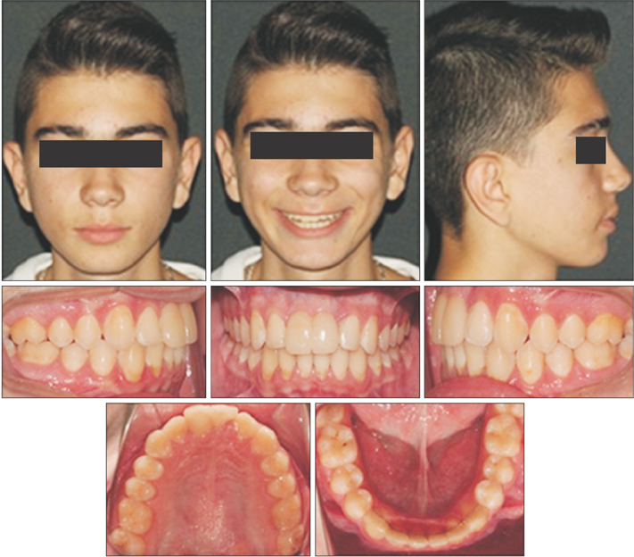
Figure 7 Post-treatment photographs.
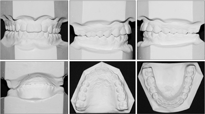
Figure 8 Post-treatment dental casts.
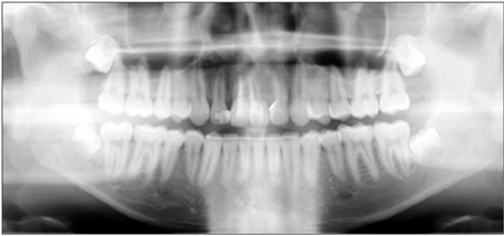
Figure 9 Post-treatment panoramic radiograph.
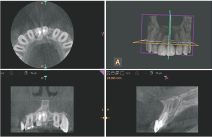
Figure 10 Post-treatment cone-beam computed tomography image.
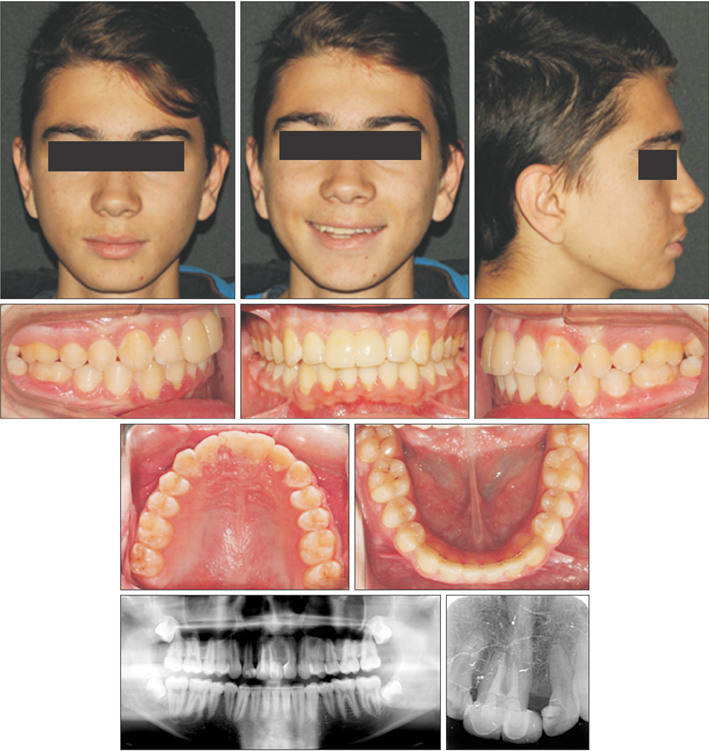
Figure 11 Photographs, panoramic radiograph, and occlusal radiograph at 1-year retention.
Reference
-
1. Brook AH, Winter GB. Double teeth. A retrospective study of ‘geminated’ and ‘fused’ teeth in children. Br Dent J. 1970; 129:123–130.
Article2. Levitas TC. Gemination, fusion, twinning, and concrescence. J Dent Child (Chic). 1965; 32:93–100.3. Foster TD. Fusion and duplication: orthodontic treatment of a developmental anomaly. Eur J Orthod. 1987; 9:240–242.
Article4. Sivolella S, Bressan E, Mirabal V, Stellini E, Berengo M. Extraoral endodontic treatment, odontotomy and intentional replantation of a double maxillary lateral permanent incisor: case report and 6-year follow-up. Int Endod J. 2008; 41:538–546.
Article5. Ekambaram M, Yiu CK, King NM. An unusual case of double teeth with facial and lingual talon cusps. Oral Surg Oral Med Oral Pathol Oral Radiol Endod. 2008; 105:e63–e67.
Article6. Mitchell WH. Letter to the editor. Dent Cosmos. 1892; 34:1036.7. Altug-Atac AT, Erdem D. Prevalence and distribution of dental anomalies in orthodontic patients. Am J Orthod Dentofacial Orthop. 2007; 131:510–514.
Article8. Zachrisson BU. Esthetics in tooth display and smile design. In : Nanda R, editor. Biomechanics and esthetics strategies in clinical orthodontics. Philedelphia: Elsevier Saunders;2005. p. 110–130.9. Primosch RE. Anterior supernumerary teeth--assessment and surgical intervention in children. Pediatr Dent. 1981; 3:204–215.10. Demircioglu Guler D, Sen Tunc E, Arici N, Ozkan N. Multidisciplinary management of a fused tooth: a case report. Case Rep Dent. 2013; 2013:634052.
Article11. Sammartino G, Cerone V, Gasparro R, Riccitiello F, Trosino O. Multidisciplinary approach to fused maxillary central incisors: a case report. J Med Case Rep. 2014; 8:398.
Article12. Ozden B, Gunduz K, Ozer S, Oz A, Otan Ozden F. The multidisciplinary management of a fused maxillary central incisor with a talon cusp. Aust Dent J. 2012; 57:98–102.
Article13. Garattini G, Crozzoli P, Brenna F. Bilateral dental fusion of the upper central incisors: a multi-disciplinary approach. J Esthet Dent. 1999; 11:149–154.
Article14. Follin M, Ericsson I, Thilander B. Orthodontic movement of maxillary incisors through the midpalatal suture area--an experimental study in dogs. Eur J Orthod. 1984; 6:237–246.
Article15. Bosio JA, Bradley TG, Hefti AF. Moving an incisor across the midline: a treatment alternative in an adolescent patient. Am J Orthod Dentofacial Orthop. 2011; 139:533–543.
Article16. Pair J. Movement of a maxillary central incisor across the midline. Angle Orthod. 2011; 81:341–349.
Article17. Garib DG, Janson G, dos Santos PB, de Oliveira Baldo T, de Oliveira GU, Ishikiriama SK. Orthodontic movement of a maxillary incisor through the midpalatal suture: a case report. Angle Orthod. 2012; 82:370–379.
Article18. Indra R, Srinivasan MR, Farzana H, Karthikeyan K. Endodontic management of a fused maxillary lateral incisor with a supernumerary tooth: a case report. J Endod. 2006; 32:1217–1219.
Article19. Yanikoğlu F, Kartal N. Endodontic treatment of a fused maxillary lateral incisor. J Endod. 1998; 24:57–59.20. Tsurumachi T, Kuno T. Endodontic and orthodontic treatment of a cross-bite fused maxillary lateral incisor. Int Endod J. 2003; 36:135–142.
Article21. Kato H, Kamio T. Diagnosis and endodontic management of fused mandibular second molar and paramolar with concrescent supernumerary tooth using cone-beam CT and 3-D printing technology: a case report. Bull Tokyo Dent Coll. 2015; 56:177–184.
Article22. Thilander B, Odman J, Lekholm U. Orthodontic aspects of the use of oral implants in adolescents: a 10-year follow-up study. Eur J Orthod. 2001; 23:715–731.
Article23. Misawa M, Lindhe J, Araújo MG. The alveolar process following single-tooth extraction: a study of maxillary incisor and premolar sites in man. Clin Oral Implants Res. 2016; 27:884–889.
Article24. Naraghi S, Andrén A, Kjellberg H, Mohlin BO. Relapse tendency after orthodontic correction of upper front teeth retained with a bonded retainer. Angle Orthod. 2006; 76:570–576.25. Brook AH. A unifying aetiological explanation for anomalies of human tooth number and size. Arch Oral Biol. 1984; 29:373–378.
Article26. Hamasha AA, Al-Khateeb T. Prevalence of fused and geminated teeth in Jordanian adults. Quintessence Int. 2004; 35:556–559.27. Cardoso JA, Almeida PJ, Fischer A, Phaxay SL. Clinical decisions for anterior restorations: the concept of restorative volume. J Esthet Restor Dent. 2012; 24:367–383.
Article28. Netter FH. Atlas of human anatomy. 6th ed. Philadelphia: Saunders/Elsevier Inc.;2014.
- Full Text Links
-
- Actions
-
Cited
- CITED
-
- Close
- Share
-
- Similar articles
-
- An experimental study on gross reactions of surrounding maxillary sutures to the widening of midpalatal suture in the dog
- A comparative experimental study on gross reactions of surounding maxillary sutures to the widening of midpalatal suture in young and adult dog
- Evaluation of the Midpalatal Suture Maturation in Young Koreans Using Cone-Beam Computed Tomography
- An experimantal study on histologic changes of surrounding maxillary sutures to the widening of midpalatal suture in the dog
- A study on the bone thickness of midpalatal suture area for miniscrew insertion
