A three-dimensional finite element analysis of the relationship between masticatory performance and skeletal malocclusion
- Affiliations
-
- 1Department of Periodontology, Dankook University College of Dentistry, Cheonan, Korea.
- 2Department of Orthodontics, Yonsei University College of Dentistry, Seoul, Korea.
- 3Department of Oral Anatomy, Dankook University College of Dentistry, Cheonan, Korea. jongta2@dankook.ac.kr
- KMID: 2027827
- DOI: http://doi.org/10.5051/jpis.2015.45.1.8
Abstract
- PURPOSE
The aim of this study was to evaluate the transfer of different occlusal forces in various skeletal malocclusions using finite element analysis (FEA).
METHODS
Three representative human cone-beam computed tomography (CBCT) images of three skeletal malocclusions were obtained from the Department of Orthodontics, Yonsei University Dental Hospital, Seoul, South Korea. The CBCT scans were read into the visualization software after separating bones and muscles by uploading the CBCT images into Mimics (Materialise). Two separate three-dimensional (3D) files were exported to visualize the solid morphology of skeletal outlines without considering the inner structures. Individual dental impressions were taken and stone models were scanned with a 3D scanner. These images were integrated and occlusal motions were simulated. Displacement and Von Mises stress were measured at the nodes of the FEA models. The displacement and stress distribution were analyzed. FEA was performed to obtain the 3D deformation of the mandibles under loads of 100, 150, 200, and 225 kg.
RESULTS
The distortion in all three skeletal malocclusions was comparable. Greater forces resulted in observing more distortion in FEA.
CONCLUSIONS
Further studies are warranted to fully evaluate the impact of skeletal malocclusion on masticatory performance using information on muscle attachment and 3D temporomandibular joint movements.
MeSH Terms
Figure
-
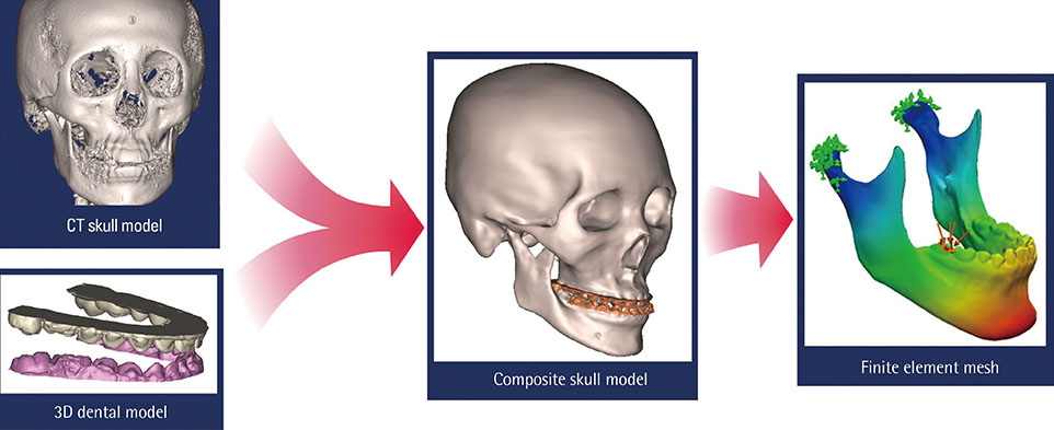
Figure 1 Dental modeling procedures used in this study. Cone-beam computed tomography (CT) images were obtained and integrated with three-dimensional (3D) information scanned from the dental model.
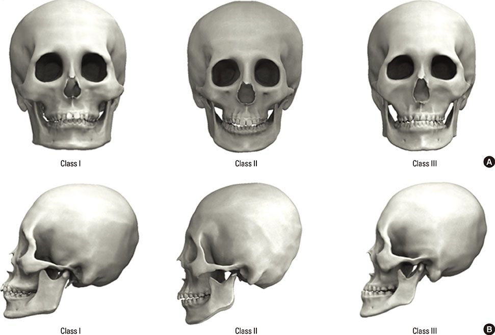
Figure 2 Three-dimensional reconstruction of human skull cone-beam computed tomography images from three representative individuals with skeletal malocclusions of classes I, II, and III. (A) Frontal views of three individuals. (B) Lateral views of three individuals with distinctive occlusal relationships.

Figure 3 Three-dimensional reconstructions of these images were produced with a surface triangularization technique. The mechanical properties assigned are shown in the figure.
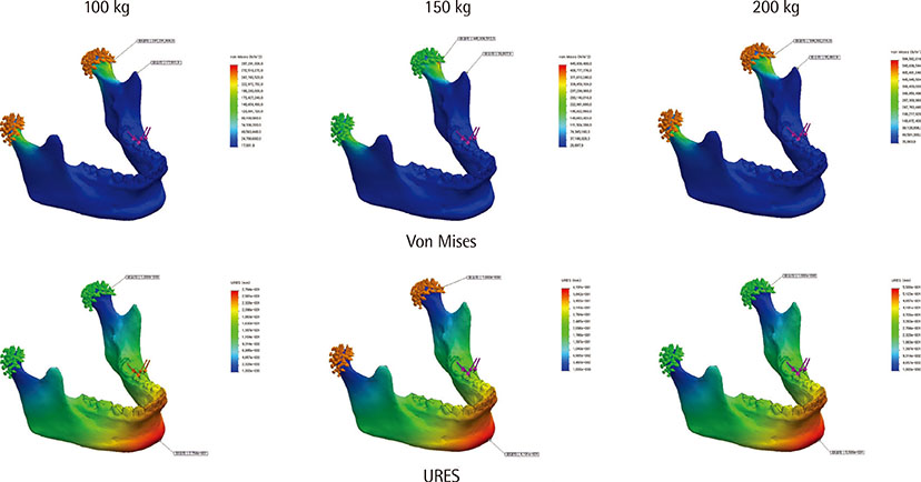
Figure 4 Pattern of Von Mises stress and maximum part deflection (URES; mm). Color-coded areas indicate the amount of distortion as shown in the inset index bar.
Reference
-
1. Sonnesen L, Bakke M. Molar bite force in relation to occlusion, craniofacial dimensions, and head posture in pre-orthodontic children. Eur J Orthod. 2005; 27:58–63.
Article2. Takeuchi N, Ekuni D, Yamamoto T, Morita M. Relationship between the prognosis of periodontitis and occlusal force during the maintenance phase: a cohort study. J Periodontal Res. 2010; 45:612–617.
Article3. Takeuchi N, Yamamoto T. Correlation between periodontal status and biting force in patients with chronic periodontitis during the maintenance phase of therapy. J Clin Periodontol. 2008; 35:215–220.
Article4. Misch CE. The effect of bruxism on treatment planning for dental implants. Dent Today. 2002; 21:76–81.5. Duyck J, Van Oosterwyck H, Vander Sloten J, De Cooman M, Puers R, Naert I. Magnitude and distribution of occlusal forces on oral implants supporting fixed prostheses: an in vivo study. Clin Oral Implants Res. 2000; 11:465–475.
Article6. Davies SJ, Gray RJ, Linden GJ, James JA. Occlusal considerations in periodontics. Br Dent J. 2001; 191:597–604.
Article7. Sassouni V. A classification of skeletal facial types. Am J Orthod. 1969; 55:109–123.
Article8. Arnett GW, Bergman RT. Facial keys to orthodontic diagnosis and treatment planning--Part II. Am J Orthod Dentofacial Orthop. 1993; 103:395–411.
Article9. English JD, Buschang PH, Throckmorton GS. Does malocclusion affect masticatory performance? Angle Orthod. 2002; 72:21–27.10. Choi DS, Cha BK, Jang I, Kang KH, Kim SC. Three-dimensional finite element analysis of occlusal stress distribution in the human skull with premolar extraction. Angle Orthod. 2013; 83:204–211.
Article11. Geng JP, Tan KB, Liu GR. Application of finite element analysis in implant dentistry: a review of the literature. J Prosthet Dent. 2001; 85:585–598.
Article12. Eskitascioglu G, Usumez A, Sevimay M, Soykan E, Unsal E. The influence of occlusal loading location on stresses transferred to implant-supported prostheses and supporting bone: a three-dimensional finite element study. J Prosthet Dent. 2004; 91:144–150.
Article13. Park JT, Lee JG, Won SY, Lee SH, Cha JY, Kim HJ. Realization of masticatory movement by 3-dimensional simulation of the temporomandibular joint and the masticatory muscles. J Craniofac Surg. 2013; 24:e347–e351.
Article14. Gateno J, Xia J, Teichgraeber JF, Rosen A. A new technique for the creation of a computerized composite skull model. J Oral Maxillofac Surg. 2003; 61:222–227.
Article15. Santler G, Karcher H, Gaggl A, Kern R. Stereolithography versus milled three-dimensional models: comparison of production method, indication, and accuracy. Comput Aided Surg. 1998; 3:248–256.
Article16. Gateno J, Xia JJ, Teichgraeber JF, Christensen AM, Lemoine JJ, Liebschner MA, et al. Clinical feasibility of computer-aided surgical simulation (CASS) in the treatment of complex cranio-maxillofacial deformities. J Oral Maxillofac Surg. 2007; 65:728–734.
Article17. Noh H, Nabha W, Cho JH, Hwang HS. Registration accuracy in the integration of laser-scanned dental images into maxillofacial cone-beam computed tomography images. Am J Orthod Dentofacial Orthop. 2011; 140:585–591.
Article18. Rangel FA, Maal TJ, Berge SJ, Kuijpers-Jagtman AM. Integration of digital dental casts in cone-beam computed tomography scans. ISRN Dent. 2012; 2012:949086.
Article19. Swennen GR, Mollemans W, De Clercq C, Abeloos J, Lamoral P, Lippens F, et al. A cone-beam computed tomography triple scan procedure to obtain a three-dimensional augmented virtual skull model appropriate for orthognathic surgery planning. J Craniofac Surg. 2009; 20:297–307.
Article20. Koc D, Dogan A, Bek B. Bite force and influential factors on bite force measurements: a literature review. Eur J Dent. 2010; 4:223–232.
Article21. Braun S, Bantleon HP, Hnat WP, Freudenthaler JW, Marcotte MR, Johnson BE. A study of bite force, part 2: relationship to various cephalometric measurements. Angle Orthod. 1995; 65:373–377.22. Braun S, Bantleon HP, Hnat WP, Freudenthaler JW, Marcotte MR, Johnson BE. A study of bite force, part 1: relationship to various physical characteristics. Angle Orthod. 1995; 65:367–372.23. Brudevold F. A basic study of the chewing forces of a denture wearer. J Am Dent Assoc. 1951; 43:45–51.
Article24. Anderson DJ. Measurement of stress in mastication. II. J Dent Res. 1956; 35:671–673.
Article25. Floystrand F, Kleven E, Oilo G. A novel miniature bite force recorder and its clinical application. Acta Odontol Scand. 1982; 40:209–214.
Article26. Gibbs CH, Mahan PE, Lundeen HC, Brehnan K, Walsh EK, Holbrook WB. Occlusal forces during chewing and swallowing as measured by sound transmission. J Prosthet Dent. 1981; 46:443–449.
Article27. Harada K, Watanabe M, Ohkura K, Enomoto S. Measure of bite force and occlusal contact area before and after bilateral sagittal split ramus osteotomy of the mandible using a new pressure-sensitive device: a preliminary report. J Oral Maxillofac Surg. 2000; 58:370–373.
Article28. Throckmorton GS, Finn RA, Bell WH. Biomechanics of differences in lower facial height. Am J Orthod. 1980; 77:410–420.
Article29. Furtado GC, Furtado A, Abu El, Butignon LE, Pesqueira AA, Paranhos LR. Relationship between the morphology of the maxillary central incisor and horizontal and vertical measurements of the face. Indian J Dent Res. 2014; 25:178–183.
Article
- Full Text Links
-
- Actions
-
Cited
- CITED
-
- Close
- Share
-
- Similar articles
-
- Three dimensional finite element analysis of mandibular stresses of complete denture occlusion
- Three-dimensional finite element analysis of unilateral mastication in malocclusion cases using cone-beam computed tomography and a motion capture system
- A study on the effect of the chincap by finite element analysis in juvenile skeletal Class III patients
- Three dimensional finite element analysis of mandibular stresses under complete dentures with variant artificial teeth forms and occlusal patterns
- ANALYSIS OF THE FIT IN THE IMPLANT PROSTHESIS USING LASER DISPLACEMENT METER AND THREE-DIMENSIONAL FINITE ELEMENT METHOD
