Korean J Orthod.
2015 May;45(3):121-129. 10.4041/kjod.2015.45.3.121.
Three-dimensional assessment of the temporomandibular joint and mandibular dimensions after early correction of the maxillary arch form in patients with Class II division 1 or division 2 malocclusion
- Affiliations
-
- 1Department of Orthodontics, Faculty of Dentistry, Hacettepe University, Ankara, Turkey. hande.gorucu@hotmail.com
- KMID: 1974803
- DOI: http://doi.org/10.4041/kjod.2015.45.3.121
Abstract
OBJECTIVE
This study aimed to assess three-dimensional changes in the temporomandibular joint positions and mandibular dimensions after correction of dental factors restricting mandibular growth in patients with Class II division 1 or division 2 malocclusion in the pubertal growth period.
METHODS
This prospective clinical study included 14 patients each with Class II division 1 (group I) and Class II division 2 (group II) malocclusions. The quad-helix was used for maxillary expansion, while utility arches were used for intrusion (group I) or protrusion and intrusion (group II) of the maxillary incisors. After approximately 2 months of treatment, an adequate maxillary arch width and acceptable maxillary incisor inclination were obtained. The patients were followed for an average of 6 months. Intraoral and extraoral photographs, plaster models, and cone-beam computed tomography (CBCT) images were obtained before and after treatment. Lateral cephalometric and temporomandibular joint measurements were made from the CBCT images.
RESULTS
The mandibular dimensions increased in both groups, although mandibular positional changes were also found in group II. There were no differences in the condylar position within the mandibular fossa or the condylar dimensions. The mandibular fossa depth and condylar positions were symmetrical at treatment initiation and completion.
CONCLUSIONS
Class II malocclusion can be partially corrected by achieving an ideal maxillary arch form, particularly in patients with Class II division 2 malocclusion. Restrictions of the mandible in the transverse or sagittal plane do not affect the temporomandibular joint positions in these patients because of the high adaptability of this joint.
MeSH Terms
Figure
-
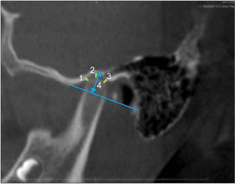
Figure 1 Anterior (1), superior (2), and posterior (3) joint spaces and the mandibular fossa depth (4) on a sagittal image of the right temporomandibular joint.
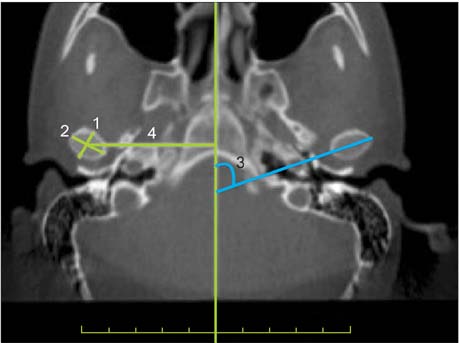
Figure 2 The greatest anteroposterior (1) and mediolateral (2) diameters of the mandibular condyle, vertical distance from the geometric centers of the condyles to the midsagittal plane (4), and the angle between the long axis of the mandibular condyle and the midsagittal plane (3) on an axial image of the temporomandibular joint.
Cited by 1 articles
-
Reliability of cone-beam computed tomography for temporomandibular joint analysis
Hande Gorucu-Coskuner, Ezgi Atik, Hakan El
Korean J Orthod. 2019;49(2):81-88. doi: 10.4041/kjod.2019.49.2.81.
Reference
-
1. Bishara SE. Class II Malocclusions: Diagnostic and clinical considerations with and without treatment. Semin Orthod. 2006; 12:11–24.
Article2. Yousefian J, Trimble D, Folkman G. A new look at the treatment of Class II Division 2 malocclusions. Am J Orthod Dentofacial Orthop. 2006; 130:771–778.
Article3. McNamara JA, Brudon WL. Orthodontic and orthopedic treatment in the mixed dentition. Ann Arbor, Mi: Needham Press;1993. p. 365.4. Gianelly AA. Boston University School of Dental Medicine. Rapid palatal expansion in the absence of crossbites: added value? Am J Orthod Dentofacial Orthop. 2003; 124:362–365.
Article5. Reichenbach E, Bruückl H, Taatz H. Kieferorthopädische klinik und therapie. Leipzig: J.A. Barth;1971. p. 248.6. Lima Filho RM, Lima AC, de Oliveira Ruellas AC. Spontaneous correction of Class II malocclusion after rapid palatal expansion. Angle Orthod. 2003; 73:745–752.7. Marshall SD, Southard KA, Southard TE. Early transverse treatment. Semin Orthod. 2005; 11:130–139.
Article8. Guest SS, McNamara JA Jr, Baccetti T, Franchi L. Improving Class II malocclusion as a side-effect of rapid maxillary expansion: a prospective clinical study. Am J Orthod Dentofacial Orthop. 2010; 138:582–591.
Article9. Volk T, Sadowsky C, Begole EA, Boice P. Rapid palatal expansion for spontaneous Class II correction. Am J Orthod Dentofacial Orthop. 2010; 137:310–315.
Article10. McNamara JA Jr. Early intervention in the transverse dimension: is it worth the effort? Am J Orthod Dentofacial Orthop. 2002; 121:572–574.
Article11. Sayin MO, Türkkahraman H. Malocclusion and crowding in an orthodontically referred Turkish population. Angle Orthod. 2004; 74:635–639.12. Ricketts RM. Bioprogressive therapy. Denver, CO: Rocky Mountain/Orthodontics;1979. p. 141.13. Pancherz H, Zieber K, Hoyer B. Cephalometric characteristics of Class II division 1 and Class II division 2 malocclusions: a comparative study in children. Angle Orthod. 1997; 67:111–120.14. Hedges R. A cephalometric evaluation of class II, Division 2. Angle Orthod. 1958; 28:191–197.15. Sabri R. Treatment of a Class II Division 2 malocclusion with space reopening for a single-tooth implant. Am J Orthod Dentofacial Orthop. 2001; 119:135–142.
Article16. Cleall JF, BeGole EA. Diagnosis and treatment of class II division 2 malocclusion. Angle Orthod. 1982; 52:38–60.17. Timmons LS. Induced change in the anteroposterior relationship of the jaws. Angle Orthod. 1972; 42:245–251.18. Vitral RW, Fraga MR, de Oliveira RS, de Andrade Vitral JC. Temporomandibular joint alterations after correction of a unilateral posterior crossbite in a mixed-dentition patient: a computed tomography study. Am J Orthod Dentofacial Orthop. 2007; 132:395–399.
Article19. Rodrigues AF, Fraga MR, Vitral RW. Computed tomography evaluation of the temporomandibular joint in Class I malocclusion patients: condylar symmetry and condyle-fossa relationship. Am J Orthod Dentofacial Orthop. 2009; 136:192–198.
Article20. Vitral RW, Telles Cde S. Computed tomography evaluation of temporomandibular joint alterations in class II Division 1 subdivision patients: condylar symmetry. Am J Orthod Dentofacial Orthop. 2002; 121:369–375.
Article21. Vitral RW, Telles Cde S, Fraga MR, de Oliveira RS, Tanaka OM. Computed tomography evaluation of temporomandibular joint alterations in patients with class II division 1 subdivision malocclusions: condyle-fossa relationship. Am J Orthod Dentofacial Orthop. 2004; 126:48–52.
Article22. Hassel B, Farman AG. Skeletal maturation evaluation using cervical vertebrae. Am J Orthod Dentofacial Orthop. 1995; 107:58–66.
Article23. Leonardi R, Caltabiano M, Cavallini C, Sicurezza E, Barbato E, Spampinato C, et al. Condyle fossa relationship associated with functional posterior crossbite, before and after rapid maxillary expansion. Angle Orthod. 2012; 82:1040–1046.
Article24. Aras A, Ada E, Saracoğlu H, Gezer NS, Aras I. Comparison of treatments with the Forsus fatigue resistant device in relation to skeletal maturity: a cephalometric and magnetic resonance imaging study. Am J Orthod Dentofacial Orthop. 2011; 140:616–625.
Article25. Ruf S, Pancherz H. Temporomandibular joint growth adaptation in Herbst treatment: a prospective magnetic resonance imaging and cephalometric roentgenographic study. Eur J Orthod. 1998; 20:375–388.
Article26. Grünheid T, Langenbach GE, Korfage JA, Zentner A, van Eijden TM. The adaptive response of jaw muscles to varying functional demands. Eur J Orthod. 2009; 31:596–612.
Article27. Du X, Hägg U. Muscular adaptation to gradual advancement of the mandible. Angle Orthod. 2003; 73:525–531.28. Aggarwal P, Kharbanda OP, Mathur R, Duggal R, Parkash H. Muscle response to the twin-block appliance: an electromyographic study of the masseter and anterior temporal muscles. Am J Orthod Dentofacial Orthop. 1999; 116:405–414.
Article29. Sood S, Kharbanda OP, Duggal R, Sood M, Gulati S. Muscle response during treatment of Class II Division 1 malocclusion with Forsus Fatigue Resistant Device. J Clin Pediatr Dent. 2011; 35:331–338.
Article
- Full Text Links
-
- Actions
-
Cited
- CITED
-
- Close
- Share
-
- Similar articles
-
- A study of the etiology of unilateral Class II, division 1 malocclusion
- A roentgenocephalomtric study on maxillary protrusion
- Expansion of the mandibular arch using a trombone appliance
- The study of craniofacial skeletal characteristics in Class II division 1 malocclusion
- An experimental study of ideal arch form of the Class I malocclusion patients with first premolar extraction
