Brain MRI Findings of Nitrogen Gas Inhalation for Suicide Attempt: a Case Report
- Affiliations
-
- 1Department of Radiology, Seoul Medical Center, Seoul, Korea. jnoon276@gmail.com
- KMID: 2400381
- DOI: http://doi.org/10.13104/imri.2017.21.4.264
Abstract
- South Korea has the highest reported suicide rate among all countries belonging to the Organization for Economic Cooperation and Development. Nitrogen is a colorless, odorless and nontoxic gas. Nitrogen gas has, however, been recently used as a method of attempted suicide, its nontoxity notwithstanding. We herein report on an unusual case involving a 30-year-old male who presented with symptoms after a suicide attempt by nitrogen inhalation. Diffusion-weighted imaging of his brain was showed curvilinear high signal intensity in the bilateral frontal and right occipital cortices, with subtle low apparent diffusion coefficient value. In addition, T2-weighted images and fluid attenuated inversion recovery images revealed subtle high signal intensity in the bilateral frontal cortices, basal ganglia and occipital cortices with contrast enhancement.
Keyword
MeSH Terms
Figure
-
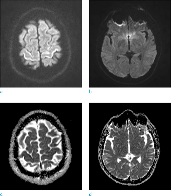
Fig. 1 A 30-year-old man after suicide attempt by nitrogen inhalation through a plastic bag. (a,b) Diffusion-weighted image shows curvilinear high signal intensity (SI) in the bilateral frontal and right occipital cortices. (c, d) Apparent diffusion coefficient map shows low value in the bilateral frontal and right occipital cortices.
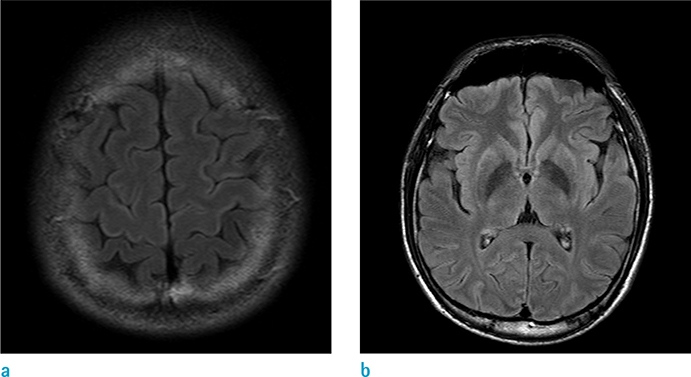
Fig. 2 Follow-up MRI (ten days later), FLAIR image shows curvilinear high signal intensity in the bilateral frontal and bilateral basal ganglia. FLAIR = fluidattenuated inversion recovery
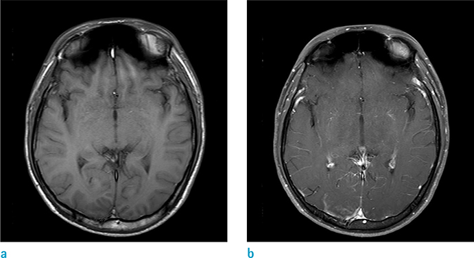
Fig. 3 Follow-up MRI (ten days later). (a) Axial T1-weighted MR image shows iso-signal intensity in the bilateral occipital cortices. (b) Axial contrast-enhanced T1-weighted MR image shows irregular enhancement in the bilateral occipital cortices.
Reference
-
1. OECD. Health at a Glance 2015: OECD indicators. Paris: OECD Publishing;2015.2. Madentzoglou MS, Kastanaki AE, Nathena D, Kranioti EF, Michalodimitrakis M. Nitrogen-plastic bag suicide: a case report. Am J Forensic Med Pathol. 2013; 34:311–314.3. USCSB. Safety Bulletin: Hazards of Nitrogen Asphyxiation, No. 2003-10-B. June 2003; 2003.4. Harding BE, Wolf BC. Case report of suicide by inhalation of nitrogen gas. Am J Forensic Med Pathol. 2008; 29:235–237.
Article5. DiPoce J, Guelfguat M, DiPoce J. Radiologic findings in cases of attempted suicide and other self-injurious behavior. Radiographics. 2012; 32:2005–2024.
Article6. Kamtchum Tatuene J, Pignel R, Pollak P, Lovblad KO, Kleinschmidt A, Vargas MI. Neuroimaging of diving-related decompression illness: current knowledge and perspectives. AJNR Am J Neuroradiol. 2014; 35:2039–2044.
Article7. White ML, Zhang Y, Helvey JT, Omojola MF. Anatomical patterns and correlated MRI findings of non-perinatal hypoxic-ischaemic encephalopathy. Br J Radiol. 2013; 86:20120464.
Article8. Huang BY, Castillo M. Hypoxic-ischemic brain injury: imaging findings from birth to adulthood. Radiographics. 2008; 28:417–439.
Article9. Maurya VK, Ravikumar R, Bhatia M, Rai R. Hypoxicischemic brain injury in an adult: magnetic resonance imaging findings. Med J Armed Forces India. 2016; 72:75–77.
Article10. Tur FC, Aksay E. Asphyxia due to accidental nitrogen gas inhalation: a case report. Hong Kong J Emerg Med. 2012; 19:46–48.
- Full Text Links
-
- Actions
-
Cited
- CITED
-
- Close
- Share
-
- Similar articles
-
- Attempted Suicide by Nitrogen Gas Asphyxiation: A Case Report
- Suicide Attempt by Inhalation of Argon Gas
- Case of Chemical Pneumonitis and Cellulitis after Suicide Attempt by Arteriovenous Fistula Injection of Gasoline
- Two cases of acute lung injury caused by nitrogen dioxide inhalation
- Attitude toward Suicide in the Elderly Suicide Attempter
