Calcifying Aponeurotic Fibroma of the Knee: a Case Report with Radiographic and MRI Finding
- Affiliations
-
- 1Department of Radiology, Pusan National University Hospital, Busan, Korea. lis@pusan.ac.kr
- 2Pusan National University School of Medicine, Busan, Korea.
- 3Department of Pathology, Pusan National University Hospital, Busan, Korea.
- 4Department of Orthopedic Surgery, Pusan National University Hospital, Busan, Korea.
- 5Department of Radiology, Inje University Haeundae Paik Hospital, Busan, Korea.
- KMID: 2400380
- DOI: http://doi.org/10.13104/imri.2017.21.4.259
Abstract
- Calcifying aponeurotic fibroma (CAF) is an uncommon benign tumor and a locally aggressive fibroblastic lesion. It commonly affects the palmar side of the hand and fingers, and the soles of the feet. The typical clinical manifestations are known as a poorly circumscribed, slow-growing, and asymptomatic firm mass. Most CAFs usually reveal low to intermediate or isointensity on T1-weighted images, and strong heterogeneous enhancement. However, various signal intensities on T2-weighted images have been reported depending on the degree of hypocellularity or the amount of calcification or collagen within the tumor. This report provides the radiographic and MRI finding of CAF arising in uncommon site, the knee region, of a 19-year-old male who presented with a painful and palpable mass.
MeSH Terms
Figure
-
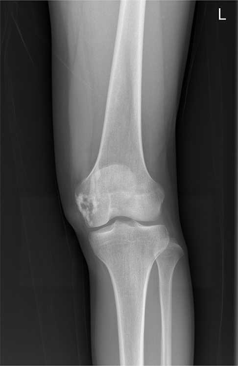
Fig. 1 A 19-year-old male patient with left knee pain. Tubular or nodular sclerotic lesions were seen in the medial femoral condyle on the anteroposterior view of the left knee radiograph. A slightly increased opacity appeared in the soft tissue area of the medial femoral condyle.
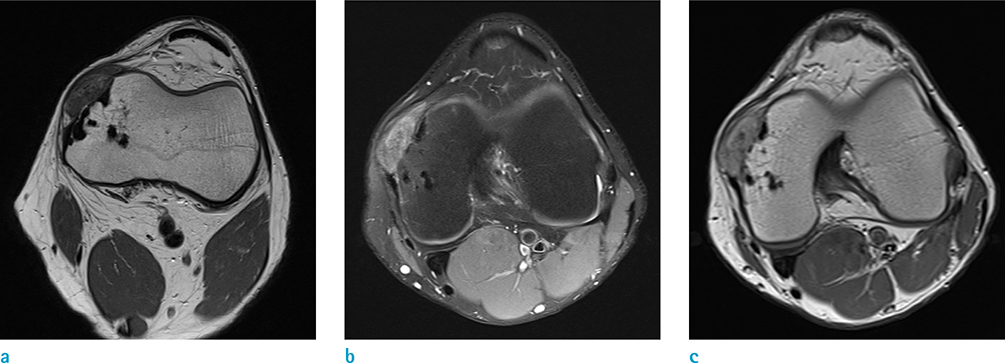
Fig. 2 (a) A soft tissue tumor about 3 cm in size was seen adjacent to the cortex of the medial femoral condyle on the axial T1-weighted MR image. The mass displayed isointensity to slightly high signal intensity compared to adjacent muscles and caused slight cortical erosion. The outer border of the tumor had a well-defined margin, but was not clearly separated from the medial patellofemoral ligament. Sclerotic lesions within the bone marrow exhibited dark signal intensities. (b) The mass displayed heterogeneously high signal intensity on the axial fat-suppressed T2-weighted image. The intermediate-to-low signal intensity portion was seen in the tumor. The posterior portion of the mass was adherent to the femoral origin of the medial collateral ligament. (c) The mass exhibited heterogeneously diffuse enhancement on the axial contrast-enhanced T1-weighted MR image. A low signal intensity region that did not enhance was observed in the mass.

Fig. 3 (a) Gross specimen of the resected tumor showed pinkish-red color and was measured at about 2.8 cm. (b) Pathologic specimen (× 100; Hematoxylin & Eosin [HE] staining) demonstrated that calcification is surrounded by fibroblastic cells, radiating from the center. (c) Cartilaginous differentiation and hyalinization in calcified areas are well seen on another slide (× 100; H&E staining).
Reference
-
1. Keasbey LE. Juvenile aponeurotic fibroma (calcifying fibroma); a distinctive tumor arising in the palms and soles of young children. Cancer. 1953; 6:338–346.
Article2. Kim OH, Kim YM. Calcifying aponeurotic fibroma: case report with radiographic and MR features. Korean J Radiol. 2014; 15:134–139.
Article3. Fetsch JF, Miettinen M. Calcifying aponeurotic fibroma: a clinicopathologic study of 22 cases arising in uncommon sites. Hum Pathol. 1998; 29:1504–1510.
Article4. Kwak HS, Lee SY, Kim JR, Lee KB. MR imaging of calcifying aponeurotic fibroma of the thigh. Pediatr Radiol. 2004; 34:438–440.
Article5. Enzinger FM, Weiss SW. Fibrous tumors of infancy and childhood: soft tissue tumors. 4th ed. St. Louis: Mosby-Year Book;2001. p. 388–395.6. Amaravati R. Rare malignant transformation of a calcifying aponeurotic fibroma. J Bone Joint Surg Am. 2002; 84-A:1889. author reply 1889.
Article7. Nishio J, Inamitsu H, Iwasaki H, Hayashi H, Naito M. Calcifying aponeurotic fibroma of the finger in an elderly patient: CT and MRI findings with pathologic correlation. Exp Ther Med. 2014; 8:841–843.
Article8. Hasegawa HK, Park S, Hamazaki M. Calcifying aponeurotic fibroma of the knee: a case report with radiological findings. J Dermatol. 2006; 33:169–173.
Article9. Parker WL, Beckenbaugh RR, Amrami KK. Calcifying aponeurotic fibroma of the hand: radiologic differentiation from giant cell tumors of the tendon sheath. J Hand Surg Am. 2006; 31:1024–1028.
Article10. Sekiguchi T, Nakagawa M, Miwa S, et al. Calcifying aponeurotic fibroma in a girl: MRI findings and their chronological changes. Radiol Case Rep. 2017; 12:620–623.
Article
