Yonsei Med J.
2009 Apr;50(2):262-265.
Measurement of Kidney Volume with Multi-Detector Computed Tomography Scanning in Young Korean
- Affiliations
-
- 1Department of Internal Medicine, The Armed Forces Yang-Ju Hospital, Yangju, Korea. chungbh@catholic.ac.kr
- 2Department of Urology, The Armed Forces Yang-Ju Hospital, Yangju, Korea.
- 3Department of Radiology, The Armed Forces Yang-Ju Hospital, Yangju, Korea.
- 4Department of Internal Medicine, The Catholic University of Korea College of Medicine, Seoul, Korea.
Abstract
- PURPOSE
Kidney volume is regarded as the most precise indicator of kidney size. However, it is not widely used clinically, because its measurement is difficult due to the complex kidney shape. We attempted to evaluate the normal kidney volume in young Korean men by using multi-detector computed tomography (MDCT). MATERIALS AND METHODS: We retrospectively reviewed MDCT data of young Korean men (113 patients). After data processing, we measured the volume and length of the kidneys. Body parameters (height, body weight, body-surface area, and total body water) and laboratory data were collected. Glomerular filtration rate (GFR) was calculated using Cockcroft-Gault (CG) equation. RESULTS: The mean kidney volume was 205.29 +/- 36.81 cm3; and mean kidney length was 10.80 +/- 0.69 cm. The former correlated significantly with height, body weight, body-surface area, and total body water (p < 0.05, correlation coefficient : gamma = 0.328, 0.649, 0.640, and 0.638, respectively). The latter also correlated significantly with all body indexes, however the correlation was weaker, except with height (p < 0.05, correlation coefficient : gamma = 0.457, 0.473, 0.505, and 0.503, respectively). Only kidney volume significantly predicted estimated GFR (adjusted R2 = 0.431, F = 85.90 and p < 0.05). CONCLUSION: The kidney volume measured with MDCT is correlated well with body parameters, and is useful to predict renal function.
Keyword
MeSH Terms
Figure
-

Fig. 1 Relationship of mean kidney length and mean kidney volume.
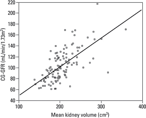
Fig. 2 Distribution of CG-GFR according to kidney volume. CG-GFR, Cockcroft-Gault-glomerular filtration rate.
Reference
-
1. Lalli AF. Renal enlargement. Radiology. 1965. 84:688–691.
Article2. Ablett MJ, Coulthard A, Lee RE, Richardson DL, Bellas T, Owen JP, et al. How reliable are ultrasound measurements of renal length in adults? Br J Radiol. 1995. 68:1087–1089.
Article3. Jones TB, Riddick LR, Harpen MD, Dubuisson RL, Samuels D. Ultrasonographic determination of renal mass and renal volume. J Ultrasound Med. 1983. 2:151–154.
Article4. Widjaja E, Oxtoby JW, Hale TL, Jones PW, Harden PN, McCall IW. Ultrasound measured renal length versus low dose CT volume in predicting single kidney glomerular filtration rate. Br J Radiol. 2004. 77:759–764.
Article5. Poggio ED, Hila S, Stephany B, Fatica R, Krishnamurthi V, del Bosque C, et al. Donor kidney volume and outcomes following live donor kidney transplantation. Am J Transplant. 2006. 6:616–624.
Article6. Bakker J, Olree M, Kaatee R, de Lange EE, Moons KG, Beutler JJ, et al. Renal volume measurements: accuracy and repeatability of US compared with that of MR imaging. Radiology. 1999. 211:623–628.
Article7. Sommer G, Bouley D, Frisoli J, Pierce L, Sandner-Porkristl D, Fahrig R. Determination of 3-dimensional zonal renal volumes using contrast-enhanced computed tomography. J Comput Assist Tomogr. 2007. 31:209–213.
Article8. Schlosser T, Mohrs OK, Magedanz A, Voigtländer T, Schmermund A, Barkhausen J. Assessment of left ventricular function and mass in patients undergoing computed tomography (CT) coronary angiography using 64-detector-row CT: comparison to magnetic resonance imaging. Acta Radiol. 2007. 48:30–35.
Article9. Mosteller RD. Simplified calculation of body-surface area. N Engl J Med. 1987. 317:1098.
Article10. Watson PE, Watson ID, Batt RD. Total body water volumes for adult males and females estimated from simple anthropometric measurements. Am J Clin Nutr. 1980. 33:27–39.
Article11. Cockcroft DW, Gault MH. Prediction of creatinine clearance from serum creatinine. Nephron. 1976. 16:31–41.
Article12. National Kidney Foundation. K/DOQI clinical practice guidelines for chronic kidney disease: evaluation, classification, and stratification. Am J Kidney Dis. 2002. 39(2):Suppl 1. S1–S266.13. Geraghty EM, Boone JM, McGahan JP, Jain K. Normal organ volume assessment from abdominal CT. Abdom Imaging. 2004. 29:482–490.
Article14. Cheong B, Muthupillai R, Rubin MF, Flamm SD. Normal values for renal length and volume as measured by magnetic resonance imaging. Clin J Am Soc Nephrol. 2007. 2:38–45.
Article15. Emamian SA, Nielsen MB, Pedersen JF, Ytte L. Kidney dimensions at sonography: correlation with age, sex, and habitus in 665 adult volunteers. AJR Am J Roentgenol. 1993. 160:83–86.
Article16. Lee BH, Ahn HJ, Kang WH, Seo GH, Kim B, Lee SG, et al. Estimation of kidney size by ultrasonography in normal Korean adults. Korean J Nephrol. 1999. 18:46–51.17. Kang KY, Lee YJ, Park SC, Yang CW, Kim YS, Moon IS, et al. A comparative study of methods of estimating kidney length in kidney transplantation donors. Nephrol Dial Transplant. 2007. 22:2322–2327.
Article18. Mahajan S, Mukhiya GK, Singh R, Tiwari SC, Kalra V, Bhowmik DM, et al. Assessing glomerular filtration rate in healthy Indian adults: a comparison of various prediction equations. J Nephrol. 2005. 18:257–261.19. Al-Khader AA, Tamim H, Sulaiman MH, Jondeby MS, Taher S, Hejaili FF, et al. What is the most appropriate formula to use in estimating glomerular filtration rate in adult Arabs without kidney disease? Ren Fail. 2008. 30:205–208.
Article20. Lerman LO, Bentley MD, Bell MR, Rumberger JA, Romero JC. Quantitation of the in vivo kidney volume with cine computed tomography. Invest Radiol. 1990. 25:1206–1211.
Article21. Kotre CJ, Owen JP. Method for the evaluation of renal parenchymal volume by X-ray computed tomography. Med Biol Eng Comput. 1994. 32:338–341.
Article22. Rao M, Stough J, Chi YY, Muller K, Tracton G, Pizer SM, et al. Comparison of human and automatic segmentations of kidneys from CT images. Int Radiat Oncol Biol Phys. 2005. 61:954–960.
Article23. Cai W, Holalkere NS, Harris G, Sahani D, Yoshida H. Dynamic-threshold level set method for volumetry of porcine kidney in CT images in vivo and ex vivo assessment of the accuracy of volume measurement. Acad Radiol. 2007. 14:890–896.
Article
- Full Text Links
-
- Actions
-
Cited
- CITED
-
- Close
- Share
-
- Similar articles
-
- Three cases of right coronary anomaly confirmed by multi-detector computed tomography
- Volumetric Measurements of Lung Nodules with Multi-Detector Row CT: Effect of Changes in Lung Volume
- Comparative evaluation of computed tomography for dental implants on the mandibular edentulous area
- Clinical Applications of Wide-Detector CT Scanners for Cardiothoracic Imaging: An Update
- MDCT Application in the Vascular System
