J Korean Med Assoc.
2007 Jan;50(1):25-32. 10.5124/jkma.2007.50.1.25.
MDCT Application in the Vascular System
- Affiliations
-
- 1Department of Radiology, Korea University College of Medicine, Korea. yhwanseok@naver.com
- KMID: 2184792
- DOI: http://doi.org/10.5124/jkma.2007.50.1.25
Abstract
- Helical CT has improved with faster gantry rotation, more powerful X-ray tubes, and improved interpolation algorithms; however, the greatest advance has been made by the recent introduction of multi detector-row computed tomography (MDCT) scanners. Fundamental advantages of MDCT include substantially shorter acquisition times, retrospective creation of thinner or thicker sections from the same raw data, and improved threedimensional (3-D) rendering with diminished helical artifacts. While these features will likely be important to many applications of CT scanning, the greatest impact has been on CT angiography. The advantages of MDCT over single detector-row CT scanners when imaging the vascular system can be broken down into three fundamental improvements, that is, speed (faster scan time), distance (longer coverage), and section thickness (better resolution). This article will focus on how the MDCT technology has substantially improved imaging of the vascular system, including pulmonary artery, aorta and extremity vessels.
Keyword
MeSH Terms
Figure
-

Figure 1 CT pulmonary angiography Pulmonary emboli are seen in left pulmonary, lobar, and segmental pulmonary arteries (A, B). Obstructed and enlarged subsegmental pulmonary arteries are seen in right lower lobe (C). Pulmonary arterial hypertension is suggested because contrast material is refluxed into azygos vein and inferior vena cava and right ventricle is enlarged
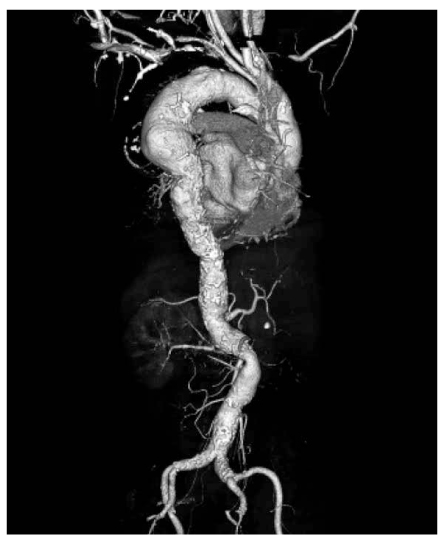
Figure 2 CT aortography. On 3D volume rendered image, atherosclerotic aortic aneurysm is seen on proximal descending thoracic aorta. Diffuse aortic calcification is also seen along the whole aorta
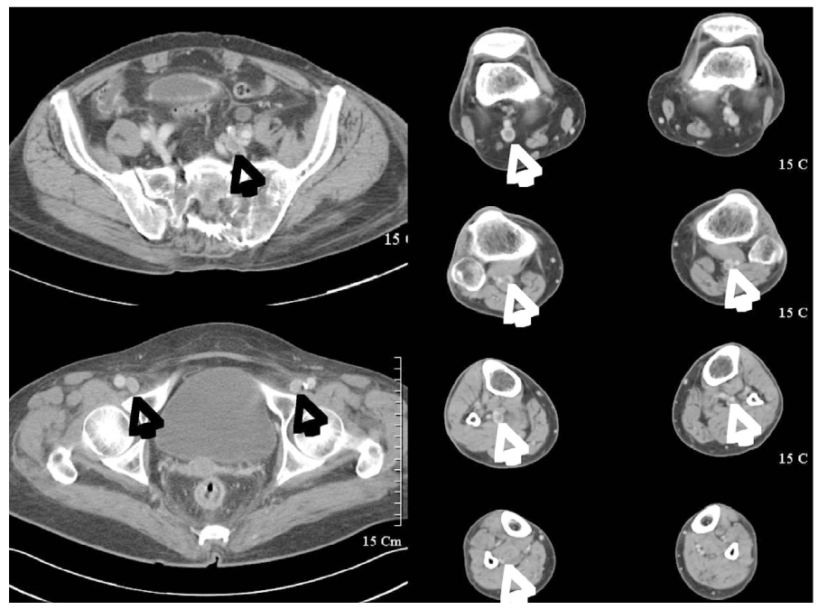
Figure 3 Indirect CT venography. Deep vein thrombosis is seen in left iliac, both femoral, right popliteal, and both peroneal veins (arrows)
Reference
-
1. Rubin GD, Schmidt AJ, Logan LJ, Sofilos MC. Multi-detector row CT angiography of lower extremity arterial inflow and runoff: initial experience. Radiology. 2001. 221:146–158.
Article2. Schoellnast H, Tillich M, Deutschmann HA, Stessel U, Schaffler GJ, Schoellnast R, Uqqowizer MM. Improvement of parenchymal and vascular enhancement using saline flush and power injection for multiple-detector-row abdominal CT. Eur Radiol. 2004. 14:659–664.
Article3. Wittram C, Meehan MJ, Halpern EF, Shepard JA, McLoud TC, Thrall JH. Trends in thoracic radiology over a decade at a large academic medical center. J Thorac Imaging. 2004. 19:164–170.
Article4. Wittram C, Maher MM, Yoo AJ, Kalra MK, Shepard JA, McLoud TC. CT angiography of pulmonary embolism: diagnostic criteria and causes of misdiagnosis. Radiographics. 2004. 24:1219–1238.
Article5. Han D, Lee KS, Franquet T, Muler NL, Kim TS, Kim H, Kwon OJ, Byun HS. Thrombotic and nonthrombotic pulmonary arterial embolism: spectrum of imaging findings. Radiographics. 2003. 23:1521–1539.
Article6. Schoepf UJ, Costello P. CT angiography for diagnosis of pulmonary embolism: state of the art. Radiology. 2004. 230:329–337.
Article7. Swensen SJ, Sheedy PF 2nd, Ryu JH, Pickett DD, Schleck CD, Llstrup DM. Outcomes after withholding anticoagulation from patients with suspected acute pulmonary embolism and negative computed tomographic findings: a cohort study. Mayo Clin Proc. 2002. 77:130–138.
Article8. Fallenberg M, Juergens KU, Wichter T, Scheld HH, Fischbach R. Coronary artery aneurysm and type-A aortic dissection demonstrated by retrospectively ECG-gated multislice spiral CT. Eur Radiol. 2002. 12:201–204.
Article9. Manghat NE, Morgan-Hughes GJ, Roobottom CA. Multidetector row computed tomography: imaging in acute aortic syndrome. Clin Radiol. 2005. 60:1256–1267.
Article10. Castaner E, Andreu M, Gallardo X, Mata JM, Cabezuelo MA, Pallardo Y. CT in nontraumatic acute thoracic aortic disease: typical and atypical features and complications. Radiographics. 2003. 23:S93–S110.
Article11. Sebastia C, Quiroga S, Boye R, Perez-Lafuente M, Castella E, Alvarez-Castells A. Aortic stenosis: spectrum of diseases depicted at multisection CT. Radiographics. 2003. 23:S79–S91.
Article12. Holden A, Smith A, Dukes P, Pilmore H, Yasutomi M. Assessment of 100 live potential renal donors for laparoscopic nephrectomy with multi-detector row helical CT. Radiology. 2005. 237:973–980.
Article13. Kawamoto S, Montgomery RA, Lawler LP, Horton KM, Fishman EK. Multi-detector row CT evaluation of living renal donors prior to laparoscopic nephrectomy. Radiographics. 2004. 24:453–466.
Article14. Fleischmann D. MDCT of renal and mesenteric vessels. Eur Radiol. 2003. 13:S 5. 94–101.
Article15. Martin ML, Tay KH, Flak B. Multidetector CT angiography of the aortoiliac system and lower extremities: a prospective comparison with digital subtraction angiography. AJR Am J Roentgenol. 2003. 180:1085–1091.
Article16. Lim KE, Hsu WC, Hsu YY, Chu PH, Ng CJ. Deep venous thrombosis: comparison of indirect multidetector CT venography and sonography of lower extremities in 26 patients. Clin Imaging. 2004. 28:439–444.17. Reid JH. Multislice CT pulmonary angiography and CT venography. Br J Radiol. 2004. 77:39–45.
Article18. Stein PD, Fowler SE, Goodman LR, Gottschalk A, Hales CA, Hull RD, Lee per KV Jr, Popovich J Jr, Quinn DA, Sos TA, Sostman HD, Tapson VF, Wakefield TW, Weg JG, Woodard PK. Multidetector computed tomography for acute pulmonary embolism. N Engl J Med. 2006. 354:2317–2327.
Article19. Chung JW, Yoon CJ, Jung SI, et al. Acute iliofemoral deep vein thrombosis: evaluation of underlying anatomic abnormalities by spiral CT venography. J Vasc Interv Radiol. 2004. 15:249–256.
Article20. Uhl JF, Verdeille S, Martin-Bouyer Y. Three-dimensional spiral CT venography for the pre-operative assessment of varicose patients. Vasa. 2003. 32:91–94.
Article21. Willmann JK, Wildermuth S. Multidetector-row CT angiography of upper-and lower-extremity peripheral arteries. Eur Radiol. 2005. 15:S 4. 3–9.22. Kim H, Chung JW, Park JH, et al. Role of CT venography in the diagnosis and treatment of benign thoracic central venous obstruction. Korean J Radiol. 2003. 4:146–152.
Article
