Comparison of 2 root surface area measurement methods: 3-dimensional laser scanning and cone-beam computed tomography
- Affiliations
-
- 1Department of Orthodontics and Pediatric Dentistry, Faculty of Dentistry, Chiang Mai University, Chiang Mai, Thailand. dhirawat.j@gmail.com
- 2Department of Radiology, Faculty of Medicine Siriraj Hospital, Mahidol University, Bangkok, Thailand.
- 3Department of Clinical Dentistry - Orthodontics, Faculty of Medicine and Dentistry, University of Bergen, Bergen, Norway.
- KMID: 2389892
- DOI: http://doi.org/10.5624/isd.2017.47.2.117
Abstract
- PURPOSE
The aim of this study was to compare the use of 3-dimensional (3D) laser scanning and cone-beam computed tomography (CBCT) as methods of root surface measurement.
MATERIALS AND METHODS
Thirty teeth (15 maxillary first premolars and 15 mandibular first premolars) from 8 patients who required extractions for orthodontic treatment were selected. Before extraction, pre-treatment CBCT images of all the patients were recorded. First, a CBCT image was imported into simulation software (Mimics version 15.01; Materialise, Leuven, Belgium) and the root surface area of each tooth was calculated using 3-Matic (version 7.01, Materialise, Leuven, Belgium). After extraction, all the teeth were scanned and the root surface area of each extracted tooth was calculated. The root surface areas calculated using these 2 measurement methods were analyzed using the paired t-test (P<.05). Correlations between the 2 methods were determined by calculating the Pearson correlation coefficient. The intraclass correlation coefficient (ICC) was used to assess intraobserver reliability.
RESULTS
The root surface area measurements (230.11±41.97 mm²) obtained using CBCT were slightly greater than those (229.31±42.46 mm²) obtained using 3D laser scanning, but not significantly (P=.425). A high Pearson correlation coefficient was found between the CBCT and the 3D laser scanner measurements. The intraobserver ICC was 1.000 for 3D laser scanning and 0.990 for CBCT.
CONCLUSION
This study presents a novel CBCT approach for measuring the root surface area; this technique can be used for estimating the root surface area of non-extracted teeth.
Figure
-
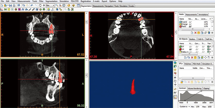
Fig. 1 Digital Imaging and Communications in Medicine (DICOM) datasets of the patients are imported into Mimics® Innovation Suite 15.01 and reconstructed.
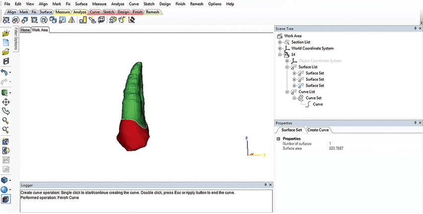
Fig. 2 The root surface area of each tooth is calculated using the 3-matic software.
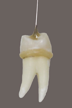
Fig. 3 Premolars are marked at the cementoenamel junction, and a resin composite is used for marking the areas above the cementoenamel junction.
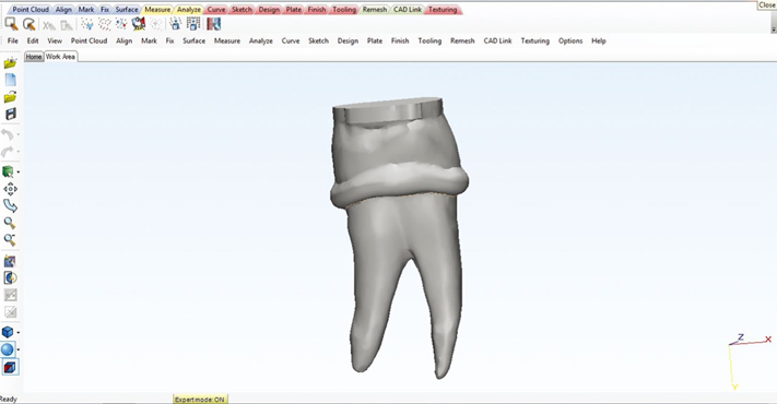
Fig. 4 The scanning range on the appropriate scope is set to cover the tooth sample using a dental laser scanner.
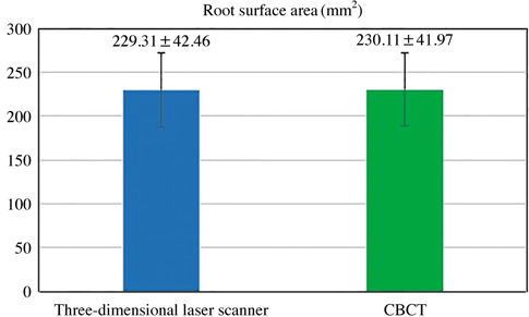
Fig. 5 Root surface area measurements obtained using 3-dimensional (3D) laser scanning and cone-beam computed tomography (CBCT)

Fig. 6 Correlation between root surface area measurements obtained using 3-dimensional (3D) laser scanning and cone-beam computed tomography (CBCT). The Pearson correlation coefficient is 0.992.
Cited by 1 articles
-
Root surface areas of maxillary permanent teeth in anterior normal overbite and anterior open bite assessed using cone-beam computed tomography
Piyadanai Suteerapongpun, Supassara Sirabanchongkran, Tanapan Wattanachai, Patiyut Sriwilas, Dhirawat Jotikasthira
Imaging Sci Dent. 2017;47(4):241-246. doi: 10.5624/isd.2017.47.4.241.
Reference
-
1. Hujoel PP. A meta-analysis of normal ranges for root surface areas of the permanent dentition. J Clin Periodontol. 1994; 21:225–229.
Article2. Mowry JK, Ching MG, Orjansen MD, Cobb CM, Friesen LR, MacNeill SR, et al. Root surface area of the mandibular cuspid and bicuspids. J Periodontol. 2002; 73:1095–1100.
Article3. Scarfe WC, Farman AG, Sukovic P. Clinical applications of cone-beam computed tomography in dental practice. J Can Dent Assoc. 2006; 72:75–80.4. Li G. Patient radiation dose and protection from cone-beam computed tomography. Imaging Sci Dent. 2013; 43:63–69.
Article5. Reuschl RP, Heuer W, Stiesch M, Wenzel D, Dittmer MP. Reliability and validity of measurements on digital study models and plaster models. Eur J Orthod. 2016; 38:22–26.
Article6. Jepsen A. Root surface measurement and a method for x-ray determination of root surface area. Acta Odontol Scand. 1963; 21:35–46.
Article7. Nicholls JI, Daly CH, Kydd WL. Root surface measurement using a digital computer. J Dent Res. 1974; 53:1338–1341.
Article8. Luthra SP, Narayan I, Subrahmanyam N. Root surface area measured by the benzene adsorption method. J Prosthet Dent. 1974; 31:185–189.
Article9. Pan JH, Chen SK, Lin CH, Leu LC, Chen CM, Jeng JY. Estimation of single-root surface area from true thickness data and from thickness derived from digital dental radiography. Dentomaxillofac Radiol. 2004; 33:312–317.
Article10. Li W, Chen F, Zhang F, Ding W, Ye Q, Shi J, et al. Volumetric measurement of root resorption following molar mini-screw implant intrusion using cone beam computed tomography. PLoS One. 2013; 8:e60962.
Article11. Gu Y, Tang Y, Zhu Q, Feng X. Measurement of root surface area of permanent teeth with root variations in a Chinese population-A micro-CT analysis. Arch Oral Biol. 2016; 63:75–81.
Article12. Taylor TT, Gans SI, Jones EM, Firestone AR, Johnston WM, Kim DG. Comparison of micro-CT and cone beam CT-based assessments for relative difference of grey level distribution in a human mandible. Dentomaxillofac Radiol. 2013; 42:25117764.13. Anderson PJ, Yong R, Surman TL, Rajion ZA, Ranjitkar S. Application of three-dimensional computed tomography in craniofacial clinical practice and research. Aust Dent J. 2014; 59:Suppl 1. 174–185.
Article14. Ludlow JB, Davies-Ludlow LE, Brooks SL, Howerton WB. Dosimetry of 3 CBCT devices for oral and maxillofacial radiology: CB Mercuray, NewTom 3G and i-CAT. Dentomaxillofac Radiol. 2006; 35:219–226.
Article15. Ludlow JB, Ivanovic M. Comparative dosimetry of dental CBCT devices and 64-slice CT for oral and maxillofacial radiology. Oral Surg Oral Med Oral Pathol Oral Radiol Endod. 2008; 106:106–114.
Article
- Full Text Links
-
- Actions
-
Cited
- CITED
-
- Close
- Share
-
- Similar articles
-
- Three-dimensional imaging modalities in endodontics
- Root surface areas of maxillary permanent teeth in anterior normal overbite and anterior open bite assessed using cone-beam computed tomography
- Linear accuracy of cone-beam computed tomography and a 3-dimensional facial scanning system: An anthropomorphic phantom study
- Detection of maxillary second molar with two palatal roots using cone beam computed tomography: a case report
- Management of root canal perforation by using cone-beam computed tomography
