Imaging Sci Dent.
2018 Jun;48(2):111-119. 10.5624/isd.2018.48.2.111.
Linear accuracy of cone-beam computed tomography and a 3-dimensional facial scanning system: An anthropomorphic phantom study
- Affiliations
-
- 1Department of Oral and Maxillofacial Radiology, Graduate School, Kyung Hee University, Seoul, Korea. hehan@khu.ac.kr
- 2Department of Dental Hygiene, College of Health, Kyungwoon University, Gumi, Korea.
- KMID: 2413922
- DOI: http://doi.org/10.5624/isd.2018.48.2.111
Abstract
- PURPOSE
This study was conducted to evaluate the accuracy of linear measurements of 3-dimensional (3D) images generated by cone-beam computed tomography (CBCT) and facial scanning systems, and to assess the effect of scanning parameters, such as CBCT exposure settings, on image quality.
MATERIALS AND METHODS
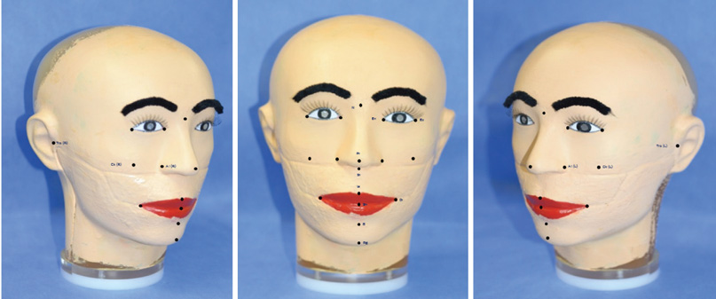
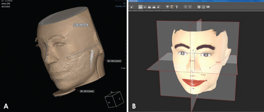
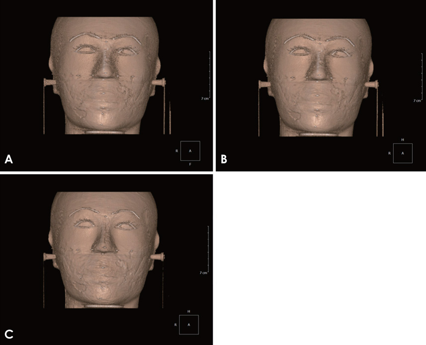
CBCT and facial scanning images of an anthropomorphic phantom showing 13 soft-tissue anatomical landmarks were used in the study. The distances between the anatomical landmarks on the phantom were measured to obtain a reference for evaluating the accuracy of the 3D facial soft-tissue images. The distances between the 3D image landmarks were measured using a 3D distance measurement tool. The effect of scanning parameters on CBCT image quality was evaluated by visually comparing images acquired under different exposure conditions, but at a constant threshold.
RESULTS
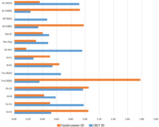
Comparison of the repeated direct phantom and image-based measurements revealed good reproducibility. There were no significant differences between the direct phantom and image-based measurements of the CBCT surface volume-rendered images. Five of the 15 measurements of the 3D facial scans were found to be significantly different from their corresponding direct phantom measurements (P < .05). The quality of the CBCT surface volume-rendered images acquired at a constant threshold varied across different exposure conditions.
CONCLUSION
These results proved that existing 3D imaging techniques were satisfactorily accurate for clinical applications, and that optimizing the variables that affected image quality, such as the exposure parameters, was critical for image acquisition.
Keyword
MeSH Terms
Figure
Reference
-
1. Swennen GR, Mollemans W, Schutyser F. Three-dimensional treatment planning of orthognathic surgery in the era of virtual imaging. J Oral Maxillofac Surg. 2009; 67:2080–2092.
Article2. McNamara JA Jr. A method of cephalometric evaluation. Am J Orthod. 1984; 86:449–469.
Article3. Kau CH, Richmond S, Incrapera A, English J, Xia JJ. Threedimensional surface acquisition systems for the study of facial morphology and their application to maxillofacial surgery. Int J Med Robot. 2007; 3:97–110.
Article4. Swennen GR, Schutyser F. Three-dimensional cephalometry: spiral multi-slice vs cone-beam computed tomography. Am J Orthod Dentofacial Orthop. 2006; 130:410–416.
Article5. Silva MA, Wolf U, Heinicke F, Bumann A, Visser H, Hirsch E. Cone-beam computed tomography for routine orthodontic treatment planning: a radiation dose evaluation. Am J Orthod Dentofacial Orthop. 2008; 133:640.e1–640.e5.
Article6. Li G, Wei J, Wang X, Wu G, Ma D, Wang B, et al. Three-dimensional facial anthropometry of unilateral cleft lip infants with a structured light scanning system. J Plast Reconstr Aesthet Surg. 2013; 66:1109–1116.
Article7. Weinberg SM, Naidoo S, Govier DP, Martin RA, Kane AA, Marazita ML. Anthropometric precision and accuracy of digital three-dimensional photogrammetry: comparing the Genex and 3dMD imaging systems with one another and with direct anthropometry. J Craniofac Surg. 2006; 17:477–483.8. Nahm KY, Kim Y, Choi YS, Lee J, Kim SH, Nelson G. Accurate registration of cone-beam computed tomography scans to 3-dimensional facial photographs. Am J Orthod Dentofacial Orthop. 2014; 145:256–264.
Article9. Schendel SA, Lane C. 3D Orthognathic surgery simulation using image fusion. Semin Orthod . 2009; 15:48–56.
Article10. Naudi KB, Benramadan R, Brocklebank L, Ju X, Khambay B, Ayoub A. The virtual human face: superimposing the simultaneously captured 3D photorealistic skin surface of the face on the untextured skin image of the CBCT scan. Int J Oral Maxillofac Surg. 2013; 42:393–400.
Article11. Katsumata A, Hirukawa A, Okumura S, Naitoh M, Fujishita M, Ariji E, et al. Effects of image artifacts on gray-value density in limited-volume cone-beam computerized tomography. Oral Surg Oral Med Oral Pathol Oral Radiol Endod. 2007; 104:829–836.
Article12. Kwong JC, Palomo JM, Landers MA, Figueroa A, Hans MG. Image quality produced by different cone-beam computed tomography settings. Am J Orthod Dentofacial Orthop. 2008; 133:317–327.
Article13. Farkas LG. Anthropometry of the head and face. 2nd ed. New York: Raven Press;1994. p. 20–26.14. Kim M, Huh KH, Yi WJ, Heo MS, Lee SS, Choi SC. Evaluation of accuracy of 3D reconstruction images using multi-detector CT and cone-beam CT. Imaging Sci Dent. 2012; 42:25–33.
Article15. Hassan B, van der Stelt P, Sanderink G. Accuracy of three-dimensional measurements obtained from cone beam computed tomography surface-rendered images for cephalometric analysis: influence of patient scanning position. Eur J Orthod. 2009; 31:129–134.
Article16. Periago DR, Scarfe WC, Moshiri M, Scheetz JP, Silveira AM, Farman AG. Linear accuracy and reliability of cone beam CT derived 3-dimensional images constructed using an orthodontic volumetric rendering program. Angle Orthod. 2008; 78:387–395.
Article17. Liang X, Lambrichts I, Sun Y, Denis K, Hassan B, Li L, et al. A comparative evaluation of cone beam computed tomography (CBCT) and multi-slice CT (MSCT). Part II: On 3D model accuracy. Eur J Radiol. 2010; 75:270–274.
Article18. Cavalcanti MG, Vannier MW. Quantitative analysis of spiral computed tomography for craniofacial clinical applications. Dentomaxillofac Radiol. 1998; 27:344–350.
Article19. Kim SH, Jung WY, Seo YJ, Kim KA, Park KH, Park YG. Accuracy and precision of integumental linear dimensions in a three-dimensional facial imaging system. Korean J Orthod. 2015; 45:105–112.
Article20. Liang X, Jacobs R, Hassan B, Li L, Pauwels R, Corpas L, et al. A comparative evaluation of cone beam computed tomography (CBCT) and multi-slice CT (MSCT): Part I. On subjective image quality. Eur J Radiol. 2010; 75:265–269.21. Brown AA, Scarfe WC, Scheetz JP, Silveira AM, Farman AG. Linear accuracy of cone beam CT derived 3D images. Angle Orthod. 2009; 79:150–157.
Article22. Hassan B, Couto Souza P, Jacobs R, de Azambuja Berti S, van der Stelt P. Influence of scanning and reconstruction parameters on quality of three-dimensional surface models of the dental arches from cone beam computed tomography. Clin Oral Investig. 2010; 14:303–310.
Article
- Full Text Links
- Actions
-
Cited
- CITED
-
- Close
- Share
- Similar articles
-
- Three-dimensional imaging modalities in endodontics
- Intraobserver and interobserver reproducibility in linear measurements on axial images obtained by cone-beam computed tomography
- Basic principle of cone beam computed tomography
- Validity of Three-dimensional Facial Scan Taken with Facial Scanner and Digital Photo Wrapping on the Cone-beam Computed Tomography: Comparison of Soft Tissue Parameters
- Comparison of conventional lateral cephalograms with corresponding CBCT radiographs


