Biomechanical analysis of distalization of mandibular molars by placing a mini-plate: A finite element study
- Affiliations
-
- 1Department of Orthodontics, Hallym University Dongtan Sacred Heart Hospital, Hwaseong, Korea.
- 2AbleMAX Inc., Seoul, Korea.
- 3Private Practice, Seoul, Korea.
- 4Department of Orthodontics, Hallym University Kangnam Sacred Heart Hospital, Seoul, Korea. ajh0225@hallym.or.kr
- KMID: 2389144
- DOI: http://doi.org/10.4041/kjod.2017.47.5.289
Abstract
OBJECTIVE
The objective of this study was to analyze the patterns of tooth movements when distalization of mandibular molars using a mini-plate took place. A finite element analysis was applied to analyze patterns of tooth movements.
METHODS
The model of the mandible and teeth were used to build a finite element analysis model, and a mini-plate was inserted in the mandibular ramus. Two different orthodontic forces were established for displacement of mandibular molars. Orthodontic forces were applied at the level of the bracket and at the level of the cemento-enamel junction in the mandibular canine respectively.
RESULTS
orthodontic forces at the level of the cemento-enamel junction resulted in a greater biomechanical bodily movement in distalization of the mandibular molars compared to when the orthodontic forces were applied at the level of the bracket. Applying orthodontic forces to the cemento-enamel junction also resulted in unwanted greater extrusive movements in distalization of the mandibular molars compared to the bracket level.
CONCLUSIONS
With considering the mode of orthodontic teeth movement, applying different vertical orthodontic forces for distalization of mandibular molars can lead to more effective distalization of teeth.
Figure
-
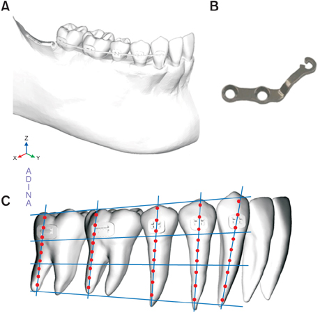
Figure 1 Construction of the finite element mode. A, Teeth and mandibular model with brackets and an archwire. B, A modified L type mini-plate. C, Axes of the finite element model and selected points (1–9) of displacement values evaluation. Y axis, anterior (+) to posterior (−) direction; X axis, right (+) to left (−) direction; Z axis, superior (+) to inferior (−) direction.
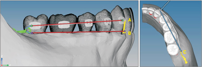
Figure 2 The vertical and horizontal position and direction of force vector. A, The bracket level force was engaged from the center of the mandibular canine bracket slot to the disto-center of the mandibular second molar bracket slot (green point). B, The cemento-enamel junction level force was engaged from the vertical arm of the archwire to the cement-enamel junction level of the mandibular second molar (purple point). C, The horizontal force vector on occlusal view was engaged from the center of the mandibular canine bracket slot to the distal end of the mandibular second molar bracket slot.

Figure 3 Teeth movements according to force application at the bracket level. A, Displacement of Y-axis. B, Displacement of X-axis. C, The superimposition of displacement of Z-axis has been magnified 10,000 times (gray, before force application; color, after force application). D, Displacement magnitude on occlusal view.

Figure 4 Teeth movements according to force application at the cemento-enamel junction level. A, Displacement of Y-axis. B, Displacement of X-axis. C, The superimposition of displacement of Z-axis has been magnified 10,000 times (gray, before force application; color, after force application). D, Displacement magnitude on occlusal view.
Cited by 1 articles
-
Total arch distalization with interproximal stripping in a patient with severe crowding
Min-Ho Jung
Korean J Orthod. 2019;49(3):194-201. doi: 10.4041/kjod.2019.49.3.194.
Reference
-
1. Henriques FP, Janson G, Henriques JF, Pupulim DC. Effects of cervical headgear appliance: a systematic review. Dental Press J Orthod. 2015; 20:76–81.
Article2. Hilgers JJ. The pendulum appliance for Class II non-compliance therapy. J Clin Orthod. 1992; 26:706–714.3. Bolla E, Muratore F, Carano A, Bowman SJ. Evaluation of maxillary molar distalization with the distal jet: a comparison with other contemporary methods. Angle Orthod. 2002; 72:481–494.4. Bondemark L, Karlsson I. Extraoral vs intraoral appliance for distal movement of maxillary first molars: a randomized controlled trial. Angle Orthod. 2005; 75:699–706.5. Caprioglio A, Cafagna A, Fontana M, Cozzani M. Comparative evaluation of molar distalization therapy using pendulum and distal screw appliances. Korean J Orthod. 2015; 45:171–179.
Article6. Nishimura M, Sannohe M, Nagasaka H, Igarashi K, Sugawara J. Nonextraction treatment with temporary skeletal anchorage devices to correct a Class II Division 2 malocclusion with excessive gingival display. Am J Orthod Dentofacial Orthop. 2014; 145:85–94.
Article7. Kim SJ, Choi TH, Baik HS, Park YC, Lee KJ. Mandibular posterior anatomic limit for molar distalization. Am J Orthod Dentofacial Orthop. 2014; 146:190–197.
Article8. Poletti L, Silvera AA, Ghislanzoni LT. Dentoalveolar class III treatment using retromolar miniscrew anchorage. Prog Orthod. 2013; 14:7.
Article9. Farret MM, Farret MM, Farret AM. Orthodontic camouflage of skeletal Class III malocclusion with miniplate: a case report. Dental Press J Orthod. 2016; 21:89–98.
Article10. Roberts WE, Viecilli RF, Chang C, Katona TR, Paydar NH. Biology of biomechanics: Finite element analysis of a statically determinate system to rotate the occlusal plane for correction of a skeletal Class III open-bite malocclusion. Am J Orthod Dentofacial Orthop. 2015; 148:943–955.
Article11. Castro LO, Castro IO, de Alencar AH, Valladares-Neto J, Estrela C. Cone beam computed tomography evaluation of distance from cementoenamel junction to alveolar crest before and after nonextraction orthodontic treatment. Angle Orthod. 2016; 86:543–549.
Article12. Ercan E, Celikoglu M, Buyuk SK, Sekerci AE. Assessment of the alveolar bone support of patients with unilateral cleft lip and palate: a cone-beam computed tomography study. Angle Orthod. 2015; 85:1003–1008.
Article13. Roth RH. The straight-wire appliance 17 years later. J Clin Orthod. 1987; 21:632–642.14. Viazis AD. Bioefficient therapy. J Clin Orthod. 1995; 29:552–568.15. Sung SJ, Kim IT, Kook YA, Chun YS, Kim SH, Mo SS. Finite-element analysis of the shift in center of resistance of the maxillary dentition in relation to alveolar bone loss. Korean J Orthod. 2009; 39:278–288.
Article16. Arreghini A, Trigila S, Lombardo L, Siciliani G. Objective assessment of compliance with intra- and extraoral removable appliances. Angle Orthod. 2017; 87:88–95.
Article17. Kook YA, Park JH, Kim Y, Ahn CS, Bayome M. Orthodontic treatment of skeletal class II adolescent with anterior open bite using mini-screws and modified palatal anchorage plate. J Clin Pediatr Dent. 2015; 39:187–192.
Article18. Bilodeau JE. Retreatment of a transfer patient with bialveolar protrusion with mini bone-plate anchorage. Am J Orthod Dentofacial Orthop. 2014; 146:506–513.
Article19. Cha BK, Choi DS, Ngan P, Jost-Brinkmann PG, Kim SM, Jang IS. Maxillary protraction with miniplates providing skeletal anchorage in a growing Class III patient. Am J Orthod Dentofacial Orthop. 2011; 139:99–112.
Article20. Jo AR, Mo SS, Lee KJ, Sung SJ, Chun YS. Finite-element analysis of the center of resistance of the mandibular dentition. Korean J Orthod. 2017; 47:21–30.
Article21. Sung EH, Kim SJ, Chun YS, Park YC, Yu HS, Lee KJ. Distalization pattern of whole maxillary dentition according to force application points. Korean J Orthod. 2015; 45:20–28.
Article22. Sohn HS. Finite element analysis of the retraction movement of maxillary and mandibular dentition using miniscrew [master's thesis]. Seoul: The Catholic University of Korea;2010.23. Chung KR, Kim SH, Choo H, Kook YA, Cope JB. Distalization of the mandibular dentition with mini-implants to correct a Class III malocclusion with a midline deviation. Am J Orthod Dentofacial Orthop. 2010; 137:135–146.
Article24. Tai K, Park JH, Tatamiya M, Kojima Y. Distal movement of the mandibular dentition with temporary skeletal anchorage devices to correct a Class III malocclusion. Am J Orthod Dentofacial Orthop. 2013; 144:715–725.
Article25. Sugawara J, Daimaruya T, Umemori M, Nagasaka H, Takahashi I, Kawamura H, et al. Distal movement of mandibular molars in adult patients with the skeletal anchorage system. Am J Orthod Dentofacial Orthop. 2004; 125:130–138.
Article26. Nakamura A, Teratani T, Itoh H, Sugawara J, Ishikawa H. Photoelastic stress analysis of mandibular molars moved distally with the skeletal anchorage system. Am J Orthod Dentofacial Orthop. 2007; 132:624–629.
Article27. Lai EH, Yao CC, Chang JZ, Chen I, Chen YJ. Three-dimensional dental model analysis of treatment outcomes for protrusive maxillary dentition: comparison of headgear, miniscrew, and miniplate skeletal anchorage. Am J Orthod Dentofacial Orthop. 2008; 134:636–645.
Article28. Consolaro A. Mini-implants and miniplates generate sub-absolute and absolute anchorage. Dental Press J Orthod. 2014; 19:20–23.
Article
- Full Text Links
-
- Actions
-
Cited
- CITED
-
- Close
- Share
-
- Similar articles
-
- Biomechanical analysis for different mandibular total distalization methods with clear aligners: A finite element study
- En masse maxillary dentition distalization with clear aligners using infrazygomatic mini-implants: A finite element study
- Evaluation of the effects of the third molar on distalization and the effects of attachments on distalization and expansion with clear aligners: Three-dimensional finite element study
- Effect of bite force on orthodontic mini-implants in the molar region: Finite element analysis
- A three-dimensional finite element analysis of molar distalization with a palatal plate, pendulum, and headgear according to molar eruption stage
