Effect of bite force on orthodontic mini-implants in the molar region: Finite element analysis
- Affiliations
-
- 1Department of Orthodontics, School of Dentistry, Chonnam National University, Gwangju, Korea.
- 2Department of Orthodontics, Graduate School of Clinical Dentistry, Ewha Womans University, Seoul, Korea. yschun@ewha.ac.kr
- KMID: 2273413
- DOI: http://doi.org/10.4041/kjod.2013.43.5.218
Abstract
OBJECTIVE
To examine the effect of bite force on the displacement and stress distribution of orthodontic mini-implants (OMIs) in the molar region according to placement site, insertion angle, and loading direction.
METHODS
Five finite element models were created using micro-computed tomography (microCT) images of the maxilla and mandible. OMIs were placed at one maxillary and two mandibular positions: between the maxillary second premolar and first molar, between the mandibular second premolar and first molar, and between the mandibular first and second molars. The OMIs were inserted at angles of 45degrees and 90degrees to the buccal surface of the cortical bone. A bite force of 25 kg was applied to the 10 occlusal contact points of the second premolar, first molar, and second molar. The loading directions were 0degrees, 5degrees, and 10degrees to the long axis of the tooth.
RESULTS
With regard to placement site, the displacement and stress were greatest for the OMI placed between the mandibular first molar and second molar, and smallest for the OMI placed between the maxillary second premolar and first molar. In the mandibular molar region, the angled OMI showed slightly less displacement than the OMI placed at 90degrees. The maximum Von Mises stress increased with the inclination of the loading direction.
CONCLUSIONS
These results suggest that placement of OMIs between the second premolar and first molar at 45degrees to the cortical bone reduces the effect of bite force on OMIs.
Figure
-
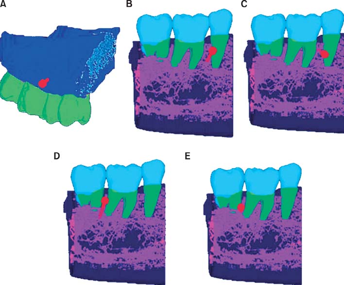
Figure 1 The five finite element models. A, Orthodontic mini-implant (OMI) placed between the maxillary second premolar and first molar at 45°; B, OMI placed between the mandibular second premolar and first molar at 45°; C, OMI placed between the mandibular second premolar and first molar at 90°; D, OMI placed between the mandibular first and second molars at 45°; E, OMI placed between the mandibular first and second molars at 90°.
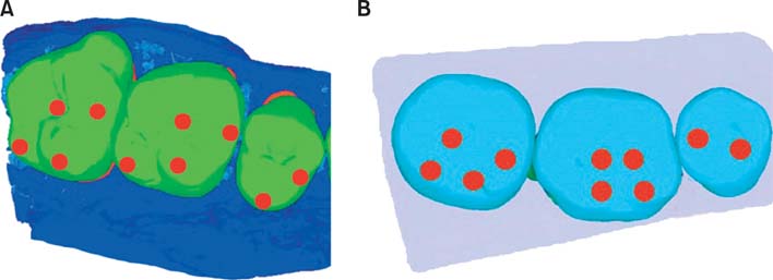
Figure 2 The 10 occlusal contact points used in the current study. A, Maxilla; B, mandible.
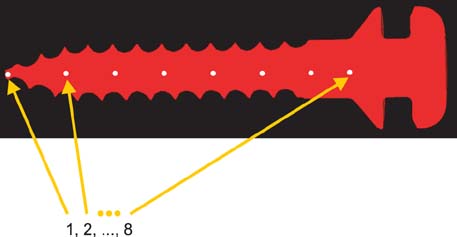
Figure 3 The eight reference nodes of the orthodontic mini-implant.

Figure 4 Displacements (A) and Von Mises stress distributions (B) of orthodontic mini-implants (OMIs) in models A, B, and D. The X-axis indicates sequential points along the OMI from the tip to the head. A, Positive Y-axis values indicate occlusal movement of the OMI, and negative Y-axis values indicate apical movement.

Figure 5 Displacements (A) and Von Mises stress distributions (B) of orthodontic mini-implants (OMIs) in models B, C, D, and E. The X-axis indicates sequential points along the OMI from the tip to the head. A, Positive Y-axis values indicate occlusal movements of the OMI, and negative Y-axis values indicate apical movements.

Figure 6 Maximum Von Mises stress of the orthodontic mini-implants in the five models according to different loading directions.
Cited by 1 articles
-
Analysis of time to failure of orthodontic mini-implants after insertion or loading
Jong-Wha Jeong, Jong-Wan Kim, Nam-Ki Lee, Young-Kyun Kim, Jong-Ho Lee, Tae-Woo Kim
J Korean Assoc Oral Maxillofac Surg. 2015;41(5):240-245. doi: 10.5125/jkaoms.2015.41.5.240.
Reference
-
1. Kanomi R. Mini-implant for orthodontic anchorage. J Clin Orthod. 1997; 31:763–767.2. Bae SM, Park HS, Kyung HM, Kwon OW, Sung JH. Clinical application of micro-implant anchorage. J Clin Orthod. 2002; 36:298–302.3. Miyawaki S, Koyama I, Inoue M, Mishima K, Sugahara T, Takano-Yamamoto T. Factors associated with the stability of titanium screws placed in the posterior region for orthodontic anchorage. Am J Orthod Dentofacial Orthop. 2003; 124:373–378.
Article4. Cheng SJ, Tseng IY, Lee JJ, Kok SH. A prospective study of the risk factors associated with failure of mini-implants used for orthodontic anchorage. Int J Oral Maxillofac Implants. 2004; 19:100–106.5. Lim HJ, Eun CS, Cho JH, Lee KH, Hwang HS. Factors associated with initial stability of miniscrews for orthodontic treatment. Am J Orthod Dentofacial Orthop. 2009; 136:236–242.
Article6. Kuroda S, Sugawara Y, Deguchi T, Kyung HM, Takano-Yamamoto T. Clinical use of miniscrew implants as orthodontic anchorage: success rates and postoperative discomfort. Am J Orthod Dentofacial Orthop. 2007; 131:9–15.
Article7. Deguchi T, Takano-Yamamoto T, Kanomi R, Hartsfield JK Jr, Roberts WE, Garetto LP. The use of small titanium screws for orthodontic anchorage. J Dent Res. 2003; 82:377–381.
Article8. Chen YJ, Chang HH, Huang CY, Hung HC, Lai EH, Yao CC. A retrospective analysis of the failure rate of three different orthodontic skeletal anchorage systems. Clin Oral Implants Res. 2007; 18:768–775.
Article9. Motoyoshi M, Yoshida T, Ono A, Shimizu N. Effect of cortical bone thickness and implant placement torque on stability of orthodontic mini-implants. Int J Oral Maxillofac Implants. 2007; 22:779–784.10. Kuroda S, Yamada K, Deguchi T, Hashimoto T, Kyung HM, Takano-Yamamoto T. Root proximity is a major factor for screw failure in orthodontic anchorage. Am J Orthod Dentofacial Orthop. 2007; 131:S68–S73.
Article11. Cattaneo PM, Dalstra M, Melsen B. The transfer of occlusal forces through the maxillary molars: a finite element study. Am J Orthod Dentofacial Orthop. 2003; 123:367–373.
Article12. Tanne K, Sakuda M, Burstone CJ. Three-dimensional finite element analysis for stress in the periodontal tissue by orthodontic forces. Am J Orthod Dentofacial Orthop. 1987; 92:499–505.
Article13. Miyaura K, Morita M, Matsuka Y, Yamashita A, Watanabe T. Rehabilitation of biting abilities in patients with different types of dental prostheses. J Oral Rehabil. 2000; 27:1073–1076.
Article14. Molly L. Bone density and primary stability in implant therapy. Clin Oral Implants Res. 2006; 17:Suppl 2. 124–135.
Article15. Javed F, Romanos GE. The role of primary stability for successful immediate loading of dental implants. A literature review. J Dent. 2010; 38:612–620.
Article16. Provatidis CG. A comparative FEM-study of tooth mobility using isotropic and anisotropic models of the periodontal ligament. Finite Element Method. Med Eng Phys. 2000; 22:359–370.
Article17. Qian H, Chen J, Katona TR. The influence of PDL principal fibers in a 3-dimensional analysis of orthodontic tooth movement. Am J Orthod Dentofacial Orthop. 2001; 120:272–279.
Article18. Toms SR, Eberhardt AW. A nonlinear finite element analysis of the periodontal ligament under orthodontic tooth loading. Am J Orthod Dentofacial Orthop. 2003; 123:657–665.
Article19. Farnsworth D, Rossouw PE, Ceen RF, Buschang PH. Cortical bone thickness at common miniscrew implant placement sites. Am J Orthod Dentofacial Orthop. 2011; 139:495–503.
Article20. Motoyoshi M, Ueno S, Okazaki K, Shimizu N. Bone stress for a mini-implant close to the roots of adjacent teeth- 3D finite element analysis. Int J Oral Maxillofac Surg. 2009; 38:363–368.
Article21. Meredith N. Assessment of implant stability as a prognostic determinant. Int J Prosthodont. 1998; 11:491–501.22. Byoun NY, Nam EH, Yoon YA, Kim IK. Three-dimensional finite element analysis for stress distribution on the diameter of orthodontic mini-implants and insertion angle to the bone surface. Korean J Orthod. 2006; 36:178–187.23. Lee NK, Choi DS, Jang IS, Cha BK. Factors influencing primary stability of miniplate anchorage: a three-dimensional finite element analysis. Korean J Orthod. 2008; 38:304–313.
Article24. Sugiura T, Horiuchi K, Sugimura M, Tsutsumi S. Evaluation of threshold stress for bone resorption around screws based on in vivo strain measurement of miniplate. J Musculoskelet Neuronal Interact. 2000; 1:165–170.25. Li J, Li H, Shi L, Fok AS, Ucer C, Devlin H, et al. A mathematical model for simulating the bone remodeling process under mechanical stimulus. Dent Mater. 2007; 23:1073–1078.
Article26. Canabarro Sde A, Shinkai RS. Medial mandibular flexure and maximum occlusal force in dentate adults. Int J Prosthodont. 2006; 19:177–182.27. Goodkind RJ, Heringlake CB. Mandibular flexure in opening and closing movements. J Prosthet Dent. 1973; 30:134–138.
Article28. Korioth TW, Hannam AG. Deformation of the human mandible during simulated tooth clenching. J Dent Res. 1994; 73:56–66.
Article29. Yoon HJ, Lim YK, Lee DY, Jo YS. Three-dimensional finite element analysis on the effect of maxillary incisor torque. Korean J Orthod. 2005; 35:137–147.
- Full Text Links
-
- Actions
-
Cited
- CITED
-
- Close
- Share
-
- Similar articles
-
- Three-dimensional finite element analysis of the deformation of the human mandible: a preliminary study from the perspective of orthodontic mini-implant stability
- Three-dimensional finite element analysis for stress distribution on the diameter of orthodontic mini-implants and insertion angle to the bone surface
- Biomechanical analysis of distalization of mandibular molars by placing a mini-plate: A finite element study
- The pattern of movement and stress distribution during retraction of maxillary incisors using a 3-D finite element method
- Effects of orthodontic mini-implant position in the dragon helix appliance on tooth displacement and stress distribution: a three-dimensional finite element analysis
