Ann Dermatol.
2017 Oct;29(5):626-629. 10.5021/ad.2017.29.5.626.
Histiocytoid Sweet Syndrome in a Child without Underlying Systemic Disease
- Affiliations
-
- 1Department of Dermatology, Inha University School of Medicine, Incheon, Korea. mikie2001@hanmail.net
- KMID: 2388917
- DOI: http://doi.org/10.5021/ad.2017.29.5.626
Abstract
- Sweet syndrome (acute, febrile, neutrophilic dermatosis) is characterized by the acute onset of an eruption of painful nodules or erythematous or violaceous plaques on the limbs, face and neck. These symptoms are accompanied by fever. The diagnostic features include histopathological findings of dermal neutrophilic infiltration without leukocytoclastic vasculitis or peripheral blood leukocytosis. Sweet syndrome is associated with infection, malignancies, autoimmune disease, pregnancy, and drugs. Patients with Sweet syndrome demonstrate a complete and rapid response to systemic steroid administration. Recently, a distinct variant of Sweet syndrome was reported, termed "histiocytoid Sweet syndrome", in which the infiltration of myeloperoxidase-positive histiocytoid mononuclear cells are observed (in contrast to the infiltration of neutrophils). The other clinical features are similar to those of classic Sweet syndrome. Pediatric Sweet syndrome is uncommon, and the histiocytoid type is even rarer. To date, four cases of histiocytoid Sweet syndrome have been reported in children. Herein, we describe a case of histiocytoid Sweet syndrome in an otherwise healthy 10-year-old boy with no underlying systemic disease in whom non-steroidal, anti-inflammatory drug treatment was successful.
MeSH Terms
Figure
-
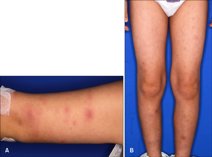
Fig. 1 Nontender nonpruritic erythematous macules and nodule
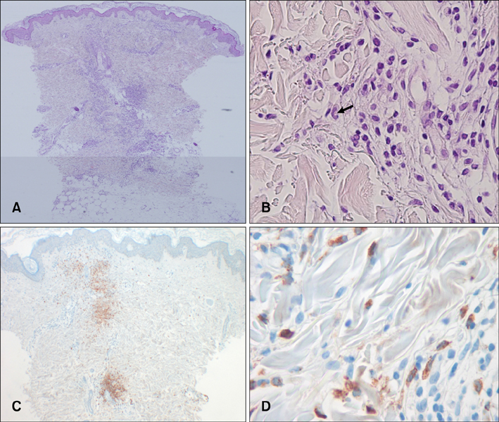
Fig. 2 (A, B) Infiltration of perivascular lymphocytes and large mononuclear cells with kidney shaped nuclei (arrow) in the dermis (H&E; A: ×40, B: ×400). (C, D) The presence of myeloperoxidase (MPO)-positive mononuclear cells in the dermis (MPO; C: ×40, D: ×400).
Reference
-
1. Sweet RD. An acute febrile neutrophilic dermatosis. Br J Dermatol. 1964; 76:349–356.2. Cohen PR, Kurzrock R. Sweet's syndrome revisited: a review of disease concepts. Int J Dermatol. 2003; 42:761–778.
Article3. Jeanfils S, Joly P, Young P, Le Corvaisier-Pieto C, Thomine E, Lauret P. Indomethacin treatment of eighteen patients with Sweet's syndrome. J Am Acad Dermatol. 1997; 36:436–439.
Article4. Liu CI, Hsiao CH, Wu JT, Tsai TF. Sweet syndrome with histiocytoid infiltrate and neutropenia: a rare combination. J Am Acad Dermatol. 2009; 61:882–884.
Article5. Cooper PH, Innes DJ Jr, Greer KE. Acute febrile neutrophilic dermatosis (Sweet's syndrome) and myeloproliferative disorders. Cancer. 1983; 51:1518–1526.
Article6. Su WP, Liu HN. Diagnostic criteria for Sweet's syndrome. Cutis. 1986; 37:167–174.7. von den Driesch P. Sweet's syndrome (acute febrile neutrophilic dermatosis). J Am Acad Dermatol. 1994; 31:535–556.
Article8. Chavan RN, Cappel MA, Ketterling RP, Wada DA, Rochet NM, Knudson R, et al. Histiocytoid Sweet syndrome may indicate leukemia cutis: a novel application of fluorescence in situ hybridization. J Am Acad Dermatol. 2014; 70:1021–1027.
Article9. Fernández-Torres RM, Castro S, Moreno A, Alvarez R, Fonseca E. Subcutaneous histiocytoid sweet syndrome associated with crohn disease in an adoloscent. Case Rep Dermatol Med. 2014; 2014:954254.10. Uihlein LC, Brandling-Bennett HA, Lio PA, Liang MG. Sweet syndrome in children. Pediatr Dermatol. 2012; 29:38–44.
Article11. Srisuttiyakorn C, Reeve J, Reddy S, Imaeda S, Lazova R. Subcutaneous histiocytoid Sweet's syndrome in a patient with myelodysplastic syndrome and acute myeloblastic leukemia. J Cutan Pathol. 2014; 41:475–479.
Article12. Kawakami T, Ohashi S, Kawa Y, Takahama H, Ito M, Soma Y, et al. Elevated serum granulocyte colony-stimulating factor levels in patients with active phase of sweet syndrome and patients with active behcet disease: implication in neutrophil apoptosis dysfunction. Arch Dermatol. 2004; 140:570–574.13. Peroni A, Colato C, Schena D, Rongioletti F, Girolomoni G. Histiocytoid Sweet syndrome is infiltrated predominantly by M2-like macrophages. J Am Acad Dermatol. 2015; 72:131–139.
Article14. Llamas-Velasco M, Concha-Garzón MJ, Fraga J, Aragüés M. Histiocytoid Sweet syndrome related to bortezomib: a mimicker of cutaneous infiltration by myeloma. Indian J Dermatol Venereol Leprol. 2015; 81:305–306.
Article15. Huang CF, Wu BY, Liaw FY, Wang WM, Chiang CP. Histiocytoid Sweet syndrome: report of two cases and review of the literature. Dermatologica Sinica. 2012; 30:71–74.
Article16. Anzalone CL, Cohen PR. Acute febrile neutrophilic dermatosis (Sweet's syndrome). Curr Opin Hematol. 2013; 20:26–35.
Article17. Hospach T, von den Driesch P, Dannecker GE. Acute febrile neutrophilic dermatosis (Sweet's syndrome) in childhood and adolescence: two new patients and review of the literature on associated diseases. Eur J Pediatr. 2009; 168:1–9.
Article18. Cho-Vega JH, Medeiros LJ, Prieto VG, Vega F. Leukemia cutis. Am J Clin Pathol. 2008; 129:130–142.
Article19. Camarillo D, McCalmont TH, Frieden IJ, Gilliam AE. Two pediatric cases of nonbullous histiocytoid neutrophilic dermatitis presenting as a cutaneous manifestation of lupus erythematosus. Arch Dermatol. 2008; 144:1495–1498.
Article
- Full Text Links
-
- Actions
-
Cited
- CITED
-
- Close
- Share
-
- Similar articles
-
- A Case of Histiocytoid Sweet's Syndrome with Myelodysplastic Syndrome
- A Case of Malignancy-associated Histiocytoid Sweet Syndrome in a Patient with AML
- Bortezomib-induced Histiocytoid Sweet Syndrome
- A Case of Histiocytoid Sweet Syndrome Developed in the Patient with Pure White Cell Aplasia
- A Case Report of Sweet's Syndrome with Parotitis
