Diagnosis of Lymphoid Malignancy by PCR for Analysis of Antigen Receptor Rearrangement after Blood Transfusion in a Dog with Acute Lymphocytic Leukemia
- Affiliations
-
- 1Animal Diseases and Biosecurity Team, National Institute of Animal Science, Rural Development Administration, Wanju 55365, Korea.
- 2Department of Veterinary Laboratory Medicine, College of Veterinary Medicine, Chonnam National University, Gwangju 61186, Korea. dyu@jnu.ac.kr
- 3Department of Companion & Laboratory Animal Science, College of Industrial Science, Kongju National University, Yesan 32439, Korea.
- 4Department of Veterinary Medical Imaging, College of Veterinary Medicine, Chonnam National University, Gwangju 61186, Korea.
- 5Department of Veterinary Internal Medicine, College of Veterinary Medicine, Chonbuk National University, Iksan 54596, Korea.
- KMID: 2388081
- DOI: http://doi.org/10.4110/in.2017.17.4.269
Abstract
- Acute lymphocytic leukemia (ALL) is uncommon lymphoid malignancy in dogs, and its diagnosis is challenging. A 14-year-old spayed female mixed breed dog was transferred to a veterinary medical teaching hospital for an immediate blood transfusion. The dog showed lethargy, pale mucous membranes, and a weak femoral pulse. Complete blood count revealed non-regenerative anemia and severe leukopenia with thrombocytopenia. ALL was tentatively diagnosed based on the predominance of immature lymphoblasts on blood film examination. For confirmation of lymphoid malignancy, PCR for antigen receptor rearrangement (PARR) on a peripheral blood sample and flow cytometry analysis were performed after blood transfusion. Flow cytometry analysis revealed that lymphocyte subsets were of normal composition, but PARR detected a T-cell malignancy. The dog was diagnosed with ALL and survived 1 wk after diagnosis. In conclusion, after blood transfusion, flow cytometry was not a reliable diagnostic method for an ALL dog, whereas PARR could detect lymphoid malignancy. Our results suggest that PARR should be the first-line diagnostic tool to detect canine lymphoid malignancy after a blood transfusion.
MeSH Terms
-
Adolescent
Anemia
Animals
Blood Cell Count
Blood Transfusion*
Diagnosis*
Dogs*
Female
Flow Cytometry
Hospitals, Teaching
Humans
Lethargy
Leukopenia
Lymphocyte Subsets
Methods
Mucous Membrane
Polymerase Chain Reaction*
Precursor Cell Lymphoblastic Leukemia-Lymphoma*
Receptors, Antigen*
T-Lymphocytes
Thrombocytopenia
Receptors, Antigen
Figure
-
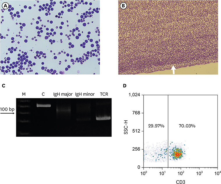
Figure 1 (A) Microscopic examination of peripheral blood prior to administration. (B) Microscopic examination of feathered edge of blood film on admission. (C) PCR for analysis of antigen receptor rearrangement from peripheral blood. First lane ‘M’ shows a 25 bp size marker, lane ‘C’ indicates positive control for DNA, lane 3 and 4 detect “major” and “minor” immunoglobulin rearrangements, respectively, and lane 5 (TCR) detects TCRγ gene rearrangement. (D) A dot plot from flow cytometry analysis. Immature lymphoid cells were prominent prior to administration but microscopic examination was not easy because of severe leukopenia on admission. PARR revealed positive for clonal rearrangement of TCRγ gene but flow cytometry did not show overt T cell predominance after blood transfusion in this dog. TCR, T-cell receptor; PARR, PCR for antigen receptor rearrangement.
Reference
-
1. Leifer CE, Matus RE. Lymphoid leukemia in the dog. Acute lymphoblastic leukemia and chronic lymphocytic leukemia. Vet Clin North Am Small Anim Pract. 1985; 15:723–739.
Article2. Adam F, Villiers E, Watson S, Coyne K, Blackwood L. Clinical pathological and epidemiological assessment of morphologically and immunologically confirmed canine leukaemia. Vet Comp Oncol. 2009; 7:181–195.
Article3. Tasca S, Carli E, Caldin M, Menegazzo L, Furlanello T, Gallego LS. Hematologic abnormalities and flow cytometric immunophenotyping results in dogs with hematopoietic neoplasia: 210 cases (2002–2006). Vet Clin Pathol. 2009; 38:2–12.
Article4. Yu DH, Li YH, Lee JH, Noh DH, Song RH, Lee MJ, Choi US, Park JH. Detection of canine lymphoma by the amplification of antigen receptor gene rearrangements. J Vet Clin. 2009; 26:419–422.5. Burnett RC, Vernau W, Modiano JF, Olver CS, Moore PF, Avery AC. Diagnosis of canine lymphoid neoplasia using clonal rearrangements of antigen receptor genes. Vet Pathol. 2003; 40:32–41.
Article6. Lana SE, Jackson TL, Burnett RC, Morley PS, Avery AC. Utility of polymerase chain reaction for analysis of antigen receptor rearrangement in staging and predicting prognosis in dogs with lymphoma. J Vet Intern Med. 2006; 20:329–334.
Article7. Morris J, Dobson JM. Haematopoietic system. In : Morris J, Dobson JM, editors. Small Animal Oncology. Malden, MA: Blackwell Science;2001. p. 228–251.8. Young KM, Vail DM. Canine acute myeloid leukemia, chronic myeloproliferative diseases, and myelodysplasia. In : Withrow SJ, MacEwen EG, Vail DM, editors. Withrow & MacEwen's Small Animal Clinical Oncology. 4th ed. St. Louis, MO: Saunders Elsevier;2007. p. 756–768.9. Avery AC, Avery PR. Determining the significance of persistent lymphocytosis. Vet Clin North Am Small Anim Pract. 2007; 37:267–282. vi
Article10. Vernau W, Moore PF. An immunophenotypic study of canine leukemias and preliminary assessment of clonality by polymerase chain reaction. Vet Immunol Immunopathol. 1999; 69:145–164.
Article11. Villiers E, Baines S, Law AM, Mallows V. Identification of acute myeloid leukemia in dogs using flow cytometry with myeloperoxidase, MAC387, and a canine neutrophil-specific antibody. Vet Clin Pathol. 2006; 35:55–71.
Article
- Full Text Links
-
- Actions
-
Cited
- CITED
-
- Close
- Share
-
- Similar articles
-
- Immunoglobulin and T-cell receptor gene rearrangement analysis in malignant lymphoid neoplasms
- Acute lymphoblastic leukemia and chronic lymphocytic leukemia in dogs
- Simultaneous Reverse Transcription-Polymerase Chain Reaction for Detection of 7 Gene Rearrangements in Acute Leukemia
- A Case of CD7+, CD4-, CD8-, CD3-acute T cell lymphoblastic leukemia
- A case of biphenotypic acute leukemia with expression of the AML1-ETO gene rearrangement
