Investig Clin Urol.
2017 Sep;58(5):324-330. 10.4111/icu.2017.58.5.324.
Does T1- and diffusion-weighted magnetic resonance imaging give value-added than bone scintigraphy in the follow-up of vertebral metastasis of prostate cancer?
- Affiliations
-
- 1Department of Urology, Pusan National University Yangsan Hospital, Pusan National University School of Medicine, Yangsan, Korea. psw@pusan.ac.kr
- 2Department of Radiology, Pusan National University Yangsan Hospital, Pusan National University School of Medicine, Yangsan, Korea.
- 3Department of Nuclear Medicine, Pusan National University Yangsan Hospital, Pusan National University School of Medicine, Yangsan, Korea.
- 4Research Institute for Convergence of Biomedical Science and Technology, Pusan National University Yangsan Hospital, Yangsan, Korea.
- KMID: 2388048
- DOI: http://doi.org/10.4111/icu.2017.58.5.324
Abstract
- PURPOSE
To evaluate the effectiveness of limited Magnetic Resonance (MR) images including T1- and diffusion-weighted image (DWI) for monitoring vertebral metastasis in patients with prostate cancer.
MATERIALS AND METHODS
From July 2014 to November 2016, patients diagnosed with spinal metastasis from prostate cancer using 99mTc bone scintigraphy were enrolled. Regardless of the primary local therapy, the changes in spinal metastasis were followed up using bone scan and biparametric MR (T1+DWI). All tests were followed up for more than 3 months.
RESULTS
Among the 14 follow-ups of 10 patients, 6 and 10 (including all progressed cases on bone scan) follow-ups were determined to show progressive disease using bone scan and biparametric MR, respectively. Otherwise, we could have predicted neurologic sequela earlier using biparametric MR. Examination time for biparametric MR was 15 minutes, and it was 4 hours for bone scan, respectively.
CONCLUSIONS
Although bone scan has been considered the standard test for bony metastasis in men with prostate cancer, limited MR including T1 and DWI has an additional benefit in monitoring spinal metastasis in patients who are already diagnosed as having spinal metastasis. The limited MR is more sensitive in detecting progressive disease. In addition, it can reduce neurologic complications caused by spinal metastasis.
MeSH Terms
Figure
-
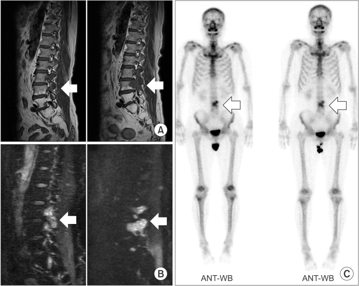
Fig. 1 The images of the 1st–2nd follow-up in the Table 1. Each right figure shows the 3-month follow-up images. T1-weighted image (A), diffusion-weighted image (B), and bone scan (C). ANT-WB, anterior view-whole-body.
Reference
-
1. Johansson JE, Andrén O, Andersson SO, Dickman PW, Holmberg L, Magnuson A, et al. Natural history of early, localized prostate cancer. JAMA. 2004; 291:2713–2719. PMID: 15187052.
Article2. Heidenreich A, Bastian PJ, Bellmunt J, Bolla M, Joniau S, van der Kwast T, et al. EAU guidelines on prostate cancer. part 1: screening, diagnosis, and local treatment with curative intent-update 2013. Eur Urol. 2014; 65:124–137. PMID: 24207135.
Article3. Kirby M, Hirst C, Crawford ED. Characterising the castration-resistant prostate cancer population: a systematic review. Int J Clin Pract. 2011; 65:1180–1192. PMID: 21995694.
Article4. Zacho HD, Barsi T, Mortensen JC, Mogensen MK, Bertelsen H, Josephsen N, et al. Prospective multicenter study of bone scintigraphy in consecutive patients with newly diagnosed prostate cancer. Clin Nucl Med. 2014; 39:26–31. PMID: 24217537.
Article5. Berg KD, Thomsen FB, Mikkelsen MK, Ingimarsdóttir IJ, Hansen RB, Kejs AM, et al. Improved survival for patients with de novo metastatic prostate cancer in the last 20 years. Eur J Cancer. 2017; 72:20–27. PMID: 28024263.
Article6. Beer TM, Armstrong AJ, Rathkopf DE, Loriot Y, Sternberg CN, Higano CS, et al. Enzalutamide in metastatic prostate cancer before chemotherapy. N Engl J Med. 2014; 371:424–433. PMID: 24881730.
Article7. Ryan CJ, Smith MR, de Bono JS, Molina A, Logothetis CJ, de Souza P, et al. Abiraterone in metastatic prostate cancer without previous chemotherapy. N Engl J Med. 2013; 368:138–148. PMID: 23228172.
Article8. Heidenreich A, Bastian PJ, Bellmunt J, Bolla M, Joniau S, van der Kwast T, et al. EAU guidelines on prostate cancer. Part II: Treatment of advanced, relapsing, and castration-resistant prostate cancer. Eur Urol. 2014; 65:467–479. PMID: 24321502.
Article9. Loblaw A, Mitera G. Malignant extradural spinal cord compression in men with prostate cancer. Curr Opin Support Palliat Care. 2011; 5:206–210. PMID: 21725245.
Article10. Bayley A, Milosevic M, Blend R, Logue J, Gospodarowicz M, Boxen I, et al. A prospective study of factors predicting clinically occult spinal cord compression in patients with metastatic prostate carcinoma. Cancer. 2001; 92:303–310. PMID: 11466683.
Article11. Venkitaraman R, Sohaib SA, Barbachano Y, Parker CC, Khoo V, Huddart RA, et al. Detection of occult spinal cord compression with magnetic resonance imaging of the spine. Clin Oncol (R Coll Radiol). 2007; 19:528–531. PMID: 17499490.
Article12. Gabriele D, Collura D, Oderda M, Stura I, Fiorito C, Porpiglia F, et al. Is there still a role for computed tomography and bone scintigraphy in prostate cancer staging? An analysis from the EUREKA-1 database. World J Urol. 2016; 34:517–523. PMID: 26276152.
Article13. Pasoglou V, Larbi A, Collette L, Annet L, Jamar F, Machiels JP, et al. One-step TNM staging of high-risk prostate cancer using magnetic resonance imaging (MRI): toward an upfront simplified "all-in-one" imaging approach? Prostate. 2014; 74:469–477. PMID: 24375774.
Article14. Lecouvet FE, El Mouedden J, Collette L, Coche E, Danse E, Jamar F, et al. Can whole-body magnetic resonance imaging with diffusion-weighted imaging replace Tc 99m bone scanning and computed tomography for single-step detection of metastases in patients with high-risk prostate cancer? Eur Urol. 2012; 62:68–75. PMID: 22366187.
Article15. Tombal B, Rezazadeh A, Therasse P, Van Cangh PJ, Vande Berg B, Lecouvet FE. Magnetic resonance imaging of the axial skeleton enables objective measurement of tumor response on prostate cancer bone metastases. Prostate. 2005; 65:178–187. PMID: 15948151.
Article16. Daldrup-Link HE, Franzius C, Link TM, Laukamp D, Sciuk J, Jürgens H, et al. Whole-body MR imaging for detection of bone metastases in children and young adults: comparison with skeletal scintigraphy and FDG PET. AJR Am J Roentgenol. 2001; 177:229–236. PMID: 11418435.17. Gosfield E 3rd, Alavi A, Kneeland B. Comparison of radionuclide bone scans and magnetic resonance imaging in detecting spinal metastases. J Nucl Med. 1993; 34:2191–2198. PMID: 8254410.18. Eisenhauer EA, Therasse P, Bogaerts J, Schwartz LH, Sargent D, Ford R, et al. New response evaluation criteria in solid tumours: revised RECIST guideline (version 1.1). Eur J Cancer. 2009; 45:228–247. PMID: 19097774.
Article19. Halabi S, Kelly WK, Ma H, Zhou H, Solomon NC, Fizazi K, et al. Meta-analysis evaluating the impact of site of metastasis on overall survival in men with castration-resistant prostate cancer. J Clin Oncol. 2016; 34:1652–1659. PMID: 26951312.
Article20. Freedland SJ, Humphreys EB, Mangold LA, Eisenberger M, Dorey FJ, Walsh PC, et al. Risk of prostate cancer-specific mortality following biochemical recurrence after radical prostatectomy. JAMA. 2005; 294:433–439. PMID: 16046649.
Article21. Heidenreich A, Aus G, Bolla M, Joniau S, Matveev VB, Schmid HP, et al. EAU guidelines on prostate cancer. Eur Urol. 2008; 53:68–80. PMID: 17920184.
Article22. Briganti A, Passoni N, Ferrari M, Capitanio U, Suardi N, Gallina A, et al. When to perform bone scan in patients with newly diagnosed prostate cancer: external validation of the currently available guidelines and proposal of a novel risk stratification tool. Eur Urol. 2010; 57:551–558. PMID: 20034730.
Article23. Smith MR, Kabbinavar F, Saad F, Hussain A, Gittelman MC, Bilhartz DL, et al. Natural history of rising serum prostate-specific antigen in men with castrate nonmetastatic prostate cancer. J Clin Oncol. 2005; 23:2918–2925. PMID: 15860850.
Article24. Mottet N, Bellmunt J, Bolla M, Briers E, Cumberbatch MG, De Santis M, et al. EAU-ESTRO-SIOG guidelines on prostate cancer. Part 1: Screening, diagnosis, and local treatment with curative intent. Eur Urol. 2017; 71:618–629. PMID: 27568654.
Article25. Cornford P, Bellmunt J, Bolla M, Briers E, De Santis M, Gross T, et al. EAU-ESTRO-SIOG guidelines on prostate cancer. Part II: Treatment of relapsing, metastatic, and castration-resistant prostate cancer. Eur Urol. 2017; 71:630–642. PMID: 27591931.
Article26. Lecouvet FE, Simon M, Tombal B, Jamart J, Vande Berg BC, Simoni P. Whole-body MRI (WB-MRI) versus axial skeleton MRI (AS-MRI) to detect and measure bone metastases in prostate cancer (PCa). Eur Radiol. 2010; 20:2973–2982. PMID: 20661742.
Article27. Venkitaraman R, Cook GJ, Dearnaley DP, Parker CC, Khoo V, Eeles R, et al. Whole-body magnetic resonance imaging in the detection of skeletal metastases in patients with prostate cancer. J Med Imaging Radiat Oncol. 2009; 53:241–247. PMID: 19624290.
Article28. Schaefer JF, Schlemmer HP. Total-body MR-imaging in oncology. Eur Radiol. 2006; 16:2000–2015. PMID: 16622688.
Article
- Full Text Links
-
- Actions
-
Cited
- CITED
-
- Close
- Share
-
- Similar articles
-
- RE: Diffusion-Weighted Imaging of Prostate Cancer: How Can We Use It Accurately?
- Diffusion-Weighted Magnetic Resonance Imaging of Spine
- Diagnosis of Spinal Metastasis: Usefulness of Additional Diffusion-Weighted Imaging
- Giant Vertebral Notochordal Rest: Magnetic Resonance and Diffusion Weighted Imaging Findings
- Bilateral Medial Medullary Infarction Demonstrated by Diffusion-Weighted Imaging: Case Report
