In vitro evaluation methods on adaptation of fixed dental prosthesis
- Affiliations
-
- 1Department of Prosthodontics, College of Dentistry, Kyungpook National University, Daegu, Republic of Korea. kblee@knu.ac.kr
- 2Advanced Dental Device Development Institute, Kyungpook National University, Daegu, Republic of Korea.
- KMID: 2388011
- DOI: http://doi.org/10.14368/jdras.2017.33.2.63
Abstract
- Clinically, the fit of fixed prosthesis is an essential element for successful restoration. The fit of prosthesis is largely classified into marginal fit and internal fit, and various methods to assess these have been introduced including microscopic margin measurement, cross-sectional measurement, silicone replica technique, 3-dimensional scanning data superposition, weight technique and micro CT scanning. Thus, this study is aimed at proposing a more convenient and accurate measurement method of fits in a digital environment by comparatively analyzing the advantages and disadvantages of each known method based on existing literature.
Keyword
MeSH Terms
Figure
-

Fig. 1 Casting misfit terminology.
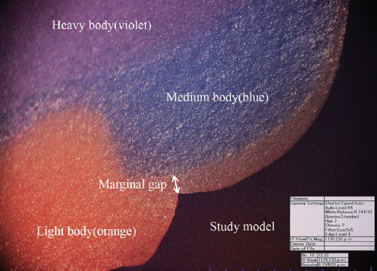
Fig. 2 Measurement of marginal gap by digital microscope at ×160 magnification (orange color: light body silicone; blue color: medium body silicon; violet color: heavy body silicone).
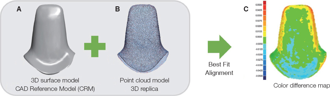
Fig. 3 Three-dimensional replica technique. (A) The 3D surface model from the digitization of the study model used as the control model (CAD reference model; CRM), (B) The point cloud model is the digitization of the light body silicone, (C) The point cloud model is projected onto the surface of the CRM. The distribution of the internal gaps was measured and depicted on the color difference map.
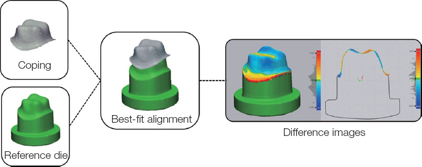
Fig. 4 Three-dimensional analysis procedure. For three- dimensional qualitative evaluation of the discrepancies in the difference between CAD-reference die and the digitized coping data, best-fit alignment was conducted in the program (Control X, Geomagic GmbH, Stuttgart, Germany) and a color-difference map was made with the superimposition process.
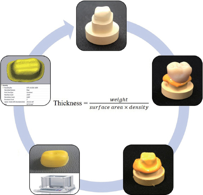
Fig. 5 Weight technique procedure. (A) Abutment, (B) Fitting of the crown to the abutment after application of light body silicon, (C) Crown removed from B, (D) Measurement of the weight corresponding to the internal gap.
Cited by 2 articles
-
Comparative evaluation of marginal and internal fit of metal copings fabricated by various CAD/CAM methods
Seung-Jin Jeong, Hye-Won Cho, Ji-Hye Jung, Jeong-Mi Kim, Yu-Lee Kim
J Korean Acad Prosthodont. 2019;57(3):211-218. doi: 10.4047/jkap.2019.57.3.211.Effect of abutment superimposition process of dental model scanner on final virtual model
Beom-Young Yu, Keunbada Son, Kyu-Bok Lee
J Korean Acad Prosthodont. 2019;57(3):203-210. doi: 10.4047/jkap.2019.57.3.203.
Reference
-
References
1. Kim MY, Lee KW, Moon HS, Chung MK. A study on the gratification of the patient in the Dental Hospital. J Korean Acad Prosthodont. 2008; 46:6582. PMID: 8349002.2. Rekow ED. High-technology innovations-and limitationsfor restorative dentistry. Dent Clin North Am. 1993; 37:513–24.3. Hung SH, Hung KS, Eick JD, Chappell RP. Marginal fit of porcelain-fused-to-metal and two types of ceramic crown. J Prosthet Dent. 1990; 63:26–31. DOI: 10.1016/0022-3913(90)90260-J.4. Schwartz NL, Whitsett LD, Berry TG, Stewart JL. Unserviceable crowns and fixed partial dentures:life-span and causes for loss of serviceability. J Am Dent Assoc. 1970; 81:1395–401. DOI: 10.14219/jada.archive.1970.0398. PMID: 5273607.5. Grasso JE, Nalbandian J, Sanford C, Bailit H. Effect of restoration quality on periodontal health. J Prosthet Dent. 1985; 53:14–9. DOI: 10.1016/0022-3913(85)90056-3.6. Holmes JR, Bayne SC, Holland GA, Sulik WD. Considerations in measurement of marginal fit. J Prosthet Dent. 1989; 62:405–8. DOI: 10.1016/0022-3913(89)90170-4.7. Suárez MJ, González de Villaumbrosia P, Pradíes G, Lozano JF. Comparison of the marginal fit of Procera AllCeram crowns with two finish lines. Int J Prosthodont. 2003; 16:229–32.8. Pelekanos S, Koumanou M, Koutayas SO, Zinelis S, Eliades G. Micro-CT evaluation of the marginal fit of different In-Ceram alumina copings. Eur J Esthet Dent. 2009; 4:278–92.9. Christensen GJ. Marginal fit of gold inlay casting. J Prosthet Dent. 1966; 16:297–305. DOI: 10.1016/0022-3913(66)90082-5.10. Sorensen SE, Larsen IB, Jörgensen KD. Gingival and alveolar bone reaction to marginal fit of subgingival crown margins. Scand J Dent Res. 1986; 94:109–14. DOI: 10.1111/j.1600-0722.1986.tb01373.x.11. Assif D, Rimer Y, Aviv I. The flow of zinc phosphate cement under a full-coverage restoration and its effect on marginal adaptation according to the location of cement application. Quitessence Int. 1987; 18:765–74.12. McLean JW, von Fraunhofer JA. The estimation of cement film thickness by an in vivo technique. Br Dent J. 1971; 131:107–11. DOI: 10.1038/sj.bdj.4802708.13. McLean JW. Polycarboxylate cements. Five years’experience in general practice. Br Dent J. 1972; 132:9–15. DOI: 10.1038/sj.bdj.4802795.14. Grey NJ, Piddock V, Wilson MA. In vitro comparison of conventional crowns and a new all-ceramic system. J Dent. 1993; 21:47–51. DOI: 10.1016/0300-5712(93)90051-Q. DOI: 10.1016/0300-5712(93)90031-K.15. Tuntiprawon M, Wilson PR. The effect of cement thickness on the fracture strength of all-ceramic crowns. Aust Dent J. 1995; 40:17–21. DOI: 10.1111/j.1834-7819.1995.tb05607.x.16. Jorgensen KD, Esbensen AL. The relationship between the film thickness of zinc phosphate cement and the retention of veneer crowns. Acta Odontol Scand. 1968; 26:169–75. DOI: 10.3109/00016356809026130.17. Passon C, Lambert RH, Lambert RL, Newman S. The effect of multiple layers of die-spacer on crown retention. Oper Dent. 1992; 17:42–9.18. Sorensen JA, Munksgaard EC. Interfacial gaps of resin cemented ceramic inlays. Eur J Oral Sci. 1995; 103:116–20. DOI: 10.1111/j.1600-0722.1995.tb00128.x.19. Molin MK, Karlsson SL, Kristiansen MS. Influence of film thickness on joint bend strength of a ceramic/resin composite joint. Dent Mater. 1996; 12:245–9. DOI: 10.1016/S0109-5641(96)80030-3.20. Kunii J, Hotta Y, Tamaki Y, Ozawa A, Kobayashi Y, Fugishima A, Miyazaki T, Fujiwara T. Effect of sintering on the marginal and internal fit of CAD/CAM-fabricated zirconia frameworks. Dent Mater J. 2007; 26:820–6. DOI: 10.4012/dmj.26.820. PMID: 18203487.21. Mitchell CA, Pintado MR, Douglas WH. Nondestructive, in vitro quantification of crown margins. J Prosthet Dent. 2001; 85:575–84. DOI: 10.1067/mpr.2001.114268. PMID: 11404758.22. Shearer B, Gough MB, Setchell DJ. Influence of marginal configuration and porcelain addition on the fit of In-Ceram crowns. Biomaterials. 1996; 17:1891–5. DOI: 10.1016/0142-9612(95)00302-9.23. Rahme HY, Tehini GE, Adib SM, Ardo AS, Rifai KT. In vitro evaluation of “replica technique” in the measurement of the fit of Procera crowns. J Contemp Dent Pract. 2008; 9:25–32.24. Tsitrou EA, Northeast SE, van Noort R. Evaluation of the marginal fit of three margin designs of resin composite crowns using CAD/CAM. J Dent. 2007; 35:68–73. DOI: 10.1016/j.jdent.2006.04.008. PMID: 16781043.25. Tjan AH, Li T, Logan GI, Baum L. Marginal accuracy of complete crowns made from alternative casting alloys. J Prosthet Dent. 1991; 66:157–64. DOI: 10.1016/S0022-3913(05)80041-1.26. Stappert CF, Dai M, Chitmongkolsuk S, Gerds T, Strub JR. Marginal adaptation of three-unit fixed partial dentures constructed from pressed ceramic systems. Br Dent J. 2004; 196:766–70. DOI: 10.1038/sj.bdj.4811390. PMID: 15220983.27. Wolfart S, Wegner SM, Al-Halabi A, Kern M. Clinical evaluation of marginal fit of a new experimental all-ceramic system before and after cementation. Int J Prosthodont. 2003; 16:587–92.28. Okutan M, Heydecke G, Butz F, Strub JR. Fracture load and marginal fit of shrinkage-free ZrSiO4 allceramic crowns after chewing simulation. J Oral Rehabil. 2006; 33:827–32. DOI: 10.1111/j.1365-2842.2006.01637.x. PMID: 17002742.29. Att W, Komine F, Gerds T, Strub JR. Marginal adaptation of three different zirconium dioxide three-unit fixed dental prostheses. J Prosthet Dent. 2009; 101:239–47. DOI: 10.1016/S0022-3913(09)60047-0.30. Tinschert J, Natt G, Mautsch W, Spiekermann H, Anusavice KJ. Marginal fit of alumina- and zirconia-based fixed partial dentures produced by a CAD/CAM system. Oper Dent. 2001; 26:367–74.31. Beschnidt SM, Strub JR. Evaluation of the marginal accuracy of different all-ceramic crown systems after simulation in the artificial mouth. J Oral Rehabil. 1999; 26:582–93. DOI: 10.1046/j.1365-2842.1999.00449.x.32. Kern M, Schaller HG, Strub JR. Marginal fit of restorations before and after cementation in vivo. Int J Prosthodont. 1993; 6:585–91.33. Assif D, Antopolski B, Helft M, Kaffe I. Comparison of methods of clinical evaluation of the marginal fit of complete cast gold crowns. J Prosthet Dent. 1985; 54:20–4. DOI: 10.1016/S0022-3913(85)80062-7.34. Groten M, Axmann D, Pröbster L, Weber H. Determination of the minimum number of marginal gap measurements required for practical in-vitro testing. J Prosthet Dent. 2000; 83:40–9. DOI: 10.1016/S0022-3913(00)70087-4.35. Luthardt RG, Bornemann G, Lemelson S, Walter MH, Hüls A. An innovative method for evaluation of the 3-D internal fit of CAD/CAM crowns fabricated after direct optical versus indirect laser scan digitizing. Int J Prosthodont. 2004; 17:680–5.36. Moldovan O, Luthardt RG, Corcodel N, Rudolph H. Three-dimensional fit of CAD/CAM-made zirconia copings. Dent Mater. 2011; 27:1273–8. DOI: 10.1016/j.dental.2011.09.006. PMID: 21983002.37. Kim KB, Kim JH, Kim WC, Kim HY, Kim JH. Evaluation of the marginal and internal gap of metal-ceramic crown fabricated with a selective laser sintering technology:two- and threedimensional replica techniques. J Adv Prosthodont. 2013; 5:179–86. DOI: 10.4047/jap.2013.5.2.179. PMID: 23755345. PMCID: PMC3675292.38. Coli P, Karlsson S. Fit of a new pressure-sintered zirconium dioxide coping. Int J Prosthodont. 2004; 17:59–64.39. Laurent M, Scheer P, Dejou J, Laborde G. Clinical evaluation of the marginal fit of cast crowns-validation of the silicon replica method. J Oral Rehabil. 2008; 35:116–22. DOI: 10.1111/j.1365-2842.2003.01203.x. PMID: 18197844.40. Kim JH, Kim KB. Evaluation of dimensional stability of digital dental model fabricated by impression scanning method. J Dent Hyg Sci. 2014; 14:1521.41. DeLong R, Heinzen M, Hodges JS, Ko CC, Douglas WH. Accuracy of a system for creating 3D computer models of dental arches. J Dent Res. 2003; 82:438–42. DOI: 10.1177/154405910308200607. PMID: 12766195.42. Bae SY, Park JY, Jeong ID, Kim JH, Kim WC. Three-dimensional analysis of marginal and internal fit of copings fabricated with polyetherketoneketone (PEKK) and zirconia. J Prosthodont Res. 2017; 61:106–12. DOI: 10.1016/j.jpor.2016.07.005. PMID: 27484816.43. Schaefer O, Watts DC, Sigusch BW, Kuepper H, Guentsch A. Marginal and internal fit of pressed lithium disilicate partial crowns in vitro:a three-dimensional analysis of accuracy and reproducibility. Dent Mater. 2012; 28:320–6. DOI: 10.1016/j.dental.2011.12.008. PMID: 22265824.44. Kim KB, Jung JK, Kim JH. Accuracy of digital impression made from different elastomeric impression materials:three-dimensional superimpositional analysis. J Dent Hyg Sci. 2014; 14:94–100.45. Nakamura T, Dei N, Kojima T, Wakabayashi K. Marginal and internal fit of Cerec 3 CAD/CAM all-ceramic crowns. Int J Prosthodont. 2003; 16:2448.46. Lee KB, Park CW, Kim KH, Kwon TY. Marginal and internal fit of all-ceramic crowns fabricated with two different CAD/CAM systems. Dent Mater J. 2008; 27:422–6. DOI: 10.4012/dmj.27.422. PMID: 18717171.47. Colpani JT, Borba M, Della Bona A. Evaluation of marginal and internal fit of ceramic crown copings. Dent Mater. 2013; 29:174–80. DOI: 10.1016/j.dental.2012.10.012. PMID: 23218747.48. Borba M, Cesar PF, Griggs JA, Della Bona Á. Adaptation of all-ceramic fixed partial dentures. Dent Mater. 2011; 27:1119–26. DOI: 10.1016/j.dental.2011.08.004. PMID: 21920595. PMCID: PMC3189283.49. Seo D, Yi Y, Roh B. The effect of preparation designs on the marginal and internal gaps in Cerec3 partial ceramic crowns. J Dent. 2009; 37:374–82. DOI: 10.1016/j.jdent.2009.01.008. PMID: 19282083.50. Alfaro DP, Ruse ND, Carvalho RM, Wyatt CC. Assessment of the internal fit of lithium disilicate crowns using micro-CT. J Prosthodont. 2015; 24:381–6. DOI: 10.1111/jopr.12274. PMID: 25753858.51. Rungruanganunt P, Kelly JR, Adams DJ. Two imaging techniques for 3D quantification of precementation space for CAD/CAM crowns. J Dent. 2010; 38:995–1000. DOI: 10.1016/j.jdent.2010.08.015. PMID: 20801185.52. Kim JH, Jeong JH, Lee JH, Cho HW. Fit of lithium disilicate crowns fabricated from conventional and digital impressions assessed with micro-CT. J Prosthet Dent. 2016; 116:551–7. DOI: 10.1016/j.prosdent.2016.03.028. PMID: 27422237.53. Nawafleh NA, Mack F, Evans J, Mackay J, Hatamleh MM. Accuracy and reliability of methods to measure marginal adaptation of crowns and FDPs:a literature review. J Prosthodont. 2013; 22:419–28. DOI: 10.1111/jopr.12006. PMID: 23289599 .
- Full Text Links
-
- Actions
-
Cited
- CITED
-
- Close
- Share
-
- Similar articles
-
- Study on methodology for the assessment of internal and marginal adaptation on fixed dental prosthesis
- Occlusion for implant-supported fixed dental prostheses in partially edentulous patients: a literature review and current concepts
- Clinical cases of implant-supported fixed dental prosthesis using modified lingual screw system (T-screw system)
- A Digitally Designed All-on-4 Restoration with Screwmentable Concept
- Trend analysis of prosthodontic treatment modality between 2005 and 2008 in Seoul National University Dental Hospital
