J Dent Rehabil Appl Sci.
2016 Sep;32(3):158-168. 10.14368/jdras.2016.32.3.158.
Study on methodology for the assessment of internal and marginal adaptation on fixed dental prosthesis
- Affiliations
-
- 1Department of Prosthodontics and Dental Research Institute, School of Dentistry, Seoul National University, Seoul, Republic of Korea. limdds@snu.ac.kr
- KMID: 2357843
- DOI: http://doi.org/10.14368/jdras.2016.32.3.158
Abstract
- This article is to review various methods used to investigate internal and marginal adaptation of fixed dental prostheses, and to summarize a merit, worth, and limitation of each method, using some results of previous studies. The methods of measuring internal and marginal gap are divided into two categories in this study; in vivo and in vitro. In vivo methods are clinical evaluations, including exploration, radiography, and impression technique. In vitro methods are laboratory evaluations such as direct view, cross-sectioning, and silicone replica technique using microscope. Measuring by micro computed tomography (CT) or profilometer is also in vitro methods. In recent years, the development of scanning systems is able to analyze 3-dimensional internal and marginal space in detail. As measuring and analyzing technology become more advanced, the ability to thoroughly examine crown adaptation is becoming both simpler and more efficient.
Keyword
MeSH Terms
Figure
-
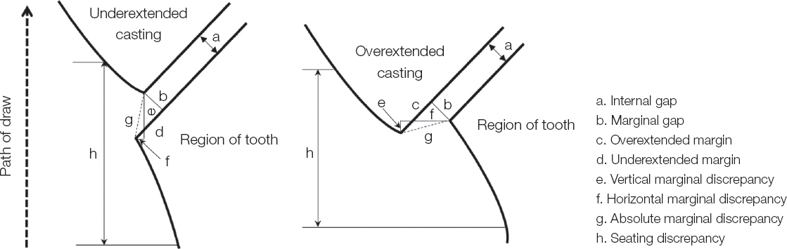
Fig. 1 Casting misfit terminology.
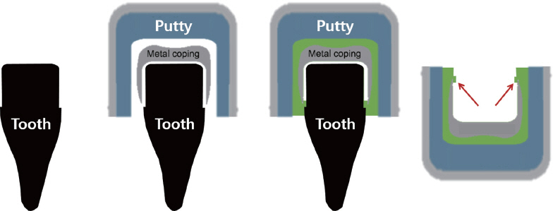
Fig. 2 The schematic of silicone putty wash impression.
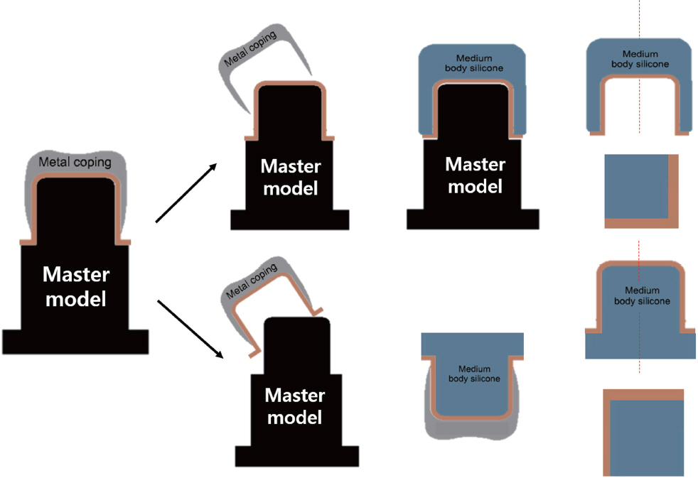
Fig. 3 2-D measurement of the thickness of the film with outer or inner coating using heavy body silicone.

Fig. 4 3-D measurement of the thickness of the film using model scanner.
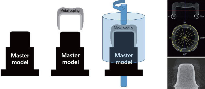
Fig. 5 The schematic of micro-CT measurement and CT image.
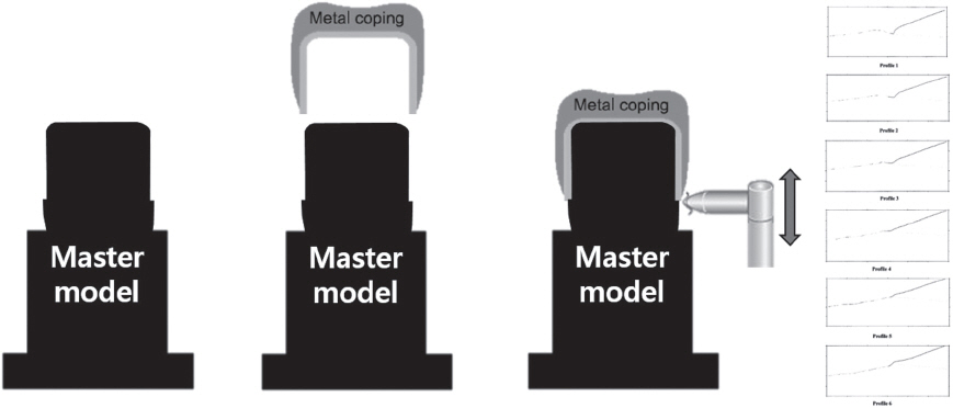
Fig. 6 The schematic of measurement using profilometry.
Reference
-
References
1. Hung SH, Hung KS, Eick JD, Chappell RP. Marginal fit of porcelain-fused-to-metal and two types of ceramic crown. J Prosthet Dent. 1990; 63:26–31. DOI: 10.1016/0022-3913(90)90260-J.2. McLean JW, von Fraunhofer JA. The estimation of cement film thickness by an in vivo technique. Br Dent J. 1971; 131:107–11. DOI: 10.1038/sj.bdj.4802708.3. Schwartz NL, Whitsett LD, Berry TG, Stewart JL. Unserviceable crowns and fixed partial dentures: life-span and causes for loss of serviceability. J Am Dent Assoc. 1970; 81:1395–401. DOI: 10.14219/jada.archive.1970.0398. PMID: 5273607.4. Grasso JE, Nalbandian J, Sanford C, Bailit H. Effect of restoration quality on periodontal health. J Prosthet Dent. 1985; 53:14–9. DOI: 10.1016/0022-3913(85)90056-3.5. Walton JN, Gardner FM, Agar JR. A survey of crown and fixed partial denture failures: length of service and reasons for replacement. J Prosthet Dent. 1986; 56:416–21. DOI: 10.1016/0022-3913(86)90379-3.6. Bader JD, Rozier RG, McFall WT Jr, Ramsey DL. Effect of crown margins on periodontal conditions in regularly attending patients. J Prosthet Dent. 1991; 65:75–9. DOI: 10.1016/0022-3913(91)90053-Y.7. Holmes JR, Bayne SC, Holland GA, Sulik WD. Considerations in measurement of marginal fit. J Prosthet Dent. 1989; 62:405–8. DOI: 10.1016/0022-3913(89)90170-4.8. A.D.A. specification No. 8 for dental zinc phosphate cement. Chilwa Kijae Hakhoe Chi. 1967; 2:64–7. PMID: 5238106.9. Ostlund LE. Cavity design and mathematics: their effect on gaps at the margins of cast restorations. Oper Dent. 1985; 10:122–37. PMID: 3913900.10. Sulaiman F, Chai J, Jameson LM, Wozniak WT. A comparison of the marginal fit of In-Ceram, IPS Empress, and Procera crowns. Int J Prosthodont. 1997; 10:478–84. PMID: 9495168.11. McLean JW, von Fraunhofer JA. The estimation of cement film thickness by an in vivo technique. Br Dent J. 1971; 131:107–11. DOI: 10.1038/sj.bdj.4802708.12. Gulker I. Margins. N Y State Dent J. 1985; 51:213–5. 217.13. Moldovan O, Rudolph H, Quaas S, Bornemann G, Luthardt RG. Internal and external fit of CAMmade zirconia bridge frameworks-a pilot study. Dtsch Zahnärztl Z. 2006; 61:38–42.14. Sorensen JA. A standardized method for determination of crown margin fidelity. J Prosthet Dent. 1990; 64:18–24. DOI: 10.1016/0022-3913(90)90147-5.15. Molin M, Karlsson S. The fit of gold inlays and three ceramic inlay systems. A clinical and in vitro study. Acta Odontol Scand. 1993; 51:201–6. DOI: 10.3109/00016359309040568. PMID: 8237304.16. Pelekanos S, Koumanou M, Koutayas SO, Zinelis S, Eliades G. Micro-CT evaluation of the marginal fit of different In-Ceram alumina copings. Eur J Esthet Dent. 2009; 4:278–92. PMID: 19704928.17. Luthardt RG, Bornemann G, Lemelson S, Walter MH, Hüls A. An innovative method for evaluation of the 3-D internal fit of CAD/CAM crowns fabricated after direct optical versus indirect laser scan digitizing. Int J Prosthodont. 2004; 17:680–5. PMID: 15686096.18. Hickel R, Roulet JF, Bayne S, Heintze SD, Mjör IA, Peters M, Rousson V, Randall R, Schmalz G, Tyas M, Vanherle G. Recommendations for conducting controlled clinical studies of dental restorative materials. Clin Oral Investig. 2007; 11:5–33. DOI: 10.1007/s00784-006-0095-7. PMID: 17262225.19. Karlsson S. A clinical evaluation of fixed bridges, 10 years following insertion. J Oral Rehabil. 1986; 13:423–32. DOI: 10.1111/j.1365-2842.1986.tb01304.x. PMID: 3534191.20. Akbar JH, Petrie CS, Walker MP, Williams K, Eick JD. Marginal adaptation of Cerec 3 CAD/CAM composite crowns using two different finish line preparation designs. J Prosthodont. 2006; 15:155–63. DOI: 10.1111/j.1532-849X.2006.00095.x. PMID: 16681497.21. Cvar JF, Ryge G. Reprint of criteria for the clinical evaluation of dental restorative materials. 1971. Clin Oral Investig. 2005; 9:215–32. DOI: 10.1007/s00784-005-0018-z. PMID: 16315023.22. Assif D, Antopolski B, Helft M, Kaffe I. Comparison of methods of clinical evaluation of the marginal fit of complete cast gold crowns. J Prosthet Dent. 1985; 54:20–4. DOI: 10.1016/S0022-3913(85)80062-7.23. Fattahi F, Giti R, Torabi K. Marginal assessment of crowns by the aid of parallel radiography. J Dent Mater Tech. 2015; 4:29–36.24. Liedke GS, Spin-Neto R, Vizzotto MB, Da Silveira PF, Silveira HE, Wenzel A. Diagnostic accuracy of conventional and digital radiography for detecting misfit between the tooth and restoration in metalrestored teeth. J Prosthet Dent. 2015; 113:39–47. DOI: 10.1016/j.prosdent.2014.08.003. PMID: 25311793.25. Groten M, Axmann D, Pröbster L, Weber H. Determination of the minimum number of marginal gap measurements required for practical in-vitro testing. J Prosthet Dent. 2000; 83:40–9. DOI: 10.1016/S0022-3913(00)70087-4.26. Groten M, Girthofer S, Pröbster L. Marginal fit consistency of copy-milled all-ceramic crowns during fabrication by light and scanning electron microscopic analysis in vitro. J Oral Rehabil. 1997; 24:871–81. DOI: 10.1046/j.1365-2842.1997.00592.x. PMID: 9467987.27. Euán R, Figueras-Álvarez O, Cabratosa-Termes J, Oliver-Parra R. Marginal adaptation of zirconium dioxide copings: influence of the CAD/CAM system and the finish line design. J Prosthet Dent. 2014; 112:155–62. DOI: 10.1016/j.prosdent.2013.10.012. PMID: 24445027.28. Beuer F, Naumann M, Gernet W, Sorensen JA. Precision of fit: zirconia three-unit fixed dental prostheses. Clin Oral Investig. 2009; 13:343–9. DOI: 10.1007/s00784-008-0224-6. PMID: 18769946.29. Tinschert J, Natt G, Mautsch W, Spiekermann H, Anusavice KJ. Marginal fit of alumina-and zirconiabased fixed partial dentures produced by a CAD/CAM system. Oper Dent. 2001; 26:367–74. PMID: 11504436.30. Kokubo Y, Nagayama Y, Tsumita M, Ohkubo C, Fukushima S, Vult von Steyern P. Clinical marginal and internal gaps of In-Ceram crowns fabricated using the GN-I system. J Oral Rehabil. 2005; 32:753–8. DOI: 10.1111/j.1365-2842.2005.01458.x. PMID: 16159354.31. Laurent M, Scheer P, Dejou J, Laborde G. Clinical evaluation of the marginal fit of cast crownsvalidation of the silicone replica method. J Oral Rehabil. 2008; 35:116–22. DOI: 10.1111/j.1365-2842.2003.01203.x. PMID: 18197844.32. Rahme HY, Tehini GE, Adib SM, Ardo AS, Rifai KT. In vitro evaluation of the “replica technique” in the measurement of the fit of Procera crowns. J Contemp Dent Pract. 2008; 9:25–32. PMID: 18264522.33. Reich S, Kappe K, Teschner H, Schmitt J. Clinical fit of four-unit zirconia posterior fixed dental prostheses. Eur J Oral Sci. 2008; 116:579–84. DOI: 10.1111/j.1600-0722.2008.00580.x. PMID: 19049530.34. Kohorst P, Brinkmann H, Li J, Borchers L, Stiesch M. Marginal accuracy of four-unit zirconia fixed dental prostheses fabricated using different computer- aided design/computer-aided manufacturing systems. Eur J Oral Sci. 2009; 117:319–25. DOI: 10.1111/j.1600-0722.2009.00622.x. PMID: 19583762.35. Kohorst P, Brinkmann H, Dittmer MP, Borchers L, Stiesch M. Influence of the veneering process on the marginal fit of zirconia fixed dental prostheses. J Oral Rehabil. 2010; 37:283–91. DOI: 10.1111/j.1365-2842.2009.02053.x. PMID: 20059649.36. Kohorst P, Junghanns J, Dittmer MP, Borchers L, Stiesch M. Different CAD/CAM-processing routes for zirconia restorations: influence on fitting accuracy. Clin Oral Investig. 2011; 15:527–36. DOI: 10.1007/s00784-010-0415-9. PMID: 20495937.37. Colpani JT, Borba M, Della Bona A. Evaluation of marginal and internal fit of ceramic crown copings. Dent Mater. 2013; 29:174–80. DOI: 10.1016/j.dental.2012.10.012. PMID: 23218747.38. Luthardt RG, Bornemann G, Lemelson S, Walter MH, Hüls A. An innovative method for evaluation of the 3-D internal fit of CAD/CAM crowns fabricated after direct optical versus indirect laser scan digitizing. Int J Prosthodont. 2004; 17:680–5. PMID: 15686096.39. Moldovan O, Luthardt RG, Corcodel N, Rudolph H. Three-dimensional fit of CAD/CAM-made zirconia copings. Dent Mater. 2011; 27:1273–8. DOI: 10.1016/j.dental.2011.09.006. PMID: 21983002.40. Kim KB, Kim JH, Kim WC, Kim HY, Kim JH. Evaluation of the marginal and internal gap of metal-ceramic crown fabricated with a selective laser sintering technology: two- and three-dimensional replica techniques. J Adv Prosthodont. 2013; 5:179–86. DOI: 10.4047/jap.2013.5.2.179. PMID: 23755345. PMCID: PMC3675292.41. Kuhn K, Ostertag S, Ostertag M, Walter MH, Luthardt RG, Rudolph H. Comparison of an analog and digital quantitative and qualitative analysis for the fit of dental copings. Comput Biol Med. 2015; 57:32–41. DOI: 10.1016/j.compbiomed.2014.11.017. PMID: 25528695.42. Pelekanos S, Koumanou M, Koutayas SO, Zinelis S, Eliades G. Micro-CT evaluation of the marginal fit of different In-Ceram alumina copings. Eur J Esthet Dent. 2009; 4:278–92. PMID: 19704928.43. Borba M, Miranda WG Jr, Cesar PF, Griggs JA, Bona AD. Evaluation of the adaptation of zirconia- based fixed partial dentures using micro-CT technology. Braz Oral Res. 2013; 27:396–402. PMID: 24036977.44. Pimenta MA, Frasca LC, Lopes R, Rivaldo E. Evaluation of marginal and internal fit of ceramic and metallic crown copings using x-ray microtomography (micro-CT) technology. J Prosthet Dent. 2015; 114:223–8. DOI: 10.1016/j.prosdent.2015.02.002. PMID: 25882975.45. Kim JH, Jeong JH, Lee JH, Cho HW. Fit of lithium disilicate crowns fabricated from conventional and digital impressions assessed with micro- CT. J Prosthet Dent. 2016; Jul. 13. doi:10.1016/j.prosdent.2016.03.028. Epub ahead of print.46. Mitchell CA, Pintado MR, Douglas WH. Nondestructive, in vitro quantification of crown margins. J Prosthet Dent. 2001; 85:575–84. DOI: 10.1067/mpr.2001.114268. PMID: 11404758.47. Limkangwalmongkol P, Kee E, Chiche GJ, Blatz MB. Comparison of marginal fit between all-porcelain margin versus alumina-supported margin on Procera Alumina crowns. J Prosthodont. 2009; 18:162–6. DOI: 10.1111/j.1532-849X.2008.00396.x. PMID: 19178619.48. Contrepois M, Soenen A, Bartala M, Laviole O. Marginal adaptation of ceramic crowns: a systematic review. J Prosthet Dent. 2013; 110:447–454. DOI: 10.1016/j.prosdent.2013.08.003. PMID: 24120071.49. Nawafleh NA, Mack F, Evans J, Mackay J, Hatamleh MM. Accuracy and reliability of methods to measure marginal adaptation of crowns and FDPs: a literature review. J Prosthodont. 2013; 22:419–28. DOI: 10.1111/jopr.12006. PMID: 23289599.
- Full Text Links
-
- Actions
-
Cited
- CITED
-
- Close
- Share
-
- Similar articles
-
- In vitro evaluation methods on adaptation of fixed dental prosthesis
- Marginal and internal discrepancy of 3-unit fixed dental prostheses fabricated by subtractive and additive manufacturing
- Comparative clinical study of the marginal discrepancy of fixed dental prosthesis fabricated by the milling-sintering method using a presintered alloy
- The use of definitive implant abutments for the fabrication of provisional crowns: a case series
- Burnishing effect on marginal misfit of implant-supported screw-and-cement retained prostheses: A case report
