J Periodontal Implant Sci.
2011 Oct;41(5):248-252. 10.5051/jpis.2011.41.5.248.
The use of definitive implant abutments for the fabrication of provisional crowns: a case series
- Affiliations
-
- 1Department of Prosthodontics, Istanbul University Faculty of Dentistry, Istanbul, Turkey. geckili@istanbul.edu.tr
- KMID: 1783620
- DOI: http://doi.org/10.5051/jpis.2011.41.5.248
Abstract
- PURPOSE
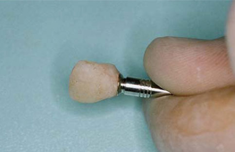
The anterior region is a challenge for most clinicians to achieve optimal esthetics with dental implants. The provisional crown is a key factor in the success of obtaining pink esthetics around restorations with single implants, by soft tissue and inter-proximal papilla shaping. Provisional abutments bring additional costs and make the treatment more expensive. Since one of the aims of the clinician is to reduce costs and find more economic ways to raise patient satisfaction, this paper describes a practical method for chair-side fabrication of non-occlusal loaded provisional crowns used by the authors for several years successfully.
METHODS
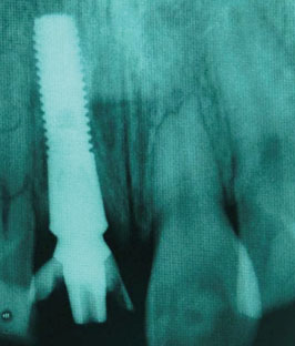
Twenty two patients (9 males, 13 females; mean age, 36,72 years) with one missing anterior tooth were treated by using the presented method. Metal definitive abutments instead of provisional abutments were used and provisional crowns were fabricated on the definitive abutments for all of the patients. The marginal fit was finished on a laboratory analogue and temporarily cemented to the abutments. The marginal adaptation of the crowns was evaluated radiographically.
RESULTS
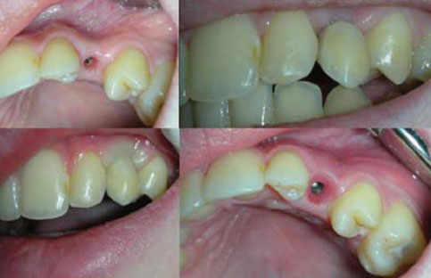
The patients were all satisfied with the final appearance and no complications occurred until the implants were loaded with permanent restorations.
CONCLUSIONS
The use of the definitive abutments for provisional crowns instead of provisional abutments reduces the costs and the same results can be obtained.
MeSH Terms
Figure
Reference
-
1. Bilhan H, Sönmez E, Mumcu E, Bilgin T. Immediate loading: three cases with up to 38 months of clinical follow-up. J Oral Implantol. 2009. 35:75–81.
Article2. Geckili O, Bilhan H, Bilgin T. A 24-week prospective study comparing the stability of titanium dioxide grit-blasted dental implants with and without fluoride treatment. Int J Oral Maxillofac Implants. 2009. 24:684–688.3. Becker W. Immediate implant placement: diagnosis, treatment planning and treatment steps/or successful outcomes. J Calif Dent Assoc. 2005. 33:303–310.4. Tarnow DP, Eskow RN. Considerations for single-unit esthetic implant restorations. Compend Contin Educ Dent. 1995. 16:778780782–784. passim.5. Belser UC, Bernard JP, Buser D. Implant-supported restorations in the anterior region: prosthetic considerations. Pract Periodontics Aesthet Dent. 1996. 8:875–883.6. Belser UC, Buser D, Hess D, Schmid B, Bernard JP, Lang NP. Aesthetic implant restorations in partially edentulous patients--a critical appraisal. Periodontol 2000. 1998. 17:132–150.
Article7. Choquet V, Hermans M, Adriaenssens P, Daelemans P, Tarnow DP, Malevez C. Clinical and radiographic evaluation of the papilla level adjacent to single-tooth dental implants. A retrospective study in the maxillary anterior region. J Periodontol. 2001. 72:1364–1371.
Article8. Kurtzman GM. In-office custom abutments and long-term provisionals. Dent Today. 2010. 29:102106108–109.9. Östman PO. A novel technique for fabrication of immediate provisional restorations. J Implant Reconstruct Dentist. 2009. 1:6–12.10. Becker W. Immediate implant placement: treatment planning and surgical steps for successful outcomes. Br Dent J. 2006. 201:199–205.
Article11. Nakamura K, Kanno T, Milleding P, Ortengren U. Zirconia as a dental implant abutment material: a systematic review. Int J Prosthodont. 2010. 23:299–309.12. Barone A, Rispoli L, Vozza I, Quaranta A, Covani U. Immediate restoration of single implants placed immediately after tooth extraction. J Periodontol. 2006. 77:1914–1920.
Article13. Hoffmann O, Beaumont C, Zafiropoulos GG. Immediate implant placement: a case series. J Oral Implantol. 2006. 32:182–189.
Article14. Chang M, Wennström JL. Peri-implant soft tissue and bone crest alterations at fixed dental prostheses: a 3-year prospective study. Clin Oral Implants Res. 2010. 21:527–534.
Article15. Park JB. Immediate placement of dental implants into fresh extraction socket in the maxillary anterior region: a case report. J Oral Implantol. 2010. 36:153–157.
Article16. Krennmair G, Seemann R, Schmidinger S, Ewers R, Piehslinger E. Clinical outcome of root-shaped dental implants of various diameters: 5-year results. Int J Oral Maxillofac Implants. 2010. 25:357–366.17. Garcia RV, Kraehenmann MA, Bezerra FJ, Mendes CM, Rapp GE. Clinical analysis of the soft tissue integration of non-submerged (ITI) and submerged (3i) implants: a prospective-controlled cohort study. Clin Oral Implants Res. 2008. 19:991–996.
Article
- Full Text Links
- Actions
-
Cited
- CITED
-
- Close
- Share
- Similar articles
-
- A comparison of retentive strength of implant cement depending on various methods of removing provisional cement from implant abutment
- Mounting and utilization of provisional prostheses on master cast for the fabrication of fixed implant-supported prostheses: a case report
- Gingival recontouring by provisional implant restoration for optimal emergence profile: report of two cases
- Implant–supported fixed prosthesis for orthognathic surgery in ectodermal dysplasia: a case report
- Conversion of implant overdenture to an implant assisted removable partial denture in maxilla: case report

