Investig Magn Reson Imaging.
2017 Jun;21(2):71-81. 10.13104/imri.2017.21.2.71.
Comparison of Three, Motion-Resistant MR Sequences on Hepatobiliary Phase for Gadoxetic Acid (Gd-EOB-DTPA)-Enhanced MR Imaging of the Liver
- Affiliations
-
- 1Department of Diagnostic Radiology, Jeju National University Hospital, Jeju, Korea. 671228kbs@naver.com
- 2Department of Internal Medicine, Jeju National University Hospital, Jeju, Korea.
- KMID: 2385603
- DOI: http://doi.org/10.13104/imri.2017.21.2.71
Abstract
- PURPOSE
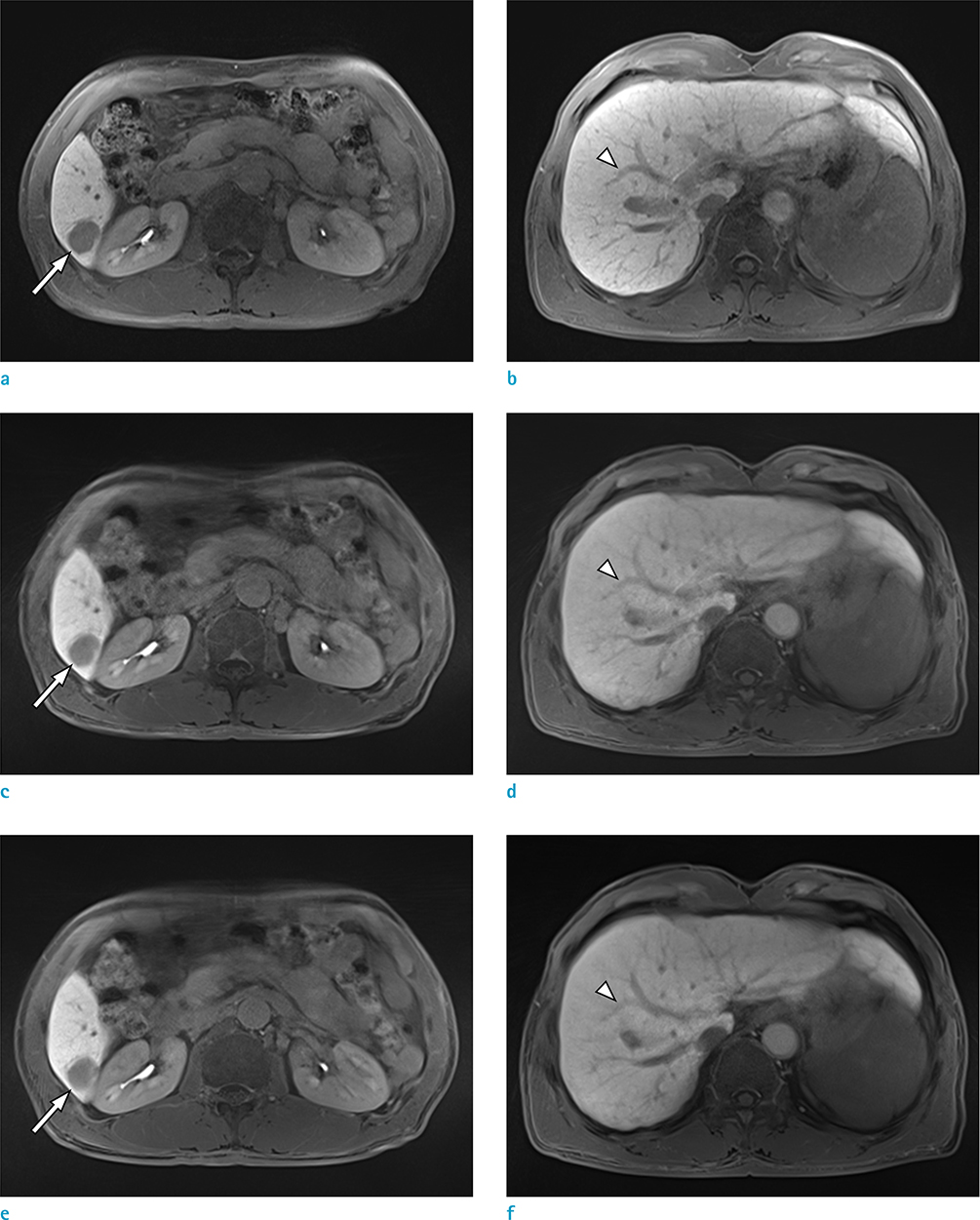
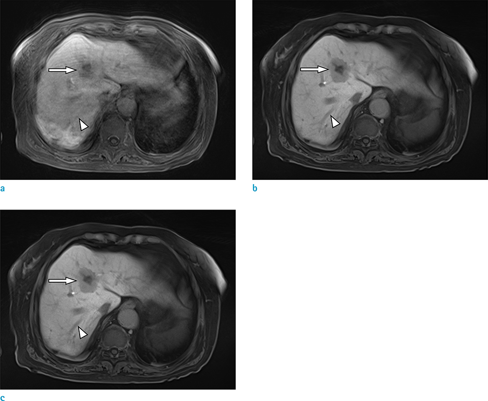
To compare three, motion-resistant, T1-weighted MR sequences on the hepatobiliary phase for gadoxetic acid-enhanced MR imaging of the liver.
MATERIALS AND METHODS
In this retrospective study, 79 patients underwent gadoxetic acid-enhanced, 3T liver MR imaging. Fifty-nine were examined using a standard protocol, and 20 were examined using a motion-resistant protocol. During the hepatocyte-specific phase, three MR sequences were acquired: 1) gradient recalled echo (GRE) with controlled aliasing in parallel imaging results in higher acceleration (CAIPIRINHA); 2) radial GRE with the interleaved angle-bisection scheme (ILAB); and 3) radial GRE with golden-angle scheme (GA). Two readers independently assessed images with motion artifacts, streaking artifacts, liver-edge sharpness, hepatic vessel clarity, lesion conspicuity, and overall image quality, using a 5-point scale. The images were assessed by measurement of liver signal-to-noise ratio (SNR), and tumor-to-liver contrast-to-noise ratio (CNR). The results were compared, using repeated post-hoc, paired t-tests with Bonferroni correction and the Wilcoxon signed rank test with Bonferroni correction.
RESULTS
In the qualitative analysis of cooperative patients, the results for CAIPIRINHA had significantly higher ratings for streak artifacts, liver-edge sharpness, hepatic vessel clarity, and overall image quality as compared to, radial GRE, (P < 0.016). In the imaging of uncooperative patients, higher scores were recorded for ILAB and GA with respect to all of the qualitative assessments, except for streak artifact, compared with CAIPIRINHA (P < 0.016). However, no significant differences were found between ILAB and GA. For quantitative analysis in uncooperative patients, the mean liver SNR and lesion-to-liver CNR with radial GRE were significantly higher than those of CAIPIRINHA (P < 0.016).
CONCLUSION
In uncooperative patients, the use of the radial GRE sequence can improve the image quality compared to GRE imaging with CAIPIRINHA, despite the data acquisition methods used. The GRE imaging with CAIPIRINHA is applicable for patients without breath-holding difficulties.
MeSH Terms
Figure
Reference
-
1. Bamrungchart S, Tantaway EM, Midia EC, et al. Free breathing three-dimensional gradient echo-sequence with radial data sampling (radial 3D-GRE) examination of the pancreas: Comparison with standard 3D-GRE volumetric interpolated breathhold examination (VIBE). J Magn Reson Imaging. 2013; 38:1572–1577.2. Birchard KR, Semelka RC, Hyslop WB, et al. Suspected pancreatic cancer: evaluation by dynamic gadolinium-enhanced 3D gradient-echo MRI. AJR Am J Roentgenol. 2005; 185:700–703.3. Kim BS, Angthong W, Jeon YH, Semelka RC. Body MR imaging: fast, efficient, and comprehensive. Radiol Clin North Am. 2014; 52:623–636.4. Rofsky NM, Lee VS, Laub G, et al. Abdominal MR imaging with a volumetric interpolated breath-hold examination. Radiology. 1999; 212:876–884.5. Semelka RC, Helmberger TK. Contrast agents for MR imaging of the liver. Radiology. 2001; 218:27–38.6. Semelka RC, Martin DR, Balci NC. Magnetic resonance imaging of the liver: how I do it. J Gastroenterol Hepatol. 2006; 21:632–637.7. Kim BS, Lee KR, Goh MJ. New imaging strategies using a motion-resistant liver sequence in uncooperative patients. Biomed Res Int. 2014; 2014:142658.8. Park YS, Lee CH, Kim IS, et al. Usefulness of controlled aliasing in parallel imaging results in higher acceleration in gadoxetic acid-enhanced liver magnetic resonance imaging to clarify the hepatic arterial phase. Invest Radiol. 2014; 49:183–188.9. Wright KL, Harrell MW, Jesberger JA, et al. Clinical evaluation of CAIPIRINHA: comparison against a GRAPPA standard. J Magn Reson Imaging. 2014; 39:189–194.10. Yu MH, Lee JM, Yoon JH, Kiefer B, Han JK, Choi BI. Clinical application of controlled aliasing in parallel imaging results in a higher acceleration (CAIPIRINHA)-volumetric interpolated breathhold (VIBE) sequence for gadoxetic acid-enhanced liver MR imaging. J Magn Reson Imaging. 2013; 38:1020–1026.11. Azevedo RM, de Campos RO, Ramalho M, Heredia V, Dale BM, Semelka RC. Free-breathing 3D T1-weighted gradient-echo sequence with radial data sampling in abdominal MRI: preliminary observations. AJR Am J Roentgenol. 2011; 197:650–657.12. Chandarana H, Block TK, Rosenkrantz AB, et al. Free-breathing radial 3D fat-suppressed T1-weighted gradient echo sequence: a viable alternative for contrast-enhanced liver imaging in patients unable to suspend respiration. Invest Radiol. 2011; 46:648–653.13. Chandarana H, Feng L, Block TK, et al. Free-breathing contrast-enhanced multiphase MRI of the liver using a combination of compressed sensing, parallel imaging, and golden-angle radial sampling. Invest Radiol. 2013; 48:10–16.14. Kim KW, Lee JM, Jeon YS, et al. Free-breathing dynamic contrast-enhanced MRI of the abdomen and chest using a radial gradient echo sequence with K-space weighted image contrast (KWIC). Eur Radiol. 2013; 23:1352–1360.15. Song HK, Dougherty L. Dynamic MRI with projection reconstruction and KWIC processing for simultaneous high spatial and temporal resolution. Magn Reson Med. 2004; 52:815–824.16. Runge VM, Richter JK, Heverhagen JT. Speed in clinical magnetic resonance. Invest Radiol. 2017; 52:1–17.17. Chandarana H, Block KT, Winfeld MJ, et al. Free-breathing contrast-enhanced T1-weighted gradient-echo imaging with radial k-space sampling for paediatric abdominopelvic MRI. Eur Radiol. 2014; 24:320–326.18. Shin HJ, Kim MJ, Lee MJ, Kim HG. Comparison of image quality between conventional VIBE and radial VIBE in free-breathing paediatric abdominal MRI. Clin Radiol. 2016; 71:1044–1049.19. Pruessmann KP, Weiger M, Scheidegger MB, Boesiger P. SENSE: sensitivity encoding for fast MRI. Magn Reson Med. 1999; 42:952–962.20. Mitchell DG, Vinitski S, Saponaro S, Tasciyan T, Burk DL Jr, Rifkin MD. Liver and pancreas: improved spin-echo T1 contrast by shorter echo time and fat suppression at 1.5 T. Radiology. 1991; 178:67–67.
- Full Text Links
- Actions
-
Cited
- CITED
-
- Close
- Share
- Similar articles
-
- Supradiaphragmatic Liver Confirmed by a Hepatocyte-specific Contrast Agent (Gd-EOB-DTPA): A Case Report
- Hepatic Lymphoma Representing Iso-Signal Intensity on Hepatobiliary Phase, in Gd-EOB-DTPA-Enhanced MRI: Case Report
- Current Limitations and Potential Breakthroughs for the Early Diagnosis of Hepatocellular Carcinoma
- Enhancement Pattern of Liver Parenchyma during Late Dynamic Phase Imaging: Comparison between Gd-EOB-DTPA and Gd-DTPA-BMA
- Image Findings of Sarcomatous Intrahepatic Cholangiocarcinoma Focused on Gd-EOB-DTPA Enhanced MRI: A Case Report


