Evaluation of the marginal and internal gaps of three different dental prostheses: comparison of the silicone replica technique and three-dimensional superimposition analysis
- Affiliations
-
- 1Institute for Health Science, Korea University, Seoul, Republic of Korea.
- 2Department of Dental Laboratory Science and Engineering, College of Health Science, Korea University, Seoul, Republic of Korea. kuc2842@korea.ac.kr
- 3Department of Public Health Sciences, Graduate School & BK21+ Program in Public Health Sciences, Korea University, Seoul, Republic of Korea.
- KMID: 2382601
- DOI: http://doi.org/10.4047/jap.2017.9.3.159
Abstract
- PURPOSE
The purposes of this study were to evaluate the marginal and internal gaps, and the potential clinical applications of three different methods of dental prostheses fabrication, and to compare the prostheses prepared using the silicone replica technique (SRT) and those prepared using the three-dimensional superimposition analysis (3DSA).
MATERIALS AND METHODS
Five Pekkton, lithium disilicate, and zirconia crowns were each manufactured and tested using both the SRT and the two-dimensional section of the 3DSA. The data were analyzed with the nonparametric version of a two-way analysis of variance using rank-transformed values and the Tukey's post-hoc test (α = .05).
RESULTS
Significant differences were observed between the fabrication methods in the marginal gap (P < .010), deep chamfer (P < .001), axial wall (P < .001), and occlusal area (P < .001). A significant difference in the occlusal area was found between the two measurement methods (P < .030), whereas no significant differences were found in the marginal gap (P > .350), deep chamfer (P > .719), and axial wall (P > .150). As the 3DSA method is three-dimensional, it allows for the measurement of arbitrary points.
CONCLUSION
All of the three fabrication methods are valid for measuring clinical objectives because they produced prostheses within the clinically acceptable range. Furthermore, a three-dimensional superimposition analysis verification method such as the silicone replica technique is also applicable in clinical settings.
Keyword
MeSH Terms
Figure
-
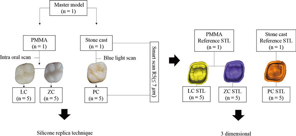
Fig. 1 Study design flowchart. PMMA, polymethyl methacrylate; LC, lithium disilicate crown; PC, Pekkton crown; STL, standard template library; ZC, zirconia crown.
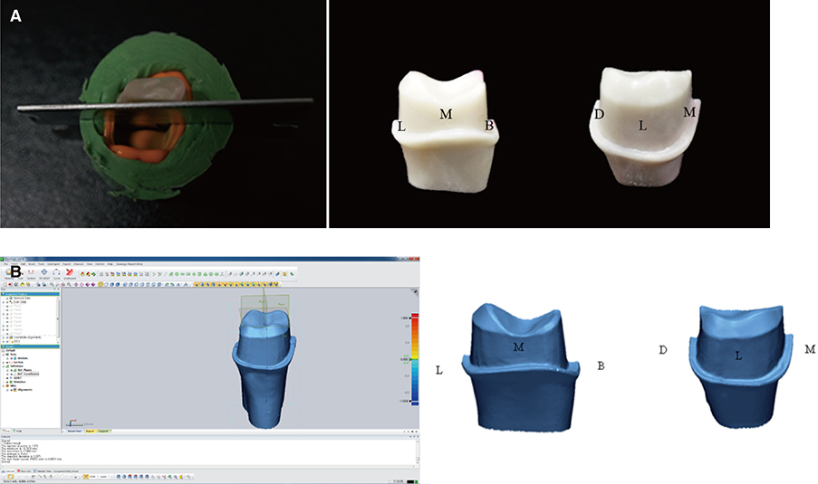
Fig. 2 Cutting regions for measurements. (A) Jig for the silicone replica technique: By the insertion of a jig for the silicone replica technique, the region was cut with the side to be measured with a constant blade in order to measure the same point, (B) Two-dimensional section of the three-dimensional superimposition analysis: the cut was made in the buccolingual and mesiodistal directions.
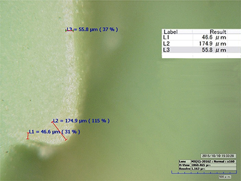
Fig. 3 Digital microscope image of the silicone replica technique (160× magnification).
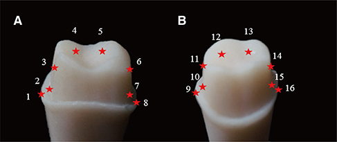
Fig. 4 Sixteen measuring points of the marginal and internal gaps. (A) Bucco-ligual view, (B) Mesio-distal view.
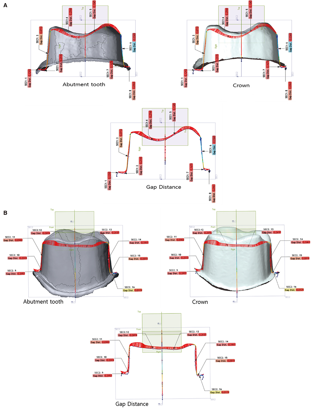
Fig. 5 Section of geometric dimensioning and tolerancing in the three-dimensional superimposition analysis. (A) Bucco-lingual direction, (B) Mesio-distal direction.

Fig. 6 Mean marginal and internal gaps according to the measurement method. SRT, silicone replicate technique; 3DSA, three-dimensional superimposition analysis.
Cited by 5 articles
-
Marginal and internal discrepancy of 3-unit fixed dental prostheses fabricated by subtractive and additive manufacturing
Jae-Won Choi
J Korean Acad Prosthodont. 2020;58(1):7-13. doi: 10.4047/jkap.2020.58.1.7.Evaluation of marginal discrepancy of pressable ceramic veneer fabricated using CAD/CAM system: Additive and subtractive manufacturing
Seen-Young Kang, Ha-Na Lee, Ji-Hwan Kim, Woong-Chul Kim
J Adv Prosthodont. 2018;10(5):347-353. doi: 10.4047/jap.2018.10.5.347.Evaluation of the reproducibility of various abutments using a blue light model scanner
Dong-Yeon Kim, Kyung-Eun Lee, Jin-Hun Jeon, Ji-Hwan Kim, Woong-Chul Kim
J Adv Prosthodont. 2018;10(4):328-334. doi: 10.4047/jap.2018.10.4.328.Accuracy of provisional crowns made using stereolithography apparatus and subtractive technique
Seen-Young Kang, Jung-Hyun Park, Ji-Hwan Kim, Woong-Chul Kim
J Adv Prosthodont. 2018;10(5):354-360. doi: 10.4047/jap.2018.10.5.354.Marginal fit of three different nanocomposite inlays fabricated with computer-aided design/computer-aided manufacturing (CAD/CAM) technology: a comparative study
Hyunsuk Choi, Jae-Young Jo, Min-Ho Hong
J Yeungnam Med Sci. 2024;41(2):80-85. doi: 10.12701/jyms.2023.00934.
Reference
-
1. Abduo J, Lyons K, Bennamoun M. Trends in computer-aided manufacturing in prosthodontics: a review of the available streams. Int J Dent. 2014; 2014:783948.2. Bosch G, Ender A, Mehl A. A 3-dimensional accuracy analysis of chairside CAD/CAM milling processes. J Prosthet Dent. 2014; 112:1425–1431.3. Fabbri G, Zarone F, Dellificorelli G, Cannistraro G, De Lorenzi M, Mosca A, Sorrentino R. Clinical evaluation of 860 anterior and posterior lithium disilicate restorations: retrospective study with a mean follow-up of 3 years and a maximum observational period of 6 years. Int J Periodontics Restorative Dent. 2014; 34:165–177.4. Piconi C, Maccauro G. Zirconia as a ceramic biomaterial. Biomaterials. 1999; 20:1–25.5. Ueda K, Güth JF, Erdelt K, Stimmelmayr M, Kappert H, Beuer F. Light transmittance by a multi-coloured zirconia material. Dent Mater J. 2015; 34:310–314.6. Thieme K, Rüssel C. Nucleation and growth kinetics and phase analysis in zirconia-containing lithium disilicate glass. J Mater Sci. 2015; 50:1488–1499.7. Almeida e Silva JS, Erdelt K, Edelhoff D, Araújo É, Stimmelmayr M, Vieira LC, Güth JF. Marginal and internal fit of four-unit zirconia fixed dental prostheses based on digital and conventional impression techniques. Clin Oral Investig. 2014; 18:515–523.8. Komine F, Blatz MB, Matsumura H. Current status of zirconia-based fixed restorations. J Oral Sci. 2010; 52:531–539.9. Kang SH, Chang J, Son HH. Flexural strength and microstructure of two lithium disilicate glass ceramics for CAD/CAM restoration in the dental clinic. Restor Dent Endod. 2013; 38:134–140.10. Oh GJ, Yun KD, Lee KM, Lim HP, Park SW. Sintering behavior and mechanical properties of zirconia compacts fabricated by uniaxial press forming. J Adv Prosthodont. 2010; 2:81–87.11. Fuhrmann G, Steiner M, Freitag-Wolf S, Kern M. Resin bonding to three types of polyaryletherketones (PAEKs)-durability and influence of surface conditioning. Dent Mater. 2014; 30:357–363.12. Schwitalla A, Müller WD. PEEK dental implants: a review of the literature. J Oral Implantol. 2013; 39:743–749.13. Kurtz SM, Devine JN. PEEK biomaterials in trauma, orthopedic, and spinal implants. Biomaterials. 2007; 28:4845–4869.14. Tetelman ED, Babbush CA. A new transitional abutment for immediate aesthetics and function. Implant Dent. 2008; 17:51–58.15. Mark HF. Encyclopedia of polymer science and technology, concise. John Wiley & Sons;2013. p. 5–1376.16. Stawarczyk B, Beuer F, Wimmer T, Jahn D, Sener B, Roos M, Schmidlin PR. Polyetheretherketone-a suitable material for fixed dental prostheses? J Biomed Mater Res B Appl Biomater. 2013; 101:1209–1216.17. Rudolph H, Luthardt RG, Walter MH. Computer-aided analysis of the influence of digitizing and surfacing on the accuracy in dental CAD/CAM technology. Comput Biol Med. 2007; 37:579–587.18. Luthardt R, Weber A, Rudolph H, Schöne C, Quaas S, Walter M. Design and production of dental prosthetic restorations: basic research on dental CAD/CAM technology. Int J Comput Dent. 2002; 5:165–176.19. Beuer F, Schweiger J, Edelhoff D. Digital dentistry: an overview of recent developments for CAD/CAM generated restorations. Br Dent J. 2008; 204:505–511.20. Garcia-Barbosa JA, Arroyo-Osorio JM, Cordoba-Nieto E. The manufacture of a maxillofacial prosthesis from an axial tomography using simulation technologies with a virtual machine tool and four-axis machining. Dyna. 2016; 83:100–105.21. Anadioti E, Aquilino SA, Gratton DG, Holloway JA, Denry I, Thomas GW, Qian F. 3D and 2D marginal fit of pressed and CAD/CAM lithium disilicate crowns made from digital and conventional impressions. J Prosthodont. 2014; 23:610–617.22. Raju SR. Comparison of marginal fit of zirconium oxide copings generated using four different CAD-CAM systems: an in vitro study. J Res Adv Dent. 2013; 3:163–171.23. Tamac E, Toksavul S, Toman M. Clinical marginal and internal adaptation of CAD/CAM milling, laser sintering, and cast metal ceramic crowns. J Prosthet Dent. 2014; 112:909–913.24. Laurent M, Scheer P, Dejou J, Laborde G. Clinical evaluation of the marginal fit of cast crowns-validation of the silicone replica method. J Oral Rehabil. 2008; 35:116–122.25. Park JY, Kim HY, Kim JH, Kim JH, Kim WC. Comparison of prosthetic models produced by traditional and additive manufacturing methods. J Adv Prosthodont. 2015; 7:294–302.26. Meng Z, Yao XS, Yao H, Liang Y, Liu T, Li Y, Wang G, Lan S. Measurement of the refractive index of human teeth by optical coherence tomography. J Biomed Opt. 2009; 14:034010.27. American National Standards Institute/American Dental Association. ANSI/ADA specification No. 19. Dental elastomeric impression materials. ISO 4823:2000. Chicago: American Dental Association;2004. Available at: http://www.iso.ch/iso/en/prods-services/ISOstore/store.html.28. Bayramoğlu E, Özkan YK, Yildiz C. Comparison of marginal and internal fit of press-on-metal and conventional ceramic systems for three- and four-unit implant-supported partial fixed dental prostheses: An in vitro study. J Prosthet Dent. 2015; 114:52–58.29. Laurent M, Scheer P, Dejou J, Laborde G. Clinical evaluation of the marginal fit of cast crowns-validation of the silicone replica method. J Oral Rehabil. 2008; 35:116–122.30. Park JY, Jeong ID, Lee JJ, Bae SY, Kim JH, Kim WC. In vitro assessment of the marginal and internal fits of interim implant restorations fabricated with different methods. J Prosthet Dent. 2016; 116:536–542.31. Meyer BJ, Mörmann WH, Lutz F. Optimization of the powder application in the Cerec method with environmentfriendly propellant systems. Schweiz Monatsschr Zahnmed. 1990; 100:1462–1468.32. Kim HH, Lee JW, Cha KS, Chung DH, Lee SM. Three-dimensional assessment of upper lip positional changes according to simulated maxillary anterior tooth movements by white light scanning. Korean J Orthod. 2014; 44:281–293.33. Kuo RF, Chen SJ, Wong TY, Lu BC, Huang ZH. Digital morphology comparisons between models of conventional intraoral casting and digital rapid prototyping. 5th International Conference on Biomedical Engineering in Vietnam;2015. 46:p. 478–480.34. Bae SY, Park JY, Jeong ID, Kim HY, Kim JH, Kim WC. Three-dimensional analysis of marginal and internal fit of copings fabricated with polyetherketoneketone (PEKK) and zirconia. J Prosthodont Res. 2017; 61:106–112.35. Persson AS, Andersson M, Odén A, Sandborgh-Englund G. Computer aided analysis of digitized dental stone replicas by dental CAD/CAM technology. Dent Mater. 2008; 24:1123–1130.36. Schaefer O, Kuepper H, Thompson GA, Cachovan G, Hefti AF, Guentsch A. Effect of CNC-milling on the marginal and internal fit of dental ceramics: a pilot study. Dent Mater. 2013; 29:851–858.37. Faul F, Erdfelder E, Lang AG, Buchner A. G*Power 3: a flexible statistical power analysis program for the social, behavioral, and biomedical sciences. Behav Res Methods. 2007; 39:175–191.38. Jeon JH, Choi BY, Kim CM, Kim JH, Kim HY, Kim WC. Three-dimensional evaluation of the repeatability of scanned conventional impressions of prepared teeth generated with white- and blue-light scanners. J Prosthet Dent. 2015; 114:549–553.39. Anadioti E, Aquilino SA, Gratton DG, Holloway JA, Denry IL, Thomas GW, Qian F. Internal fit of pressed and computer-aided design/computer-aided manufacturing ceramic crowns made from digital and conventional impressions. J Prosthet Dent. 2015; 113:304–309.40. Flügge TV, Schlager S, Nelson K, Nahles S, Metzger MC. Precision of intraoral digital dental impressions with iTero and extraoral digitization with the iTero and a model scanner. Am J Orthod Dentofacial Orthop. 2013; 144:471–478.41. Nedelcu RG, Persson AS. Scanning accuracy and precision in 4 intraoral scanners: an in vitro comparison based on 3-dimensional analysis. J Prosthet Dent. 2014; 112:1461–1471.42. Katsoulis J, Mericske-Stern R, Rotkina L, Zbären C, Enkling N, Blatz MB. Precision of fit of implant-supported screw-retained 10-unit computer-aided-designed and computer-aided-manufactured frameworks made from zirconium dioxide and titanium: an in vitro study. Clin Oral Implants Res. 2014; 25:165–174.43. Guess PC, Vagkopoulou T, Zhang Y, Wolkewitz M, Strub JR. Marginal and internal fit of heat pressed versus CAD/CAM fabricated all-ceramic onlays after exposure to thermo-mechanical fatigue. J Dent. 2014; 42:199–209.44. McLean JW, von Fraunhofer JA. The estimation of cement film thickness by an in vivo technique. Br Dent J. 1971; 131:107–111.45. Bindl A, Mörmann WH. Marginal and internal fit of all-ceramic CAD/CAM crown-copings on chamfer preparations. J Oral Rehabil. 2005; 32:441–447.46. Örtorp A, Jönsson D, Mouhsen A, Vult von Steyern P. The fit of cobalt-chromium three-unit fixed dental prostheses fabricated with four different techniques: a comparative in vitro study. Dent Mater. 2011; 27:356–363.47. Kahramanoğlu E, Kulak-Ozkan Y. Marginal and internal adaptation of different superstructure and abutment materials using two different implant systems for five-unit implant-supported fixed partial dentures: an in vitro study. Int J Oral Maxillofac Implants. 2013; 28:1207–1216.48. Hamza TA, Ezzat HA, El-Hossary MM, Katamish HA, Shokry TE, Rosenstiel SF. Accuracy of ceramic restorations made with two CAD/CAM systems. J Prosthet Dent. 2013; 109:83–87.49. Song TJ, Kwon TK, Yang JH, Han JS, Lee JB, Kim SH, Yeo IS. Marginal fit of anterior 3-unit fixed partial zirconia restorations using different CAD/CAM systems. J Adv Prosthodont. 2013; 5:219–225.50. Lins L, Bemfica V, Queiroz C, Canabarro A. In vitro evaluation of the internal and marginal misfit of CAD/CAM zirconia copings. J Prosthet Dent. 2015; 113:205–211.51. Neves FD, Prado CJ, Prudente MS, Carneiro TA, Zancopé K, Davi LR, Mendonça G, Cooper LF, Soares CJ. Microcomputed tomography evaluation of marginal fit of lithium disilicate crowns fabricated by using chairside CAD/CAM systems or the heat-pressing technique. J Prosthet Dent. 2014; 112:1134–1140.52. Jeong ID, Lee JJ, Jeon JH, Kim JH, Kim HY, Kim WC. Accuracy of complete-arch model using an intraoral video scanner: An in vitro study. J Prosthet Dent. 2016; 115:755–759.53. Ji MK, Park JH, Park SW, Yun KD, Oh GJ, Lim HP. Evaluation of marginal fit of 2 CAD-CAM anatomic contour zirconia crown systems and lithium disilicate glass-ceramic crown. J Adv Prosthodont. 2015; 7:271–277.
- Full Text Links
-
- Actions
-
Cited
- CITED
-
- Close
- Share
-
- Similar articles
-
- Verification of a computer-aided replica technique for evaluating prosthesis adaptation using statistical agreement analysis
- Comparison of the fit accuracy of zirconia-based prostheses generated by two CAD/CAM systems
- Study on methodology for the assessment of internal and marginal adaptation on fixed dental prosthesis
- In vitro evaluation methods on adaptation of fixed dental prosthesis
- Comparison of the fit of cast gold crowns fabricated from the digital and the conventional impression techniques
