Proposal of an Appropriate Decalcification Method of Bone Marrow Biopsy Specimens in the Era of Expanding Genetic Molecular Study
- Affiliations
-
- 1Department of Pathology, Yonsei University College of Medicine, Seoul, Korea. revita@naver.com
- KMID: 2381382
- DOI: http://doi.org/10.4132/jptm.2015.03.16
Abstract
- BACKGROUND
The conventional method for decalcification of bone specimens uses hydrochloric acid (HCl) and is notorious for damaging cellular RNA, DNA, and proteins, thus complicating molecular and immunohistochemical analyses. A method that can effectively decalcify while preserving genetic material is necessary.
METHODS
Pairs of bilateral bone marrow biopsies sampled from 53 patients were decalcified according to protocols of two comparison groups: EDTA versus HCl and RDO GOLD (RDO) versus HCl. Pairs of right and left bone marrow biopsy samples harvested from 28 cases were allocated into the EDTA versus HCl comparison group, and 25 cases to the RDO versus HCl comparison group. The decalcification protocols were compared with regards to histomorphology, immunohistochemistry, and molecular analysis. For molecular analysis, we randomly selected 5 cases from the EDTA versus HCl and RDO versus HCl groups.
RESULTS
The decalcification time for appropriate histomorphologic analysis was the longest in the EDTA method and the shortest in the RDO method. EDTA was superior to RDO or HCl in DNA yield and integrity, assessed via DNA extraction, polymerase chain reaction, and silver in situ hybridization using DNA probes. The EDTA method maintained intact nuclear protein staining on immunohistochemistry, while the HCl method produced poor quality images. Staining after the RDO method had equivocal results. RNA in situ hybridization using kappa and lambda RNA probes measured RNA integrity; the EDTA and RDO method had the best quality, followed by HCl.
CONCLUSIONS
The EDTA protocol would be the best in preserving genetic material. RDO may be an acceptable alternative when rapid decalcification is necessary.
Keyword
MeSH Terms
Figure
-
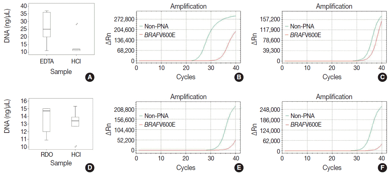
Fig. 1. The quality, quantity, and feasibility of real time PCR study is compared between EDTA, RDO, and HCl protocols. The first row demonstrates EDTA versus HCl, and the second row RDO versus HCl (A, D, DNA yield; B, PCR result of EDTA; E, PCR result of RDO; C, F, PCR results of HCl). D, PCR result of RDO. PCR, polymerase chain reaction; EDTA, ethylenediaminetetraacetic acid disodium salt dehydrate; RDO, RDO GOLD; HCl, hydrochloric acid; PNA, peptide nucleic acid.

Fig. 2. In EDTA versus HCl comparison of a pair of bone marrow sampled from the same patient (A–D, EDTA versus HCl group; A, B, EDTA protocol; C, D, HCl protocol), and in RDO versus HCl comparison of a pair of bone marrow sampled from the same patient (E–H, RDO versus HCl group; E, F, RDO protocol; G, H, HCl protocol), all the three methods of EDTA, RDO, and HCl protocols demonstrate intact and well-preserved histological features of bone marrow. EDTA, ethylenediaminetetraacetic acid disodium salt dehydrate; HCl, hydrochloric acid; RDO, RDO GOLD.

Fig. 3. In HER2 dual color silver in situ hybridization study, almost every nucleus in cases of EDTA protocol demonstrates two intact signals of HER2 and CEP17, while cases of RDO or HCl protocol barely demonstrate HER2 or CEP17 signals from the nucleus due to the severe breakdown of DNA (A, B, comparison of EDTA versus HCl in a pair of bone marrow from the same patients; C, D, comparison of RDO versus HCl in a pair of bone marrow from the same patients). EDTA, ethylenediaminetetraacetic acid disodium salt dehydrate; RDO, RDO GOLD; HCl, hydrochloric acid.

Fig. 4. The quality of RNA is compared in kappa light chain (A–D) and lambda light chain (E–H) RNA in situ hybridization using a pair of bone marrow specimens from the same patient. In a case of kappa light chain-restricted plasma cell myeloma, EDTA protocol (A) reveals intact quality while HCl (B) protocol shows poor quality in kappa light chain RNA in situ hybridization. In a case of lambda light chain-restricted plasma cell myeloma, EDTA protocol (E) reveals intact quality while HCl (F) protocol shows poor quality. In a case of polyclonal plasma cell infiltration within the bone marrow, the EDTA and RDO protocol (C and G, respectively) reveal intact quality while HCl (D, H) protocol show poor quality in kappa and lambda light chain RNA in situ hybridization, respectively. EDTA, ethylenediaminetetraacetic acid disodium salt dehydrate; HCl, hydrochloric acid; RDO, RDO GOLD.

Fig. 5. In immunohistochemistry of Ki67 (A–D), EDTA (A) shows intact quality while HCl (B) shows poor quality in a pair of bone marrow sample from the same patient. A similar result is noted in comparison of RDO (C) versus HCl (D). Nuclear staining of cyclin D1 is intact in EDTA (E), while it is not in HCl (F) of a paired bone marrow from the same patient. Nuclear staining of TdT is intact in RDO (G), while it is poor in HCl (H) of a paired bone marrow from the same patient. Cytoplasmic membrane staining of CD138 (I–L) reveals intact quality in all three protocols: EDTA (I), RDO (K), and HCl (J, L). EDTA, ethylenediaminetetraacetic acid disodium salt dehydrate; HCl, hydrochloric acid; RDO, RDO GOLD.
Cited by 3 articles
-
Extremely Well-Differentiated Papillary Thyroid Carcinoma Resembling Adenomatous Hyperplasia Can Metastasize to the Skull: A Case Report
Ju Yeon Pyo, Jisup Kim, Sung-eun Choi, Eunah Shin, Seok-Woo Yang, Cheong Soo Park, Seok-Mo Kim, SoonWon Hong
Yonsei Med J. 2017;58(1):255-258. doi: 10.3349/ymj.2017.58.1.255.Good Laboratory Standards for Clinical Next-Generation Sequencing Cancer Panel Tests
Jihun Kim, Woong-Yang Park, Nayoung K. D. Kim, Se Jin Jang, Sung-Min Chun, Chang-Ohk Sung, Jene Choi, Young-Hyeh Ko, Yoon-La Choi, Hyo Sup Shim, Jae-Kyung Won
J Pathol Transl Med. 2017;51(3):191-204. doi: 10.4132/jptm.2017.03.14.Molecular biomarker testing for non–small cell lung cancer: consensus statement of the Korean Cardiopulmonary Pathology Study Group
Sunhee Chang, Hyo Sup Shim, Tae Jung Kim, Yoon-La Choi, Wan Seop Kim, Dong Hoon Shin, Lucia Kim, Heae Surng Park, Geon Kook Lee, Chang Hun Lee
J Pathol Transl Med. 2021;55(3):181-191. doi: 10.4132/jptm.2021.03.23.
Reference
-
1. Singh VM, Salunga RC, Huang VJ, et al. Analysis of the effect of various decalcification agents on the quantity and quality of nucleic acid (DNA and RNA) recovered from bone biopsies. Ann Diagn Pathol. 2013; 17:322–6.
Article2. Reineke T, Jenni B, Abdou MT, et al. Ultrasonic decalcification offers new perspectives for rapid FISH, DNA, and RT-PCR analysis in bone marrow trephines. Am J Surg Pathol. 2006; 30:892–6.
Article3. Brown RS, Edwards J, Bartlett JW, Jones C, Dogan A. Routine acid decalcification of bone marrow samples can preserve DNA for FISH and CGH studies in metastatic prostate cancer. J Histochem Cytochem. 2002; 50:113–5.
Article4. Alers JC, Krijtenburg PJ, Vissers KJ, van Dekken H. Effect of bone decalcification procedures on DNA in situ hybridization and comparative genomic hybridization. EDTA is highly preferable to a routinely used acid decalcifier. J Histochem Cytochem. 1999; 47:703–10.
Article5. Adegboyega PA, Gokhale S. Effect of decalcification on the immunohistochemical expression of ABH blood group isoantigens. Appl Immunohistochem Mol Morphol. 2003; 11:194–7.
Article6. Castania VA, Silveira JW, Issy AC, et al. Advantages of a combined method of decalcification compared to EDTA. Microsc Res Tech. 2015; 78:111–8.
Article7. Wickham CL, Sarsfield P, Joyner MV, Jones DB, Ellard S, Wilkins B. Formic acid decalcification of bone marrow trephines degrades DNA: alternative use of EDTA allows the amplification and sequencing of relatively long PCR products. Mol Pathol. 2000; 53:336.
- Full Text Links
-
- Actions
-
Cited
- CITED
-
- Close
- Share
-
- Similar articles
-
- An Experimental Study for Minimum Level of Decalcification to Detect the Osteolytic Bone Metastasis of Long Bone on Plain Radiography
- Bone Marrow Examination: Adventures in Diagnostic Hematology
- An Effective Role Pulsed Unipolar Magnetic Field for Bony Decalcification
- A case of bone marrow necrosis due to miliary tuberculosis
- Evaluation for usefulness of bone marrow study in fever of unknown origin
