Role of Endogenous Bone Marrow Stem Cells Mobilization in Repair of Damaged Inner Ear in Rats
- Affiliations
-
- 1Department of ORL-H&N Surgery, Faculty of Medicine, University of Alexandria, Alexandria, Egypt. ahmedelbanna@yahoo.com
- 2Department of Histology, Faculty of Medicine, University of Alexandria, Alexandria, Egypt.
- KMID: 2380809
- DOI: http://doi.org/10.15283/ijsc.2015.8.2.146
Abstract
- BACKGROUND AND OBJECTIVES
The utilization of the stem cells is widely used in the last few years in different fields of medicine, either by external transplantation or endogenous mobilization, most of these studies still experimental on animals; few were tried on human as in the spinal cord injury or myocardial infarction. As regard its use in the inner ear, stem cell transplantation was examined in many previous studies, while the mobilization idea is a new method to be experimented in inner ear hair cell regeneration. The present work assessed the possibility of mobilizing endogenous bone marrow derived stem cells (SCs) in rats using granulocyte colony stimulating factor (G-CSF) to induce regeneration and repair to experimentally damaged inner ear hair cells by Amikacin injection.
METHODS
The study included thirty adult Sprague Dawley male rats. Experimental induction of inner ear damage was done by repeated intratympanic injection of amikacin sulfate. Mobilization of bone marrow SCs was provoked by subcutaneous injection of GCSF. Cochlear integrity, induction of hearing loss and functional recovery of sensory hearing loss were assessed using Distortion Product Otoacoustic Emission (DPOAEs). The morphological alteration and recovery of the organ of Corti was assessed histologically using the light and scanning electron microscopes.
RESULTS
After six month duration, there was improvement in 50% of the sensorineural DPOAE results. Functional recovery coincided with the repair of structural components of organ of Corti.
CONCLUSIONS
SCs mobilization by G-CSF is a promising alternative method for replacement therapy in sensorineural hearing loss.
Keyword
MeSH Terms
-
Adult
Amikacin
Animals
Bone Marrow*
Colony-Stimulating Factors
Ear, Inner*
Granulocyte Colony-Stimulating Factor
Granulocytes
Hair
Hearing
Hearing Loss
Hearing Loss, Sensorineural
Humans
Injections, Subcutaneous
Male
Myocardial Infarction
Organ of Corti
Rats*
Regeneration
Spinal Cord Injuries
Stem Cell Transplantation
Stem Cells*
Amikacin
Colony-Stimulating Factors
Granulocyte Colony-Stimulating Factor
Figure
-

Fig. 1 Light photomicrographs of control rat inner ear cavity demonstrating the cochlear cavity divided by Reissener’s membrane (RM) into scala media (SM) and scala vestibule(SV). The organ of Corti (OC) intervenes between the scala media and the scala tympani (ST). It is bounded laterally by the stria vascularis (St V) and medially by the spiral ganglion (SG). The semicircular duct (SD) shows the macula utriculi (U) and the crista ampullaris (CA) (H&E stain. Mic. Mag. ×40).
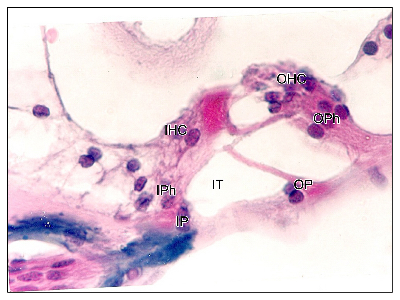
Fig. 2 Higher magnification of the cellular components of the control organ of Corti demonstrating: A single row of inner hair cells (IHC) supported by the underlying inner phalangeal cells (IPhC), and several rows of outer hair cells (OHC) supported by the outer phalangeal cells (OPhC). The inner tunnel (IT) is bounded between the inner (IP) and outer pillar (OP) cells (H&E stain. Mic. Mag. ×1000).
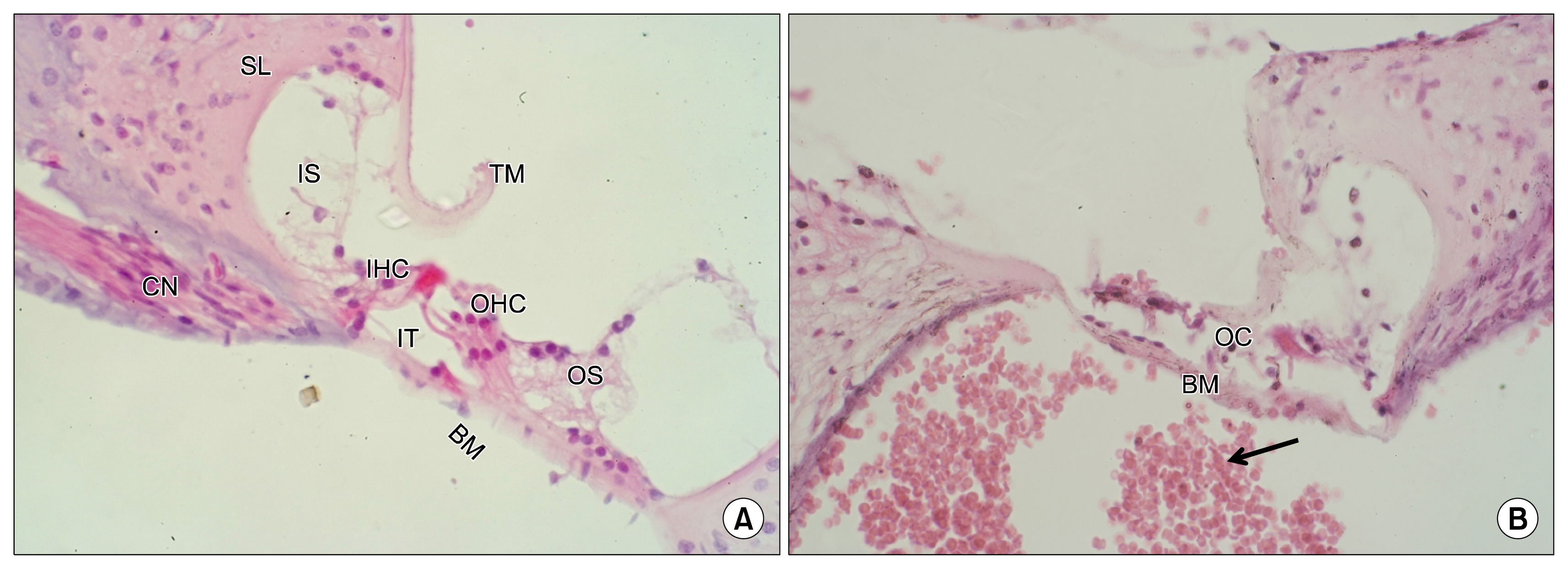
Fig. 3 (A) Light photomicrographs of control rat Organ of Corti showing: The main structural components including the spiral limbus (SL), tectorial membrane (TM), inner supporting cells (IS), inner hair cells (IHC), inner tunnel (IT), outer hair cells (OHC), outer supporting cells (OS), basilar membrane (BM) and the related cochlear nerve (CN). (B) An OAE negative rat (deaf), 6 months after administration of amikacin revealing destruction of the organ of Corti (OC), disappearance of the inner and outer supporting cells and infiltration of the cochlear cavity by red blood corpuscles (arrow). Note the intact basilar membrane (BM) (H&E stain. Mic. Mag. ×400).

Fig. 4 (A) The crista ampullaris shows an intact superficial layer of hair cells (HC) supported by an underlying layer of supporting cells (SC) and connective tissue stroma (ct). (B) The macula utriculi show an intact superficial layer of hair cells (HC) and the overlying otoconia (O). It is supported by a deeper layer of supporting cells (SC) and connective tissue stroma (ct). (C, D) Light photomicrograph of OAE negative rat, 6 months after injection with amikacin: The crista ampullaris reveals vacuolation and widening of interstitial spaces between hair cells and supporting cells (thick arrow). Note that many hair cells (HC) are still intact. The macula utriculi show intact layers of hair and supporting cells (arrow) (H&E stain. Mic. Mag. ×400).
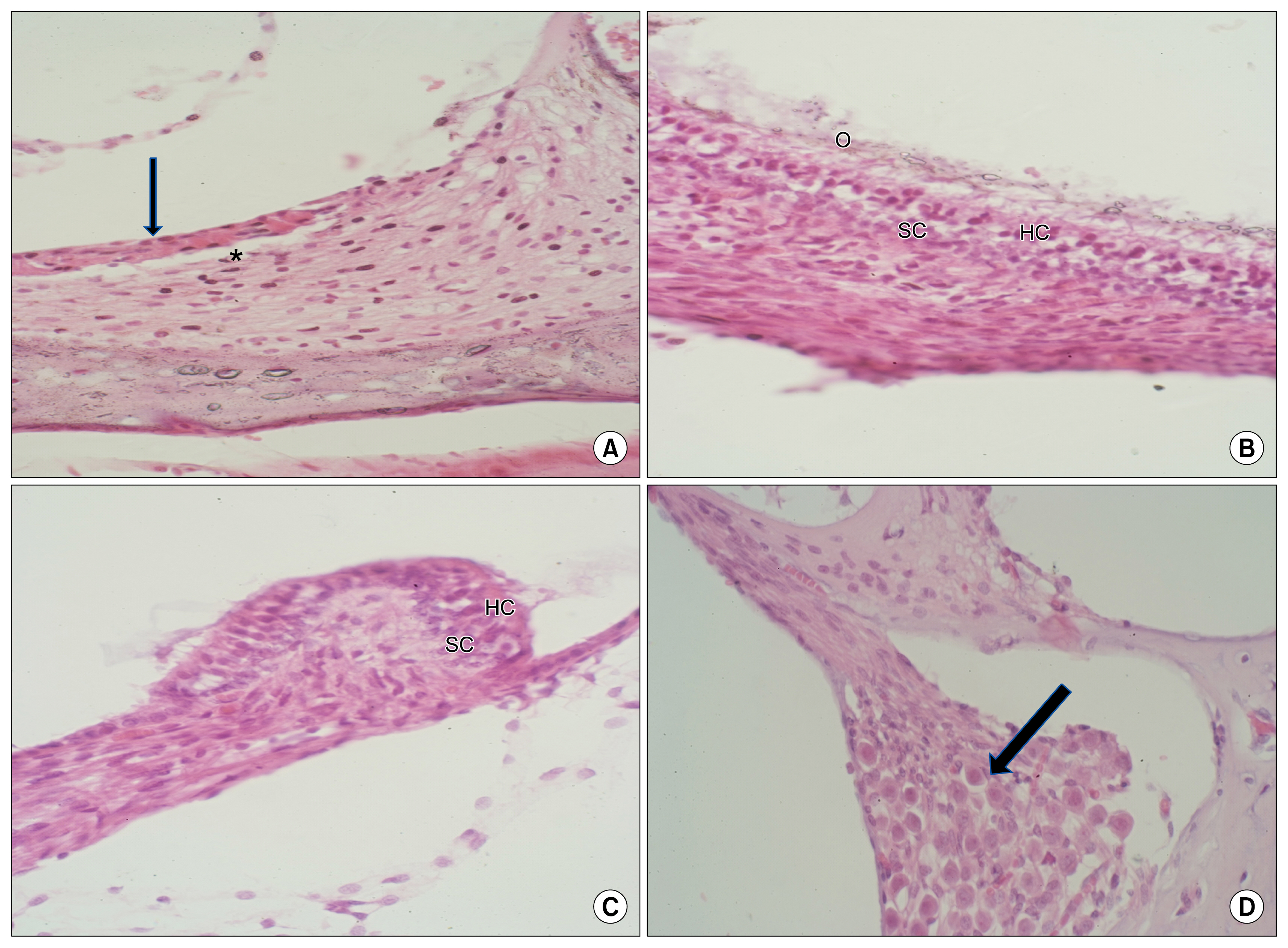
Fig. 5 (A) The stria vascularis of rat recovering after GCSF therapeutic regimen reveals a complete sheet of intact superficial epithelium (arrow) with residual interstitial vacuolation in the supporting connective tissue stroma (*). (B) The macula utriculi of a rat recovering after GCSF therapeutic regimen show intact structural components: (HC) hair cells, (SC) supporting cells, (O) otoconia. (C) The crista ampullaris of rat recovering after GCSF therapeutic regimen shows intact layers of hair cells (HC) and supporting cells (SC). (D) The spiral ganglion of rat recovering after GCSF therapeutic regimen (arrow) is composed of intact bipolar cells with apparently average cell density (H&E stain. Mic Mag. ×100).

Fig. 6 (A) Light photomicrographs of organ of Corti of OAE positive rats recovering within variable durations from receiving GCSF therapeutic regimen: After 4 weeks, the organ of Corti shows few outer hair cells (arrow). No other cellular components of the organ of Corti are identifiable. (B) After 8 weeks, the scala tympani reveals group of cells (arrow) that are apparently organized into a primitive Organ of Corti. SL=spiral limbus, BM=basilar membrane. (C) After 12 weeks, the organ of Corti is formed of well structured rows of inner supporting cells (IS), inner hair cells (IH), outer hair cells (OH) and the intervening tunnels (T). SL=spiral limbus, TM=tectorial membrane. (D) Light photomicrographs of the inner ear from OAE positive rats recovering after 24 weeks from receiving GCSF therapeutic regimen: showing a normal, fully structured organ of Corti composed of intact rows of outer hair cells (OH), outer pillar cells (OP) and an intact row of inner hair cells (IH) supported by inner pillar cells (IP). The outer (OPh) and inner (IPh) phalangeal cells border the inner tunnel (IT). (IS) inner supporting cells (H&E stain. Mic Mag. ×400).

Fig. 7 (A) Scanning electron photomicrograph of control rat organ of Corti demonstrating: Normal hair cells (HC) with intact stereocilia (*) projecting from their apices. (B) Surface view of the reticular lamina (RL) showing three parallel rows of intact stereocilia of the outer hair cells. (Ph C) bodies of supporting outer phalangeal cells. (C) Scanning electron photomicrographs of the cochlear cavity in an OAE negative (deaf) rat, 6 months after injecting amikacin: The cochlear cavity is infiltrated by many red blood cells (arrow). No details of hair cells neither of its stereocilia can be identified. (D) Surface view of the reticular lamina (RL) of the organ of Corti showing parallel rows of empty indentations (arrow) marking the lost outer hair cells (Mic. Mag. ×5000).
Reference
-
References
1. Gulya AJ, Anson BJ. Anatomy of the ear and temporal bone. Shambaugh GE, Glasscock ME, editors. Surgery of the ear. 5th ed. Hamilton: B.C. Decker Inc;2003. p. 35–57.2. Gulya AJ, Schuknecht HF. Anatomy of the temporal bone with surgical implications. 2nd ed. Pearl River, NY: Parthenon Pub Inc;1995.3. Anson BJ, Donaldson JA. Surgical anatomy of the temporal bone. 3rd ed. Philadelphia: WB Saunders;1981.4. Michaels L, Hellquist HB. Ear, nose and throat histopathology. 2nd ed. London: Springer;2001. DOI: 10.1007/978-1-4471-0235-9.5. Lim DJ. Functional structure of the organ of Corti: a review. Hear Res. 1986; 22:117–146. DOI: 10.1016/0378-5955(86)90089-4. PMID: 3525482.
Article6. Weissman IL. Stem cells: units of development, units of regeneration, and units in evolution. Cell. 2000; 100:157–168. DOI: 10.1016/S0092-8674(00)81692-X. PMID: 10647940.7. Sell S. Stem cells. Stem Cell Handbook. Sell S, editor. 2004. p. 1–18.8. Blau HM, Brazelton TR, Weimann JM. The evolving concept of a stem cell: entity or function? Cell. 2001; 105:829–841. DOI: 10.1016/S0092-8674(01)00409-3. PMID: 11439179.9. Díaz-Flores L Jr, Madrid JF, Gutiérrez R, Varela H, Valladares F, Alvarez-Argüelles H, Díaz-Flores L. Adult stem and transit-amplifying cell location. Histol Histopathol. 2006; 21:995–1027. PMID: 16763950.10. Bryder D, Rossi DJ, Weissman IL. Hematopoietic stem cells: the paradigmatic tissue-specific stem cell. Am J Pathol. 2006; 169:338–346. DOI: 10.2353/ajpath.2006.060312. PMID: 16877336. PMCID: 1698791.11. Handgretinger R, Gordon PR, Leimig T, Chen X, Buhring HJ, Niethammer D, Kuci S. Biology and plasticity of CD133+ hematopoietic stem cells. Ann N Y Acad Sci. 2003; 996:141–151. DOI: 10.1111/j.1749-6632.2003.tb03242.x. PMID: 12799292.12. Wagers AJ, Christensen JL, Weissman IL. Cell fate determination from stem cells. Gene Ther. 2002; 9:606–612. DOI: 10.1038/sj.gt.3301717. PMID: 12032706.
Article13. Cohen Y, Nagler A. Umbilical cord blood transplantation--how, when and for whom? Blood Rev. 2004; 18:167–179. DOI: 10.1016/S0268-960X(03)00064-X. PMID: 15183901.14. Nagler A, Bellehsen L, Schaffer F. Stem Cells: Clinical Applications. 7th ed. Adkinson: Middleton’s Allergy;2008. p. 259–268.15. Parker MA, Corliss DA, Gray B, Anderson JK, Bobbin RP, Snyder EY, Cotanche DA. Neural stem cells injected into the sound-damaged cochlea migrate throughout the cochlea and express markers of hair cells, supporting cells, and spiral ganglion cells. Hear Res. 2007; 232:29–43. DOI: 10.1016/j.heares.2007.06.007. PMID: 17659854. PMCID: 2032013.
Article16. Demetri GD, Griffin JD. Granulocyte colony-stimulating factor and its receptor. Blood. 1991; 78:2791–2808. PMID: 1720034.
Article17. Carletons HM, Drury RAB, Willington EA. Carleton’s histological technique. 5th ed. Oxford;Oxford University Press;1980. p. 43.18. Glauert AM. Fixation, dehydration and embedding of biological specimens. 1st ed. Amsterdam, Oxford: North Holland Pub;1986. p. 111–114.19. Ito J, Kojima K, Kawaguchi S. Survival of neural stem cells in the cochlea. Acta Otolaryngol. 2001; 121:140–142. DOI: 10.1080/000164801300043226. PMID: 11349765.
Article20. Tateya I, Nakagawa T, Iguchi F, Kim TS, Endo T, Yamada S, Kageyama R, Naito Y, Ito J. Fate of neural stem cells grafted into injured inner ears of mice. Neuroreport. 2003; 14:1677–1681. DOI: 10.1097/00001756-200309150-00004. PMID: 14512836.
Article21. Li H, Liu H, Heller S. Pluripotent stem cells from the adult mouse inner ear. Nat Med. 2003; 9:1293–1299. DOI: 10.1038/nm925. PMID: 12949502.
Article22. Li H, Roblin G, Liu H, Heller S. Generation of hair cells by stepwise differentiation of embryonic stem cells. Proc Natl Acad Sci U S A. 2003; 100:13495–13500. DOI: 10.1073/pnas.2334503100. PMID: 14593207. PMCID: 263842.
Article23. Parker MA, Cotanche DA. The potential use of stem cells for cochlear repair. Audiol Neurootol. 2004; 9:72–80. DOI: 10.1159/000075998. PMID: 14981355.
Article24. Martinez-Monedero R, Oshima K, Heller S, Edge AS. The potential role of endogenous stem cells in regeneration of the inner ear. Hear Res. 2007; 227:48–52. DOI: 10.1016/j.heares.2006.12.015. PMID: 17321086. PMCID: 2020819.
Article25. Li H, Corrales CE, Edge A, Heller S. Stem cells as therapy for hearing loss. Trends Mol Med. 2004; 10:309–315. DOI: 10.1016/j.molmed.2004.05.008. PMID: 15242678.
Article26. Pellicer M, Giráldez F, Pumarola F, Barquinero J. Stem cells for the treatment of hearing loss. Acta Otorrinolaringol Esp. 2005; 56:227–232. DOI: 10.1016/S0001-6519(05)78606-4. PMID: 15999787.27. Zohlnhöfer D, Ott I, Mehilli J, Schömig K, Michalk F, Ibrahim T, Meisetschläger G, von Wedel J, Bollwein H, Seyfarth M, Dirschinger J, Schmitt C, Schwaiger M, Kastrati A, Schömig A. REVIVAL-2 Investigators. Stem cell mobilization by granulocyte colony-stimulating factor in patients with acute myocardial infarction: a randomized controlled trial. JAMA. 2006. 295:p. 1003–1010. DOI: 10.1001/jama.295.9.1003.
Article28. Kloner RA. Attempts to recruit stem cells for repair of acute myocardial infarction: a dose of reality. JAMA. 2006; 295:1058–1060. DOI: 10.1001/jama.295.9.1058. PMID: 16507807.
Article29. Schäbitz WR, Kollmar R, Schwaninger M, Juettler E, Bardutzky J, Schölzke MN, Sommer C, Schwab S. Neuro-protective effect of granulocyte colony-stimulating factor after focal cerebral ischemia. Stroke. 2003; 34:745–751. DOI: 10.1161/01.STR.0000057814.70180.17.
Article30. Shyu WC, Lin SZ, Lee CC, Liu DD, Li H. Granulocyte colony-stimulating factor for acute ischemic stroke: a randomized controlled trial. CMAJ. 2006; 174:927–933. DOI: 10.1503/cmaj.051322. PMID: 16517764. PMCID: 1405861.
Article31. Oishi A, Otani A, Sasahara M, Kojima H, Nakamura H, Yodoi Y, Yoshimura N. Granulocyte colony-stimulating factor protects retinal photoreceptor cells against light-induced damage. Invest Ophthalmol Vis Sci. 2008; 49:5629–5635. DOI: 10.1167/iovs.08-1711. PMID: 18676635.
Article32. Meuer K, Pitzer C, Teismann P, Krüger C, Göricke B, Laage R, Lingor P, Peters K, Schlachetzki JC, Kobayashi K, Dietz GP, Weber D, Ferger B, Schäbitz WR, Bach A, Schulz JB, Bähr M, Schneider A, Weishaupt JH. Granulocyte-colony stimulating factor is neuroprotective in a model of Parkinson’s disease. J Neurochem. 2006; 97:675–686. DOI: 10.1111/j.1471-4159.2006.03727.x. PMID: 16573658.
Article
- Full Text Links
-
- Actions
-
Cited
- CITED
-
- Close
- Share
-
- Similar articles
-
- Bone marrow-derived stem cells contribute to regeneration of the endometrium
- The Effect of In Vivo Mobilization of Bone Marrow Stem Cells on the Pancreas of Diabetic Albino Rats (A Histological & Immunohistochemical Study)
- Endogenous Stem Cells in the Ear
- Endogenous Stem Cells in Homeostasis and Aging
- Neural Differentiation of Bone Marrow-Derived Mesenchymal Stem Cells: Applicability for Inner Ear Therapy
